Book - Contributions to Embryology Carnegie Institution No.56-4
| Embryology - 27 Apr 2024 |
|---|
| Google Translate - select your language from the list shown below (this will open a new external page) |
|
العربية | català | 中文 | 中國傳統的 | français | Deutsche | עִברִית | हिंदी | bahasa Indonesia | italiano | 日本語 | 한국어 | မြန်မာ | Pilipino | Polskie | português | ਪੰਜਾਬੀ ਦੇ | Română | русский | Español | Swahili | Svensk | ไทย | Türkçe | اردو | ייִדיש | Tiếng Việt These external translations are automated and may not be accurate. (More? About Translations) |
Mall FP. and Meyer AW. Studies on abortuses: a survey of pathologic ova in the Carnegie Embryological Collection. (1921) Contrib. Embryol., Carnegie Inst. Wash. Publ. 275, 12: 1-364.
- In this historic 1921 pathology paper, figures and plates of abnormal embryos are not suitable for young students.
1921 Carnegie Collection - Abnormal: Preface | 1 Collection origin | 2 Care and utilization | 3 Classification | 4 Pathologic analysis | 5 Size | 6 Sex incidence | 7 Localized anomalies | 8 Hydatiform uterine | 9 Hydatiform tubal | Chapter 10 Alleged superfetation | 11 Ovarian Pregnancy | 12 Lysis and resorption | 13 Postmortem intrauterine | 14 Hofbauer cells | 15 Villi | 16 Villous nodules | 17 Syphilitic changes | 18 Aspects | Bibliography | Figures | Contribution No.56 | Contributions Series | Embryology History
| Historic Disclaimer - information about historic embryology pages |
|---|
 |
Chapter 4. Analysis of Abortuses classes as Pathologic
| Online Editor Comment | |||||||||||||||||||||||||||||||||||||||||||||||||||||||||||||||||||||||||||||||||||||||||||||||||||||||||||||||||||||||||||||||||||||||||||||||||||||||||||||||||||||||||||||||||||||||||||||||||||||||||||||||||||||||||||||||||||||||||||||||||||||||||||||||||||||||||||||||||||||||||||||||||||||||||||||||||||||||||||||||||||||||||||||||||||||||||||||||||||||||||||||||||||||||||||||||||||||||||||||||||||||||||||||||||||||||||||||||||||||||||||||||||||||||||||||||||||||||||||||||||||||||||||||||||||||||||||||||||||||||||||||||||||||||||||||||||||||||||||||||||||||||||||||||||||||||||||||||||||||||||||||||||||||||||||||||||||||||||||||||||||||||||||||||||||||||||||||||||||||||||||||||||||||||||||||||||||||||||||||||||||||||||||||||||||||||||||||||||||||||||||||||||||||||||||||||||||||||||||||||||||||||||||||||||||||||||||||||||||||||||||||||||||||||||||||||||||||||||||||||||||||||||||||||||||||||||||||||||||||||||||||||||||||||||||||||||||||||||||||||||||||||||||||||||||||||||||||||||||||||
|---|---|---|---|---|---|---|---|---|---|---|---|---|---|---|---|---|---|---|---|---|---|---|---|---|---|---|---|---|---|---|---|---|---|---|---|---|---|---|---|---|---|---|---|---|---|---|---|---|---|---|---|---|---|---|---|---|---|---|---|---|---|---|---|---|---|---|---|---|---|---|---|---|---|---|---|---|---|---|---|---|---|---|---|---|---|---|---|---|---|---|---|---|---|---|---|---|---|---|---|---|---|---|---|---|---|---|---|---|---|---|---|---|---|---|---|---|---|---|---|---|---|---|---|---|---|---|---|---|---|---|---|---|---|---|---|---|---|---|---|---|---|---|---|---|---|---|---|---|---|---|---|---|---|---|---|---|---|---|---|---|---|---|---|---|---|---|---|---|---|---|---|---|---|---|---|---|---|---|---|---|---|---|---|---|---|---|---|---|---|---|---|---|---|---|---|---|---|---|---|---|---|---|---|---|---|---|---|---|---|---|---|---|---|---|---|---|---|---|---|---|---|---|---|---|---|---|---|---|---|---|---|---|---|---|---|---|---|---|---|---|---|---|---|---|---|---|---|---|---|---|---|---|---|---|---|---|---|---|---|---|---|---|---|---|---|---|---|---|---|---|---|---|---|---|---|---|---|---|---|---|---|---|---|---|---|---|---|---|---|---|---|---|---|---|---|---|---|---|---|---|---|---|---|---|---|---|---|---|---|---|---|---|---|---|---|---|---|---|---|---|---|---|---|---|---|---|---|---|---|---|---|---|---|---|---|---|---|---|---|---|---|---|---|---|---|---|---|---|---|---|---|---|---|---|---|---|---|---|---|---|---|---|---|---|---|---|---|---|---|---|---|---|---|---|---|---|---|---|---|---|---|---|---|---|---|---|---|---|---|---|---|---|---|---|---|---|---|---|---|---|---|---|---|---|---|---|---|---|---|---|---|---|---|---|---|---|---|---|---|---|---|---|---|---|---|---|---|---|---|---|---|---|---|---|---|---|---|---|---|---|---|---|---|---|---|---|---|---|---|---|---|---|---|---|---|---|---|---|---|---|---|---|---|---|---|---|---|---|---|---|---|---|---|---|---|---|---|---|---|---|---|---|---|---|---|---|---|---|---|---|---|---|---|---|---|---|---|---|---|---|---|---|---|---|---|---|---|---|---|---|---|---|---|---|---|---|---|---|---|---|---|---|---|---|---|---|---|---|---|---|---|---|---|---|---|---|---|---|---|---|---|---|---|---|---|---|---|---|---|---|---|---|---|---|---|---|---|---|---|---|---|---|---|---|---|---|---|---|---|---|---|---|---|---|---|---|---|---|---|---|---|---|---|---|---|---|---|---|---|---|---|---|---|---|---|---|---|---|---|---|---|---|---|---|---|---|---|---|---|---|---|---|---|---|---|---|---|---|---|---|---|---|---|---|---|---|---|---|---|---|---|---|---|---|---|---|---|---|---|---|---|---|---|---|---|---|---|---|---|---|---|---|---|---|---|---|---|---|---|---|---|---|---|---|---|---|---|---|---|---|---|---|---|---|---|---|---|---|---|---|---|---|---|---|---|---|---|---|---|---|---|---|---|---|---|---|---|---|---|---|---|---|---|---|---|---|---|---|---|---|---|---|---|---|---|---|---|---|---|---|---|---|---|---|---|---|---|---|---|---|---|---|---|---|---|---|---|---|---|---|---|---|---|---|---|---|---|---|---|---|---|---|---|---|---|---|---|---|---|---|---|---|---|---|---|---|---|---|---|---|---|---|---|---|---|---|---|---|---|---|---|---|---|---|---|---|---|---|---|---|---|---|---|---|---|---|---|---|---|---|---|---|---|---|---|---|---|---|---|---|---|---|---|---|---|---|---|---|---|---|---|---|---|---|---|---|---|---|---|---|---|---|---|---|---|---|---|---|---|---|---|---|---|---|---|---|---|---|---|---|---|---|---|---|---|---|---|---|---|---|---|---|---|---|---|---|---|---|---|---|---|---|---|---|---|---|---|---|---|---|---|---|---|---|---|---|---|---|---|---|---|---|---|---|---|---|---|---|---|---|---|---|---|---|---|---|---|---|---|---|---|---|---|---|---|---|---|---|---|---|---|---|---|---|---|---|---|---|---|---|---|---|---|---|---|---|---|---|---|---|---|---|---|---|---|---|---|---|---|---|---|---|---|---|---|---|---|---|---|---|---|---|---|---|---|---|---|---|---|---|---|---|---|---|---|---|---|---|---|---|---|---|---|---|---|---|---|---|---|---|---|---|---|---|---|---|---|---|---|
|
|
These are links to other normal Carnegie Collection numbered embryos available on this educational site.
| ||||||||||||||||||||||||||||||||||||||||||||||||||||||||||||||||||||||||||||||||||||||||||||||||||||||||||||||||||||||||||||||||||||||||||||||||||||||||||||||||||||||||||||||||||||||||||||||||||||||||||||||||||||||||||||||||||||||||||||||||||||||||||||||||||||||||||||||||||||||||||||||||||||||||||||||||||||||||||||||||||||||||||||||||||||||||||||||||||||||||||||||||||||||||||||||||||||||||||||||||||||||||||||||||||||||||||||||||||||||||||||||||||||||||||||||||||||||||||||||||||||||||||||||||||||||||||||||||||||||||||||||||||||||||||||||||||||||||||||||||||||||||||||||||||||||||||||||||||||||||||||||||||||||||||||||||||||||||||||||||||||||||||||||||||||||||||||||||||||||||||||||||||||||||||||||||||||||||||||||||||||||||||||||||||||||||||||||||||||||||||||||||||||||||||||||||||||||||||||||||||||||||||||||||||||||||||||||||||||||||||||||||||||||||||||||||||||||||||||||||||||||||||||||||||||||||||||||||||||||||||||||||||||||||||||||||||||||||||||||||||||||||||||||||||||||||||||||||||||||
Group 1. Specimens Composed of Villi Only
A. Uterine
Among the series of over 2,000 abortuses in the Carnegie Collection I have so far been able to find only a few specimens which undoubtedly fall into the category of intrauterine absorption. Among these are Nos. 698, 970, 1640, and 1926. Nevertheless, not even in these cases had total absorption occurred, and from evidence to be considered I have really come to doubt whether absolutely complete absorption occurs in man in any but the earliest stages of development or under the rarest conditions.
As stated by Mall, specimens of the first class of the pathologic division, i.e., those composed of villi only, are obtained very largely from tubal pregnancies. Nevertheless, Mall emphasized that "a very large number belonging to this group would be found in uterine pregnancies also if our methods of collection and study were as reliable as they are for tubal pregnancy." Of the 353 uterine specimens classed as pathologic among the first 1,000 accessions, only 17, or 4.8 per cent, are composed of villi only, as compared to 35, or 32.4 per cent, of 108 tubal specimens. That is, specimens composed of villi only are nearly seven times more common among tubal than among uterine gestations. However, this is due almost wholly to the fact that the uterine specimens are fairly representative of the whole period of gestation, while the tubal specimens are derived almost wholly from the first two months of pregnancy. Specimens composed of villi only would form about the same proportion among uterine abortuses contained in the first two groups of the pathologic, as among the tubal, but they form only 12.9 per cent of all uterine specimens contained in the first four groups of this division. Hence the inference that the great majority of tubal conceptuses come to an early death seems indicated by these facts alone.
That villi only are so frequently found in tubal pregnancies is probably due also to the occurrence of tubal abortion, in consequence of which the conceptus may be ejected from the tube but some of the villi left attached, and more especially to the effect of repeated hemorrhage. The conceptuses often are strangulated as a result of hemorrhage which detaches them completely and then leads to their disintegration. Although the villi may be, but probably are not, inherently more resistant than the rest of the chorionic vesicle, some of them usually survive, because in the absence of a decidua which becomes detached, they remain attached to the implantation site, thus retaining their connection with the source of nourishment. In uterine specimens this is impossible, for the entire decidua is cast off. For these, and probably also for other reasons, Mall found that villi almost always can be detected by microscopic examination of serial sections of the implantation cavity in cases which clinically are tubal pregnancies, even if they can not be detected by the most careful inspection of the gross specimen or by microscopic examination of frozen sections of portions of the implantation site.
Whether or not the surviving elements of a conceptus are villi only depends very largely upon the age of the conceptus and upon the sequence of events responsible for its death. Indeed, if the entire conceptus is erupted from its implantation cavity by a sudden severe hemorrhage, it -is very unlikely that the villi will be the surviving elements, unless the conceptus is extremely young. The same thing would be true of a conceptus which a severe general inflammatory process had suddenly detached. For in both of the assumed cases the villi would undoubtedly succumb to the destructive processes earlier than the chorionic membrane, the syncytium, or the trophoblast. If, on the other hand, the infectious process gains entrance into the cavity of the chorionic vesicle itself, the latter and the embryo rapidly disintegrate and are destroyed, while some of the villi may long remain in a state of relatively good preservation.

From these considerations it is evident that it would be possible to form three other groups of specimens in addition to those composed of villi only those composed of remnants of both trophoblast and syncytium, or of one or the other alone. That such specimens actually occur will become evident in the course of this discussion. At present they are included in group 1. However, it is not for this reason alone that the designation "villi only" does not fully describe the first group in the pathologic division. Three specimens in this group, for example, are hydatiform moles, one of which, No. 323, forms a large, compact mass, a portion of which is shown in section in figure 6. Another is composed largely of bloodclot surrounded by decidua, and still another consists largely of a decidual cast with mere traces of syncytium, trophoblast, and perhaps of portions of one or two villi. Furthermore, since it is practically impossible to examine all specimens in their entirety in a complete microscopic series, there is some possibility that the portions examined may not form an adequate basis for the correct classification of the specimen. Hence, for this reason also some specimens are bound to get into incorrect categories. Nor is it without significance that no provision is made among the normal specimens for a group of villi only. Under the present classification all of these are placed in the pathologic division. This would seem to imply that normal villi are never aborted alone in uterine pregnancy or found alone in tubal pregnancies or in tubal abortions. Yet material from curettage, or from abortions the result of interference by the patient herself, no doubt may contain none but normal villi. I have seen the question raised nowhere, but it seems doubtful whether, except perhaps in the earliest stages, villi can ever develop wholly normally in a tube. Hence the above objection to the present classification might be waived for tubal but not for uterine specimens.
Among the possible causes of the destruction of conceptuses, inflammatory conditions, as indicated by infiltration, as a rule seem largely predominant. They existed in the great majority of the uterine specimens, protocols of which are attached, and sooner or later seem to lead to fetal death. It is interesting that fetal death in these cases is not the result of invasion of the conceptus, or even of its villi, by the inflammatory process itself. It is possible that the production of toxins may be a factor, but the morphologic evidence seems to point to interference with the nutritive supply through decidual and chorionic changes. It is not difficult to see that the accumulation of pus in the region of attachment of the villi, or even the accumulation of large masses of leucocytes, must seriously interfere with the free intervillous circulation. Obstruction to the blood-current, if sufficiently severe, would also lead to the death of the cyema, and finally to that of the chorionic vesicle itself in consequence of interference with the indispensable gaseous interchanges.
No matter how severe the infection of the uterus was found to be, or how large the accumulations of pus at the region of implantation, well-preserved villi never were found infiltrated with cells of maternal origin. When infiltration was present within the stroma the latter, and especially the epithelium of the villi, could be shown to be degenerate. If, on the other hand, the infectious process was introduced directly into the chorionic vesicle, the latter soon disintegrated and the infection extended into the stroma of the villi also. An insuperable difficulty encountered in connection with the question of infection in many cases is the inability to determine positively whether the infection existed within the uterus before implantation occurred, or whether it was incidental to mechanical interference. An examination of the material seems to show that the final effect upon the villi, and of course also upon the membranes of the embryo, rarely may be the same in both cases. This probably is due to the fact that a young conceptus may be loosened partially only, at the time of interference or of infection, thus establishing conditions which lead to its death. As a rule, however, in these cases maceration changes are likely to be much more rapid than under conditions of a chronic endometritis pre-existent to the conception. Nevertheless, mild general or a severe local preimplantation endometritis no, doubt could produce results wholly comparable to those resulting from a mild general uterine infection incident to mechanical interference, -especially in the case of young conceptuses. It is conceivable that in the case of a low-grade endometritis, the fertilized ovum may undergo a perfectly normal development for a restricted period, and then suffer from more or less sudden interference with its development through exacerbation or extension of the infectious process, just as might be the case under other conditions. Ordinarily, however, it would seem that the changes within the conceptus should be more gradual, and also much more general under conditions of a chronic endometritis than under those of accidental or incidental infection. Nevertheless, the changes in the villi sometimes appear wholly comparable in both cases. The stroma in many of them finally undergoes what Mall has called mucoid degeneration, with complete disappearance of the mesenchyme and final disintegration of the epithelium. The blood-vessels generally become effaced at a very early day before the stroma has undergone any important changes. Rarely, as the endothelium degenerates, it leaves a faint, more or less incomplete outline marked by the degenerating nuclei. Consequently it happens that the surviving remnants of a vessel may be represented merely by a small number of poorly preserved nuclei.
The epithelium of the villi usually is preserved longer, but finally the syncytium may fuse with the Langhans layer, forming a dense coagulum. Or the pycnotic nuclei of both layers may retain their relative positions, ultimately becoming resolved into fine granules which Mall, in several of his publications and also repeatedly in the protocols, has spoken of as nuclear dust. This fine granulation seems to herald beginning calcification. Later the granules may fuse with each other and with the necrotic cytoplasm, forming a hyalin band at the periphery of the villus, which stains heavily with eosin and also with iron hematoxylin. It alone may make the outline of the pre-existent villus evident. Fibrosis of the villi is seen but rarely in these early specimens, and when it occurs, hyalin degeneration is not infrequently present in the form of trabeculse or a framework in the midportion of the villus. Remnants of the syncytial masses or of trophoblast usually survive everything else.


Since no decidua and very little trophoblast were found upon the villi of several young conceptuses, it seems doubtful in some cases whether good implantation occurred at all, as suggested by No. 1843, shown in figure 7. In these specimens the villi nevertheless seem to undergo considerable development, but the embryo, after it becomes dependent upon the circulation, finally dies, probably by asphyxiation, and then the processes of maceration, digestion, and absorption begin. In other cases it also seems likely that the young ovum becomes embedded quite normally, but that strangulation results from severe hemorrhage which loosens the attaching villi, thus interrupting the intervillous circulation. Since the resulting stagnation of the blood must make impossible the indispensable chemical interchanges upon which the life of the cyema depends, the latter probably dies first. It is decidedly interesting that considerable hemorrhage, sufficient to result in the death of both cyema and chorionic vesicle, can occur while the whole conceptus is still surrounded by the early decidua capsularis without rupture of the latter. Such a specimen was discovered in No. 698, which is in the final stages of absorption. In this unique specimen (received from Dr. N. E. B. Igelhart), which has a menstrual age of 56 days, there remains only the merest trace of a chorionic vesicle in the form of a striated coagulum, a few questionable "shadows" of villi, several small fragments of syncytium, and a few detached accumulations of trophoblast. The place of the conceptus is occupied by blood-clot formed into an elongated body 50 by 20 by 13 mm. This body is completely surrounded by an intact decidua capsularis. The latter is easily recognized, both in the gross and in the microscopic specimen, and the decidua vera, which also is intact, can be seen clearly with the unaided eye in every detail of its relations, as shown in figure 8. This indeed is a unique specimen and especially significant in connection with No. 970, to be discussed in the next group, and with certain better-preserved specimens recorded in the literature. The failure of complete absorption of the last few small remnants of this conceptus is probably due to the fact that the small remnants of degenerated trophoblast and syncytium which remained were no longer able to inhibit menstruation. Hence the decidua of pregnancy, together with these few small remnants of the conceptus, was expelled in toto at the time of onset of the next period, and it may be extremely significant that this occurred exactly two menstrual months after the beginning of the last period. Since 3 other specimens of a series of 16, composed of villi only, were aborted at the time of recurrence of the regular period, the idea that abortion occurs oftener at that than at any other time would seem to receive some confirmation. Moreover, it would appear quite natural that a decidua which has subserved its functions would be more likely to be shed at this time and that an unabsorbed conceptus which had been converted essentially into a foreign body should then be expelled.
It is impossible to decide how far the development of this conceptus had progressed before its death, but the extent of absorption shows beyond any doubt that the latter would have been absolutely complete before the advent of the next or third period had the second period also been inhibited. Since in the assumed case the decidual cast then would have been expelled after the ovum had been completely absorbed, this decidual cast might have directed attention to the possibility of the existence of a tubal rather than a uterine pregnancy. In view of the facts here revealed, such a sequence of events might well give the impression of the existence of an early tubal pregnancy which had undergone spontaneous retrogression without ever having given rise to the characteristic symptoms. In this connection I am reminded of the fact that gynecologists have been of the opinion that some tubal pregnancies undergo spontaneous cure. In many of these cases the healing probably follows tubal abortion, but specimens in this collection also indicate the possibility of another sequence of events. In some instances, for example, the small intratubal blood-clot in which a small conceptus becomes enclosed at the time of hemorrhage seems to undergo reduction within the tube. Under these circumstances the conceptus, which was separated from its implantation site, may then undergo retrogression, maceration, disintegration, and finally may be completely absorbed, and the tube heal. Nor does it seem impossible that the chorionic vesicle may remain and undergo a similar fate within the tube in cases in which the cyema alone is aborted.
Of the 16 specimens finally classed in group 1, all of which were examined both macroscopically and microscopically, 7, or 43.7 per cent, show hydatiform degeneration. In each of these specimens the abortion very probably was not induced. In 5 of these specimens in which some or all of the decidua accompanied the specimen, it showed changes indicative of endometritis. In 4 of these the infiltration was marked and in one it was slight. One specimen counted as showing hydatiform changes was extremely degenerate, however, and unaccompanied by decidua, and may therefore perhaps be rejected, thus leaving 6 specimens, or 37.5 per cent, definitely showing hydatiform degeneration.
The decidua was included in 13 of the 16 specimens in this group, but the material was very necrotic in one case and too little of it accompanied another. The infiltration was slight in one of the remaining 11, and very marked in the other 10 cases. Hence, although infiltration of the decidua was present in only 10, or 62.5 per cent, of the 16 specimens of this group, the decidua showed definite signs of inflammation in every one of the 10 in which it was present and sufficiently well preserved. Some decidua, in fact, contained considerable masses of purulent material. The infiltration was often very marked locally, small accumulations of leucocytes being scattered about more or less at random ; but this form of infiltration frequently was accompanied also by an infiltration more or less general and uniform in character, and by other changes. Whether or not these infiltrations were confined to the decidua I am not able to say, for uterine musculature was not included. In most cases, however, the process had the appearance of a low-grade chronic inflammation. In only a few was a severe infection very evidently present.
Of the abortuses composed of villi only included in the first 1,000, all but 3 had a maximum length of less than 50 mm. One measured 68 mm., another 100 mm., and a third 120 mm. However, since considerable allowance must be made for distortion, for variations in the length of villi, and for maceration, as well as for increase in size due to the surrounding decidua and blood-clot, it is evident that the measurements of the abortuses are often too large to represent, even approximately, the true age, not only of the accompanying cyema, but of the chorionic vesicle or placenta as well. It is evident also that, in case of abortuses composed of villi only, the measurements, even if not affected by the presence of blood-clot and decidua, could in no sense be more than roughly indicative of the age of the specimen unless the chorionic membrane were preserved sufficiently to retain its form and size. Furthermore, since the specimens in this group include material from curettage also, a discussion of their size as related to their anatomical or menstrual age can have no value.
The largest specimen in this group is a hydatiform degeneration, containing no trace of the cyema. Such a specimen can not with propriety be designated as villi only; nevertheless, the exigencies of the situation make its inclusion here of some practical value. In other instances one could speak of the size of the mole, perhaps, but unless composed of solid masses of villi, moles really belong in the second group. In still other instances, such as No. 698, the main mass of the abortus was composed of decidua, so that although only what was originally taken for the chorionic vesicle was measured, this measurement nevertheless is wholly erroneous, for it is impossible to exclude the blood-clot, which, in this case, very greatly affects the size of the specimen.
Unfortunately, menstrual age is not a reliable criterion of the true age of the specimens in this group, the state of preservation of which alone indicated that many of them were retained a considerable length of time after their death. The latter is indicated, not only by the disproportion between the size of the abortus and the menstrual age, but also by the degenerative changes present in the specimens themselves. The longest menstrual age (218 days) is found in specimen No. 70, in which the size of the conceptus or cyema indicates an anatomical age of only 50 days.
Upon attempting to correlate the clinical data with the objective examination, it was found that in one case in which abortifacients were held responsible for the termination of pregnancy, an intense infection was present. This was true also in four other cases, in which it was specifically stated that infection was absent, a conjunction of things to be referred to again.
B. Tubal
In contrasting the tubal specimens composed of villi only with similar specimens from the uterus, the lesser number of villi contained in the tubal cases is very striking. It may be recalled that the number of villi found in uterine specimens varies from none to large hydatiform masses weighing several pounds. But even aside from the latter, which properly do not belong in this group, villi found in uterine specimens are far more numerous, as a rule, than in the tubal cases. One of the main reasons for this difference lies in the fact that the tubal specimens as a whole undoubtedly are much younger and hence less resistant. Their youth may be explained very largely by the anatomical conditions under which development proceeds in the tube and in part probably also by the efforts at abortion which probably are inaugurated very early through the occurrence of tubal peristalsis. It is not unlikely that this peristalsis may expel most of the villi, with the surrounding blood-clot, into the peritoneal cavity, leaving behind only a few stragglers. In the absence of anything truly comparable to decidual development within the tube, the villi at best must be embedded less securely and also may degenerate faster when once detached. Moreover, in the absence of such a nidus as the'hypertrophied endometrium, the whole development of the conceptus necessarily must be retarded.
Most of the villi in tubal specimens lie isolated in the contained blood-clot; hence matting of the villi was practically absent and calcification and coagulation necroses were not seen, facts which suggest the occurrence of early interference with development. Sometimes a few villi which lay near each other were decidedly necrotic, but they were not fused into a large, solid mass by degeneration or by inflammatory products in any of the cases in this group. This fact and the appearance of the clots would seem to suggest that there often is a constant trickling of blood from the distal extremity of the tube, so that old clots form slowly, new blood being added more or less constantly, thus prolonging the life of some villi, or at least of the chorionic epithelium. In several cases the villi were quite well preserved, though fibrous, but by far the greater number were decidedly degenerate. Except in instances in which the whole villus was necrotic, the epithelium was preserved better than the stroma, a fact which probably may be explained by the presence of fresh blood. In all except the necrotic specimens, the epithelium not only was well preserved, but also was not infrequently very active, as noted in several instances by Mall (1915). Considerable masses of trophoblast were present in a number of instances, and smaller syncytial masses (or more frequently syncytial buds) also were seen. Usually some portions of both were extremely well preserved, and in one instance large masses of degenerate trophoblast completely filled the interplical spaces and the mucosal diverticulae along a considerable sector of the tube. In two other instances the degenerate trophoblast which bordered, and to some extent invaded, the musculature reminded Mall of Hofbauer cells.
The stroma of the villi was non-vascular in practically all instances, and only a few small remnants of the degenerating vessels remained in some. To some extent absence of vessels may be due to the youth of the specimens, but in the main it is probably due to other factors. Even the villi that were capped by considerable trophoblast and syncytium and which still were implanted in the musculature were often non-vascular, and their stroma, as noted by Mall, was usually mucoid. In contrasting the changes in the stroma of the villi found in tubal with that of the same group of uterine specimens, the more degenerate character of the stroma in the former is very evident. Moreover, not a single villus with a fine, clear, glassy, translucent stroma was seen in this group, nor w.ere any present which had a well-preserved young cellular stroma, or others in which the formation of more than a few Hofbauer cells was in progress. The whole appearance was rather that of a rapid destruction, although most of the clots in which the villi were embedded were relatively fresh. Considerable portions of the clots often contained a fibrin network, but all were unorganized, and no instances of an ingrowth of connective tissue from the tubal wall were seen, in spite of the fact that a few of the clots were relatively old and necrotic.
In two instances in which no portion of the tube had been cut, the presence of infection in it was made probable by the appearance of the contained clots. In most of the latter the leucocytes were congregated more or less, or were formed into small clumps even. Phagocytosis of the fetal membranes by the leucocytes or by other cells was not noticed, although leucocytes had accumulated at the periphery or even had entered into the interior of the stroma of degenerate villi. No embryonic remnants whatever were found in the sections of a portion of one tube, and degenerate trophoblast and syncytium only were present in two. This fact, however, does not imply that phagocytosis was responsible for the absence of villi.
Out of the 33 specimens originally in this group, 2 were found to contain remnants of the chorionic membrane and of the amnion, and hence were transferred to groups 2 and 3 respectively, and 10 were added. In 3, or 9 per cent, of the 42 cases remaining, no infiltration, either of the clot or the wall of the tube, was noticed. In 12, or 28.6 per cent, the infiltration was marked, and in 17, or 40.4 per cent, it was slight.

Changes simulating those of lues were noticed in no tubal conceptuses in this group, but several excellent examples of hydatiform degeneration were found in Nos. 415, 602, 686, 772, and 889. According to Seitz (19040, the occurrence of hydatiform moles was observed in connection with tubal pregnancies by Freund, Matwejew and Sykow, Otto, and Wenzel. Others no doubt have observed it since then, but as only a few villi are contained in a single cross-section of the tube, and but few cross-sections of each specimen were examined in our series, one can not be certain of one's diagnosis in every instance. If more villi were present this difficulty would be obviated, although it must be remembered that a large series of specimens necessarily supplement each other. Furthermore, the changes in many villi are so typical, both as to outward form and structure, as to be undoubted. Since many of the villi were decidedly degenerate, one could hardly expect to find much proliferation of the endothelium, but remarkable specimens, such as that in figure 9, were occasionally found. In some cases the presence of hydatiform degeneration became probable only through comparison of the villi in question with those found in many undoubted cases of hydatiform degeneration examined previously.
Two unusually fine specimens of hydatiform degeneration were transferred to this from group 2. No. 367 was a fine, clear, partly cystic specimen in which syncytial buds were invading the stroma of some of the villi. Although only vestiges of the vessels remain, the trophoblast is well preserved and syncytial buds are found on some of the trophoblastic nodules. The outlines of many villi are very sinuous and the epithelium is well preserved. In some respects this is one of the most unusual specimens I have found in the entire series, both of ectopic and uterine specimens. In the other specimen, No. 720, some of the implanted villi which remain show hydatiform degeneration, and many of them have fine, long syncytial buds. Although no vessels were seen in this specimen, the trophoblast nevertheless is abundant.
In the 5 cases above mentioned the presence of hydatiform degeneration was undoubted, and in 6 others its presence was highly probable, making 11 cases, or 26.2 per cent of the entire group. This is a somewhat lower incidence than in the uterine cases in this group, which was 37.5 per cent. Either the tube-wall or the contained clot gave evidence of the presence of infection in 8 of these 11 cases. If we exclude one case in which the tube was not included in the section, we get a percentage of infiltration of 80. Moreover, since only a few sections of each specimen wepe examined, and since the evidences of an old infection are not always easily detected in a markedly dilated and altered tube, it is not improbable that infiltration was present in more of these cases of hydatiform degeneration, as was the case in the uterine specimens. The existing infiltration was intense in one and slight in the other half, and since only one case not included among those showing hydatiform degeneration had an apparently normal tube, infiltration hence was almost constantly present also in the entire series of cases included in the group of the tubal specimens.
Although the alleged menstrual age ranged from 6 to 113 days, only a rough correspondence between it and the structure of the specimens was found to exist. The specimen with the longest duration contained only a few questionable degenerate syncytial remnants, and in No. 9000, which had a menstrual age of 69 days, no embryonic remnants whatever were found. No. 967c, which had a menstrual age of 70 to 100 days, contained only a few degenerate villi, although the same thing was true also in other instances with a much shorter duration.
Group 2. Chorion without Amnion or Cyema
- Cyema - the embryonic cells of the conceptus.
A. Uterine
The absence of the amnion in these specimens seems to be the result of destruction by lysis. However, absence of it in young specimens also might be due to failure of formation. The latter was the opinion of Giacomini (1888), who nevertheless believed that deformities of the amnion are rare. The only specimens in which the absence of the amnion could be ascribed to failure of development is No. 1843, donated to Stanford University by Dr. Falk, of Modesto, California. This is a very young specimen, however, and it is not impossible that a small cavity found in it represents the early amniotic cavity, as suggested by Meyer (191 9e). Moreover, it may be doubted whether the human embryonic disk could develop far if formation of the amnion were inhibited. It is true that Panum (1860) and Dareste (1883) both reported cases of absence of the amnion in the chick and that Dareste emphasized that all the cephalic or the caudal portions may be absent. But since it is possible that the process of formation of the amnion is a totally different one in man, the direct application of these observations upon the chick to man is open to question. Panum and Dareste both believed that the anomalies of the amnion, observed by them in the chick, were secondary and the opinion of Giacomini was based largely upon the failure to secure absorption of the amnion experimentally in rabbits.
Specimens of human abortuses in which the amnion has undergone partial destruction are very common. Moreover, all degrees of destruction are represented in these cases, and since the cyema, too, is usually lacking, the assumption that absence of the amnion is due to failure of formation naturally would necessitate a similar conclusion regarding the embryo. This would leave one in the position of assuming that a chorionic vesicle which never had contained an embryo or amnion nevertheless might develop independently and attain some size.
Since the absence of the cyema in some of these vesicles is undoubtedly due to the mechanical injury incident to interference with a purely normal gestation, it follows that some of them were unquestionably normally developed chorionic vesicles and hence do not belong in the pathologic division. The difficulty lies in identifying them. Nevertheless, the structural characteristics of some are suggestive, even if not wholly unequivocal. In one instance, for example, in which a small nodule was seen upon opening an apparently intact ovum, this nodule was found to be composed of fragments of villi which could have been introduced into the chorionic vesicle only at the time of interference, or accidentally when it was opened in the laboratory. In another instance the chorionic vesicle contained foreign material when opened. Besides, the splendid preservation of the tissues of some specimens also shows that they were obtained in a practically normal, fresh, and unmacerated condition, which rarely is the case in any but instances of induced abortions, whether they be therapeutic, accidental, or criminal.
As already indicated, there is no hard and fast line of demarcation between the first three and later groups of the pathologic division. Indeed, it is not always easy to determine, even histologically, whether or not cyemic remnants are present, for it is sometimes impossible to decide whether a small hyalin or webbed mass contained within the chorionic vesicle is or is not a remnant of the cyema itself. Then, too, some of the specimens included in this group contain clumps or small accumulations of erythroblasts, which probably had their source in the bloodvessels of the body of the embryo, although they may also have come from the vessels of the cord or of the chorionic membrane itself. In most instances these cells really are cyemic remnants, yet their presence has not excluded the specimens from this group of empty vesicles. Moreover, in another instance (No. 663), considerable remnants of the yolk-sac were found, although nothing could be positively identified as a remnant of either embryo or amnion. In other cases either of the last two structures or both may be represented by a very degenerate fragment, which is merely a so-called shadow or (better) gossamer form. Indeed, the entire chorionic vesicle is sometimes reduced to a mere gossamer form, as illustrated by No. 606 shown in figure 11.
From these things it is evident that group 2 nevertheless includes chorionic vesicles which contain free erythroblasts within their cavities or remnants of the yolk-sac and even of the amnion itself. However, since all specimens in the first three groups differ from each other only in the degree of destruction of the embryo and fetal membranes, this overlapping is a matter of no serious consequence for any except statistical deductions. Moreover, since the whole of a specimen is examined only seldom in a complete series, it foUows that some of them will be classified incorrectly for this reason alone. No. 771a, for example, contains an undoubted remnant of the amnion, and hence belongs in the next group. No. 644 is composed of villi only, and therefore falls into group 1. Since No. 663 was found to contain numerous undoubted remnants of the yolk-sac, it is not at all unlikely that some embryonic masses which could not be identified certainly as such were nevertheless contained in this specimen, which would then be classified in group 4. This particular specimen is contained in a very degenerate hyalin abortus measuring 35 by 15 by 10 mm. The conceptus is composed of an extremely folded and almost structureless chorionic vesicle and of included and isolated cyemic remnants. As so often is the case, this abortus was much larger than the contained chorionic vesicle, which measured only about 5 by 3 mm. in section.
Hence, if Nos. 29, 664, and 771a are excluded from this group, only 40 uterine specimens remain, to which must be added three transferred from other groups. It is stated that in one case (No. 970) the specimen was obtained at autopsy, and in another (No. 865) at hysterectomy, and that in a third the abortion was induced.
In three instances (Nos. 71, 278, and 77 la) it was stated that the patients had a chronic endometritis, and in one case (No. 865) the patient was said to suffer from "an old pelvic inflammation, " but showed "no evidence of venereal disease. " In two cases reported as having a chronic endometritis (Nos. 71 and 278) the clinical diagnosis was confirmed by microscopic examination. In the case in which an old pelvic inflammation but no venereal disease was said to have been present, it was found that a severe endometritis with abscess formation was nevertheless indicated histologically. Only five cases (Nos. 661, 663, 753, 876, and 986) were reported as having no infection, but four of these contained evidence of the existence of an intense endometritis, accompanied in two instances (Nos. 876 and 986) by abscess formation. The specimen from the fifth case (No. 663) was so very necrotic that a positive diagnosis could not be made, but it is unlikely that a severe infection was present. Infiltration of the endometrium was present in 30 of the 48 cases, including 2 doubtful ones. This makes a percentage of 62.5, but since the decidua was not included in 20 cases, was too degenerate for study in 3 of the rest, and especially since only a relatively small portion of each abortus was examined microscopically, it is very probable that this percentage of evidence of infection, high as it is, nevertheless is entirely too low. Omitting the doubtful cases, the decidua was found infiltrated in 28 of 29 cases, or in 99.6 per cent. In 82.8 per cent of these the infiltration was marked. In 5 cases the infiltration of the endometrium was comparatively slight, but in 24 it was severe. In one case infection was found to be present within the chorionic vesicle, and although no decidua accompanied this specimen, it is unlikely that the infection was confined entirely to the vesicle. In 4 cases both the interior of the vesicle and the decidua undoubtedly were infected, and in 2 (Nos. 435a and 750) no decision could be reached.
Twenty specimens, or 41.6 per cent, were identified as showing hydatiform degeneration, 3 cases being doubtful and 2 others probable. In 15 of these cases in which the decidua was included it showed inflammatory changes. These changes were marked in 12 and slight in 3 cases.
Among the alleged causes of the termination of pregnancy in the cases not accounted for otherwise, we find that a fall was mentioned in case No. 71, in which evidences of chronic endometritis were present; anxiety is given as the proximate cause in No. 664, and fright in No. 883, both of which cases show the presence of an intense infection, which in one was so severe that the chorionic vesicle was destroyed almost completely. Two of the 3 abortions reported as having been spontaneous (Nos. 750 and 829) show the presence of a severe infection. In 4 instances in which the uterus was considered to be normal, evidence of infection was found in 2. In No. 883 it was found to be severe and in No. 978 it was mild. Although only one of these 39 specimens was said to have been induced, it is more than likely that this is true of more of them, although it seems exceedingly unlikely that an ovum could become implanted, and decidual formation actually progress in a normal way in so infiltrated and abnormal an endometrium as is present in many of these specimens. The anatomical condition of some of the chorionic vesicles among the non-infected group suggests that some of these abortions also were induced.
The histological changes found in this group differ in no essential respect from those present in group 1. Mall also found that the changes in the villi of the chorionic vesicle from an ovarian pregnancy (see No. 550 in the next group) showed essentially the same changes as uterine specimens developing "under faulty implantation or infection." Maceration changes are present in all specimens, but vary extremely in degree. Coagulation necroses are absent, except in small areas, and beginning "infarct" formation was present only in portions showing especially severe infection. In the latter the destruction apparently had been very rapid and hence the degenerative changes differ somewhat from those produced by a low-grade chronic endometritis pre-existent to the implantation.
In most of the specimens, as noted by Mall, two forms of degeneration of the villi exist side by side, a fibrous and a "mucoid" transformation of the stroma. The former usually affects but few villi, the latter affects many. Sometimes the degenerate stroma, instead of being "mucoid," was finely granular. In some cases many villi show intense so-called granular hyperplasia. Blood-vessels were absent in both the chorionic membrane and the villi of many of the specimens, a fact which is especially significant in connection with the duration of the changes under consideration. In one instance (No. 596) many of the vessels were in the last stages of degeneration; another specimen showed the presence of numerous small abscesses between the epithelium and the stroma of the chorionic membrane. In those specimens in which the cavity of the chorionic vesicles had been infected, the chorionic membrane was not only decidedly thickened as a result of the loosening up of the fibrous tissue, but also was undergoing a very rapid destruction. In these instances it is not uncommon to find the infectious process invading the stroma of the villi from that of the chorionic membrane, but as long as the chorionic epithelium was found intact an extramural infection, no matter how severe, was never noticed to have caused an inflammatory reaction in the villi or in the chorionic membrane, not even in cases in which the latter was surrounded by a wall of polymorphonuclear leucocytes.
The content of these amnionless chorionic vesicles was usually a coagulum, which was generally amorphous or finely granular, but rarely also finely webbed or reticulated, reminding one of the "corps reticule" of Velpeau (1855). The only histological elements which this magma contained were small fragments of or cells from the chorionic membrane and clumps of erythroblasts, composed either of individual or of coalescing cells, or the degeneration forms of these or of other embryonic cells. Some foreign materials and remnants of the yolk-sac rarely also were present, and fibrosis of the decidua, which is to be discussed separately, was quite common.

It was especially interesting that this group also contained specimens illustrating the process of intrauterine destruction and absorption of early conceptuses. In one specimen (No. 606) measuring 18 by 13 by 6 mm., all the tissues, even including the last or smallest remnant of the nuclei, had been destroyed completely. Not a single cell contour was preserved; not even by coagulum, as is frequently the case in non-degenerate deciduse. Yet in spite of these things, the form and relative proportions of the entire vesicle, with its surrounding villi, were preserved so well that in describing the gross specimen, Mall noted: "In appearance the specimen is normal," although under later microscopical examination he found it "difficult to make out any structure whatsoever. In fact, even the nuclei of the chorionic membrane have disappeared entirely, leaving only a fine reticular structure." One might say of this specimen, a photograph of some of the villi of which is shown in figure 11, that only an extremely finely textured, disordered web of hyalin material composes the apparently intact chorionic vesicle and the enveloping villi. What we really have here is a cast of the entire chorionic vesicle, including villi, which is formed by hyalin degeneration products that have preserved the form of the vesicle in every detail. From these things it is evident that only a little longer retention of this vesicle in utero would have sufficed to effect its complete disappearance. It does not therefore follow, however, that the disintegration products necessarily would have been completely absorbed. They might, to be sure, have been expelled, at least in part, with the decidua.
The finding of this specimen recalled a personal communication made to me by Professor Mall which suggests that a small cyema received some years ago probably was in a similar structural condition. This specimen was found apparently well-preserved and normal in form when the intact chorionic vesicle was opened in the laboratory, but had completely disintegrated a few moments later.
An exceedingly interesting specimen bearing upon this question of intrauterine destruction and absorption of conceptuses is one received from Dr. R. W. Hammack, of Manila. This specimen, No. 970, which was referred to in the preceding chapter, was found in the uterus at autopsy. The uterus contained some bloodclot, and the decidua was described by Mall as being "covered with hemorrhagic nodules measuring in general about 10 mm. in diameter. One of these, located medially, is larger than the rest, and a narrow block of tissue cut out of it and sectioned was found to contain part of an ovum. The ovum with its villi measures 3 by 5 mm. The crelom is filled with a homogeneous substance, through which are scattered individual cells and also some strands of tissue from the chorionic membrane. The villi are about 0.5 mm. in length and covered with an active trophoblast. This layer of trophoblast ramifies into the adjacent tissue, is intermingled with a great deal of fibrinoid substance and cells, and penetrates the blood-sinuses. There are many buds of syncytium and considerable inflammatory reaction in the surrounding tissues. Towards the lumen, the ovum is covered with decidua reflexa, marked off with a layer of fibrinoid substance. The sections examined show no trace of an embryo." On account of the swollen condition of the chorionic membrane and the lack of sharpness of the mesenchyme, it is evident that pronounced degeneration of this conceptus has occurred. However, the internal limits of the chorionic membrane are well defined and the mesenchyme has become decidedly loosened, disordered, and degenerated. Groups of mesenchyme cells have wandered out into the magma, which contains cellular detritus also. The stroma of most of the villi is decidedly degenerate and in some cases is represented by a coagulum containing cellular remnants surrounded by necrotic epithelium. The syncytial islands are ill preserved and the trophoblast also is degenerate. The entire ovum is surrounded by a decidedly hemorrhagic, degenerate, and inflammatory decidua and mucosa. Although some evidences of maceration seem to be present in the latter, they are relatively slight and can in no way account for the condition of the chorionic vesicle, the whole appearance of which suggests rapid degeneration. The extent of this degeneration is indicated by the entire absence of the cyema itself, by the appearance of the remaining tissues, and by the absence of remnants of the amnion and yolk-sac. All these things make it impossible that the embryo was well preserved up to the time of the death of the patient and the subsequent autopsy.
The presence of a severe infection in the endometrium naturally directs attention to the possibility of the destruction of the amnion and cyema of this specimen directly from this cause. However, since there is no infection of the cavity of the chorionic vesicle, such an assumption becomes untenable. Besides, the character of the changes in the chorionic vesicle itself makes it quite evident that this degeneration was produced by other conditions than a severe sudden infection. A low-grade endometritis may have been not only a contributory but the prime factor, but, in view of the severe hemorrhage which surrounds the vesicle and which must have produced rapid stasis and hence asphyxiation of the conceptus, it is unnecessary to assume any other contributory cause whatsoever; for, as No. 698 so well illustrates, hemorrhage alone, no matter what its cause, is entirely sufficient to effect the complete destruction of either embryo or vesicle, or of both. Nor is it necessary to assume that such a severe outpouring of blood from the tapped vessels in the uterine mucosa is necessarily or even probably pathologic. Indeed, it may be purely accidental and the result of a number of physical factors, none of which necessarily is related to diseases of either the ovum or the endometrium, or of the maternal organism as a whole.

A very interesting specimen belonging to this group is No. 1224, a portion of which is represented in figure 10. This specimen was received in an unopened uterus removed by hysterectomy for cervical myoma. The conceptus, which measures 36 by 25 by 13 mm., was collapsed, free in the uterus, and embedded in mucus. The only content of the chorionic vesicle was a dark-grayish coagulum which contained no remnant of the embryo or of the amnion. This amorphous magma included only a few isolated cells, yet in spite of this fact the trophoblast, which had markedly proliferated, was well preserved over large areas and many of the vessels in the chorionic membrane were filled completely with erythroblasts. A few degenerate masses of trophoblast and fused degenerate villi also were present. Some of the villi show evidences of maceration, others of mucoid hydatiform degeneration, as shown in figure 12, although they still may contain vessels. Some, however, are represented by a hyalin outline only. Both the stroma and the epithelium of many of the villi are well preserved, however, and the same thing holds for the chorionic membrane.
The decidua shows slight general and very marked local infiltration. Some remarkably dense periglandular and peri vascular zones of infiltration are present.
The regenerated mucosa, too, is infiltrated and contains islands composed exclusively of round cells. In view of the condition of the decidua, the clinical observation of the presence of a weakly positive Wasserman reaction may have special significance. Besides maceration effects evident in the chorionic vesicle, many of the villi show changes undoubtedly hydatiform in character. It is decidedly unlikely that the cause of this intrauterine destruction, and probably also of the absorption of the embryo, is to be sought in the presence of the cervical myoma. Indeed, it is unlikely that the latter played any role other than that of obstruction of the cervical canal, and so furthered absorption of the conceptus. We have here, then, perhaps an evidence of the effect of endometritis upon the implanted ovum. Since the latter contained no evidence of violence, and since it was wholly unopened and noninfected, and above all, since the infiltration within the decidua suggested a chronic rather than an acute condition, such a conclusion would seem to be justified, although interference with the gestation can not be excluded absolutely. The dimensions of the abortuses in this group (in many instances, at least) convey a very incorrect idea of the actual size of the conceptuses. This is due to the fact that the main bulk of the specimen often is blood-clot and decidua. Besides, chorionic vesicles which originally were recorded as having a certain diameter, later were recorded in three dimensions, because they appeared approximately spherical. In still other cases, such as No. 71, the chorionic vesicle is folded so extensively that accurate measurements are impossible. Then, too, the increase in size of the vesicles, in consequence of maceration and infection, also must be borne in mind in considering the true size of the normal specimen from which they may have arisen. Although the large dimensions of some of the abortuses suggest that their menstrual age is considerable, most of them really are relatively young. The longest menstrual age recorded for any of those among the first 1,000 specimens is 280 days. However, an inspection of the specimen with this age, as well as a microscopic examination of it, suggests that the menstrual history is not a reliable criterion, even if we assume, as suggested by Mall, that in this instance the chorionic vesicle grew somewhat after the death of the embryo. A comparison of the histologic picture in this specimen with that found in placentae retained only approximately as long, shows a very marked contrast indeed, largely because of the absence of the inevitable age changes present in the latter.
B. Tubal
Any lingering doubts as to the correctness of the conclusion that a very large percentage of the tubal specimens composed of villi only when received really belong in the class of hydatiform degenerations were dispelled quickly by the examination of this group. This is due largely to the fact that instead of isolated or detached villi in more or less advanced stages of degeneration and embedded completely in blood-clot, the preparations contain sections of whole chorionic vesicles, sometimes entirely free from blood. Some of them were implanted almost perfectly in the wall of the tube, and although many of them were folded extremely and collapsed more or less, small areas of several were nevertheless implanted undisturbed. The villi in some of these implanted specimens were so characteristic, and the whole picture so exquisite, that these specimens rightly belong among the very finest instances of hydatiform degeneration found among all specimens, both tubal and uterine.

Many of the tubal specimens are remarkable indeed, and this is true in particular of a case of double-ovum twin pregnancy received from Dr. Cecil Vest. In this specimen the two chorionic vesicles, the intervillous spaces of which were devoid of blood, lay in almost the same transverse diameter of the tube, and hence had distended the latter considerably. Both were implanted quite well over the entire area of contact, which included the whole perimeter of the tube. The chorionic vesicles were flattened at the region of mutual contact, which divided the tube somewhat unequally, as shown in figure 13, one of the original drawings. Although the cyema and the amnion had long disintegrated completely, and although the chorionic membrane itself is thin, covered by degenerate epithelium and also disintegrating, the epithelium of the villi not only is well preserved, but is accompanied by large masses of trophoblast and considerable syncytium. Syncytial buds are found on the chorionic membrane also. The tubal mucosa is largely, and the tubal wall partly, destroyed by the invading trophoblast. Only a few small vestiges of the walls of the villous vessels remain, and the stroma of all of the villi has undergone changes characteristic of hydatiform degeneration. One villus also contains an epithelial cyst resulting from epithelial imagination with subsequent isolation of the distal extremity, a process to be referred to again in connection with the uterine specimens. Since most of these villi still are implanted in the tube, there no longer can be any question as to the conditions under which hydatiform changes in the stroma of the villi are inaugurated. As illustrated in previous instances in which isolated and small groups of villi were still implanted, the advent of degeneration of the stroma usually, if not always, occurs, in part at least, before the villus is detached. Hence it is not merely a maceration change.

As shown in section in figure 14, some exceedingly fine hydatiform villous trees were found among the specimens in this group. Scaffoldings or frameworks, formed by the proliferating sycytium arising from the epithelium of the chorionic membrane, were also seen. Since syncytial buds were found far out on proliferations of trophoblast which capped the villi, and also in the center of trophoblastic nodules, the origin of the syncytium from the Langhans layer would seem to be exceptionally well illustrated. In some cases a detached hydatiform villus was fastened to two portions of the tube-wall. It is well to remember, however, that these attachments may have been gained, and indeed probably were gained, before the separation of the particular villus from the chorionic vesicle.
In most of the cases of tubal specimens belonging to this group and not showing hydatiform change, the few isolated villi were so degenerate, or necrotic even, that no diagnosis of any kind would seem to be justified. In these instances the partly or wholly collapsed chorionic vesicle also was very degenerate and usually folded extremely, the folds radiating more or less from the unfolded portion of the vesicle. Not rarely the apposed fibrous surfaces of these folds had been fused so intimately that they simulated villi very closely indeed and could easily be mistaken for them. Both isolated villi and chorionic vesicles were almost invariably embedded in blood-clot, and in some instances hyaline outlines only remained of the villi. Villi with a dense, fibrous, non-vascular stroma were seen in a few instances only, and they usually were found in the presence of severe infections.
Infiltrations of the tube-wall or of the clot were found in 20 (or 57.1 per cent) of the specimens in which it was cut, as compared with 93.3 per cent of infiltration of the deciduaB in the uterine series. Of the former, 65 per cent showed slight and 35 per cent marked infiltration. In by far the majority of cases the picture was that of a low-grade chronic, rather than of a severe acute infection. Moreover, in several instances the infiltration was so slight that one possibly might attribute it to the effect of the pregnancy itself. Chronic changes, especially in the mucosa of the tube, were quite common, however.
Of the 37 cases in this group, 17, or 46 per cent, showed the presence of undoubted hydatiform degeneration. In one additional case its existence was doubtful. Of these 17 cases of hydatiform degeneration, 12, or 70.6 per cent, came from tubes which were infiltrated. In 4, or 23.5 per cent, of these cases, the infiltration was marked, and in the rest it was slight. Although the incidence of infiltration is high, it is decidedly lower than in the corresponding uterine group, in which it was 100 per cent. The incidence of infiltration of the tube- wall or of the clot, usually sufficiently pronounced to be indicative of infection, was 70.6 per cent, as compared with 93.3 per cent in the corresponding uterine group. Nevertheless, the incidence of hydatiform degeneration was somewhat higher in the tubal cases, in which it was 46 per cent, as compared with the uterine, in which it was 40 per cent. Furthermore, the fact that many of these tubes showed the effects of chronic rather than of acute changes seems to suggest that the mere presence of an infection is not enough to cause the advent of hydatiform degeneration. These changes would seem to result rather from the modifications produced in the decidua and in the tube by the infectious process, and it is not unlikely that the greater incidence of hydatiform degeneration in the tube may, as already suggested, be due in part to the absence of a nidus comparable to the endometrium, for it is not unlikely that even a somewhat fibrous decidua may offer better conditions for implantation than a perfectly normal tube.
Some fibrous villi were found, and matting and gluing of the villi occasionally were present. Degenerative changes in the nuclei of the syncytium, up to and beyond the stage designated by Mall as "nuclear dust," were noticed in several specimens, which probably had been retained longer after isolation within the blood-clot so as to inaugurate calcification. Contrary to what one might assume, the length of the period of recurring hemorrhages is not a reliable guide to the condition of the villi. Indeed, one can not say even that the longer the duration the greater the degeneration and necrosis, for repeated hemorrhage apparently may and does occur as a result of only partial detachment of the chorionic vesicle.
However, if the latter is detached completely by the first hemorrhage, and if the salpingectomy is not performed until weeks later, the detached villi necessarily will be found necrotic, especially if they are embedded in a clot rather than bathed in fresh blood. In one case, with a history of recurrent hemorrhages during a period of 44 days, only a few necrotic, detached villi were found in the sections examined; and in a second case in which the period of hemorrhages had lasted 18 days, none but incipent changes were present in the vessels of the villi. The chorionic vesicle was ruptured in this, as in a number of other cases, a fact which probably may be attributable, in part, at least, to tubal peristalsis.
Group 3. Chorion with Amnion
A. Uterine
As stated in Chapter III, fetal vesicles without an embryo, except a few specimens filled completely with coagulum which might make the finding of an embryonic remnant difficult or impossible, never came to the attention of His (1882). Yet Miiller (1847) had spoken of moles with a cord only or with a cord with a fringed or free end, and even of cords without a trace of an embryo. Such abortuses, according to Miiller, usually are from the second and third months. The singularity of his experience was regarded by His as noteworthy, and he added that "one would a priori expect that an embryo which has died in utero would be dissolved completely at body temperature in the fluid in which it is contained, as certainly would seem to be the case extra uterum. "
Rokitansky (1842-1846) also believed that the embryo might disappear, for he wrote:
- "The entire fetus may be atrophic, the consequence of the cachectic state of the mother; but those cases are of greater importance which result from disease of the membranes, the placenta, and the cord; and, if occurring in the earliest period of embryonic life, may cause the embryo to disappear entirely, or so far as to leave but few traces."
Seiler (1832) also reported finding an empty ovum, and Robin (1854) called attention to a "rapport sur un cas de mort et de dissolution de 1'embryon, par suite a'hemorrhagie des membranes de 1'oeuf," observed by M. Boussi and published by Robin in 1846. Hence it would seem that the experience of His was exceptional, although the report of Robin would indicate that specimens devoid of an embryo were regarded as rather rare.
It was emphasized by Miiller (1847) that the amnion often is preserved in macerated ova. Not infrequently it is covered or even completely embedded in coagulum, as stated by Rokitansky (1861). Since, as Miiller rightly emphasized, the amnion may be firmly adherent to the chorion, it is not always possible to tell by inspection of the gross specimen alone whether or not it is present. This difficulty is due also to the fact that the internal surface of the chorion is often exceedingly smooth and the chorion itself very thin. As stated further by Miiller, the amnion may be torn, fused with the embryo or with the chorion, or be destroyed completely. As illustrated by several specimens in the next group, it may also be detached completely from the chorion and bear only a small sessile embryo. Whether or not the amnion can be recognized in the gross specimen depends not alone upon age or upon whether it has fused with the chorion, but also upon the condition of the other content of the chorionic vesicle. If the contained fluid is a clear liquid of the character of the normal amniotic fluid, it is usually very easy to detect the amnion, but if the fluid is exceedingly turbid from degeneration products of the embryo and blood, the recognition of the amnion becomes much more difficult, especially if the latter is partly disintegrated. This difficulty is increased still further if the chorionic vesicle and the amnion are filled with a flocculent magma or with a dense, blood-tinged coagulum. The ease of recognition of the amnion depends also upon the degree of distension of the amnion itself. Nevertheless, even if it be distended and splendidly preserved, but fused together with the chorion, detection becomes possible only upon microscopic examination. Since fusion of the fibrous layers of both chorion and amnion in these abortuses is often so intimate that no line of demarcation can be detected between the two membranes even by means of the microscope, the presence of the amniotic epithelium remains the only criterion.
Very often, too, the amnion is not preserved in its entirety, but is represented by tags of membrane only. Whenever it is practically coextensive with, but not adherent to, the chorion it is easily recognized, because it is distended and also because of the presence of a narrow extra-amniotic or peri-amniotic space containing a clear fluid and some strands of "magma." Rarely, the amnion has collapsed completely and lies in folds forming small masses which it is not always possible to distinguish from small cyemic or cordal remnants by inspection alone.
Since, as His (1882) stated, the amnion is folded closely around embryos 1 cm. in length, remains only a few millimeters distant when an embryonic length of 15 mm. is reached, and is coextensive with the chorionic cavity at a length of about 25 mm., the ease of its recognition depends also upon the age of the particular specimen, although its relative size is subject to considerable variation. Moreover, in the case of very young conceptuses, a further difficulty in identification by the unaided eye is introduced through the presence of a thin, distended yolk-sac.
Although the amnion is an exceedingly delicate membrane, it is undoubtedly true, as stated by Miiller, that it may be preserved, for a considerable period after the death of the embryo, in conceptuses of not altogether too early an age. Nevertheless, its destruction no doubt is much more rapid before it is fused with the chorion. This is true particularly in case of intrachorionic infections which quickly lead to disintegration of the amnion if they occur in the period before fusion with the chorion has occurred ; after this period, on the contrary, the amniotic epithelium, unless degenerate, seems to act as a formidable barrier to the passage of the infection into the chorionic membrane, in the same manner as does the chorionic epithelium in cases of extramural infection. If the chorionic vesicle is infected previous to the fusion of the fetal membranes, the infection can easily enter the stroma of both the amniotic and chorionic membranes and destroy them, especially the amnion, in a relatively short time, thus leaving villi only.
That a severe endometritis might not result in thickening by fibrous proliferation of the fetal membranes is well illustrated by No. 922, in which both membranes were extremely thin, especially as compared to their size, although the infection of the endometrium was severe.
The resistance to infection, especially, perhaps, of young conceptuses, seems very striking and recalls the experimental work of Maffuci (1894), but the apparent failure of the tissues of young conceptuses to react toward infection as do the tissues of the maternal organism is still more striking. However, this apparent absence of defensive proliferative reactions on the part of embryonic tissues may be due partly to the immaturity and the inadequately differentiated nature of some of the tissues.
In two specimens included in this group (Nos. 651e and 682), Mall found remnants of the yolk-sac and the cord respectively; and in No. 645 also an epitheliumlined cavity in the chorionic membrane, probably allantoic in origin. The presence of these structures did not, however, affect the classification of the respective chorionic vesicles. Nevertheless, these observations are interesting because, like other similar observations previously mentioned, they indicate that the yolk-sac and a portion of the cord or the allantoic stalk may persist even after the destruction of the amnion and embryo has become complete. That the embryo and amnion disintegrate relatively easily was emphasized also by Mall (1908).
The amniotic conditions in some of the specimens of this group seem to imply a growth of the chorionic vesicle after the death of both embryo and amnion. Since the early amnion is related very intimately to the embryo, and since its growth probably is very largely dependent upon the maintenance of the normal composition of the amniotic fluid, a slightly continued growth of the chorion would seem to be not improbable. But since the composition of the amniotic fluid must change soon after the death of the embryo, it is probable that such an exceedingly delicate and non-vascular structure as the amnion can not long survive. None of these things is true, however, of the chorion, which often retains its connection with its nutritive supply through the fastening villi, and hence is not seriously affected at once by the degenerative changes within the contained vessels. Instances in which the amniotic vesicle is only a fraction say one-fifth or even oneeighth as large as the chorionic vesicle, are not very rare, and others in which, at the time of abortion, the relative proportions of these membranes are wholly unlike the normal, are not uncommon. In one instance, for example (No. 1962), which came under my direct observation, the dimensions of the chorionic vesicle were 61 by 44 by 34 mm., but the largest diameter of the slightly collapsed amnion was only 19 mm. In this case the peri-amniotic and intra-amniotic fluids were entirely normal in gross appearance and contained no suspended matter whatever. The embryo was detached but intact and was represented by a small irregular nodule 3.5 mm. long. From this it will be seen that although the amnion was relatively too large for the embryo, it nevertheless was too small for the chorion. That the small size of the amnion was not due to retraction seems to be indicated by the absence of any thickening in its walls. Indeed, the latter were entirely transparent, so that the whole amniotic cavity could be carefully inspected without opening the vesicle. In view of these facts, no other explanation for the abnormal proportions between the two vesicles seems possible than the assumption of the growth of the chorionic vesicle after the death of the embryo and amnion. It also is possible that both embryo and amnion may have been retarded and the chorion accelerated in its growth, but the great disproportion between the embryo and amnion makes even this assumption, as sole cause of the disproportion, rather improbable.
It is not at all surprising that the idea that the fetal membranes can continue to grow independently after the death of the embryo was once quite generally held, for the disproportion between the size of the membranes and that of the contained embryo is often very striking. Seiler (1832) also stated that the embryos in aborted ova usually are too small. In most of the cases the disproportions may be due largely or wholly to the existence of a genuine or of a pseudohydramnios, but in others this apparently is not the case. The fact that the yolk-sac, too, may be entirely too large in comparison with the embryonic disk also seems to suggest that it, too, is somewhat independent in its development, and since the amnion usually is non-vascular, it is not at all unlikely that its growth is not inhibited immediately after the death of the embryo. Yet such growth probably could not be long continued before the amnion is fused with the chorion, for with death of the embryo and consequent change in composition of the amniotic fluid further growth probably would become difficult. But after fusion of the amnion with the chorion, the former obtains a more or less independent source of nutrition and consequently is affected less by a change in composition of the amniotic fluid. That the chorion may remain well implanted some time after the death of the embryo can not be doubted, and although cessation of the fetal circulation probably would seriously interfere with its proper nutrition, this interference would not necessarily prevent some growth on its part. This would be true especially of early conceptuses in which the circulation had not as yet been long established. However, that the chorion can undergo considerable independent development is open to grave doubts, and it may be recalled that it used to be held quite generally that the placenta also grew after the death of the fetus. Schickele (1907) also doubted the occurrence of growth of the chorion after the death of the embryo.
Most of the specimens in this, as those in the two previous groups, are small . This becomes evident especially if it is recalled that, as stated in the previous chapter, the main bulk of these abortuses often is due to blood-clot, decidua, and even to the products of inflammation. This is splendidly illustrated by No. 77, which measured 70 by 40 by 30 mm., although the contained chorionic vesicle was only about 22 mm. large. Similar instances are Nos. 408 and 564.
The same pathologic conditions which were present in group 2 recur here with somewhat the same frequency. In the case of No. 813 the chorionic cavity, which measured 70 by 25 mm., had an extremely thickened wall, which, in connection with other changes present, suggested changes produced by lues, although the history carried the comment "No venereal disease." This specimen contains evidences of the presence of an old inflammatory condition also, and in spite of the small size of the conceptus has a menstrual age of 200 days. Since the coalesced fetal membranes are extremely thick indeed, the thickest of any specimens of this size that came to my attention one might assume that the chorionic vesicle probably grew after the death of the embryo. That some growth occurred is not at all impossible, but it does not seem likely that this growth was considerable. The decidua is very necrotic and a considerable number of inflammatory areas are present. Consequently, it seems more likely that the fibrosis of the membranes and of some of the villi is the result of other things than retention only. As is well known, extremely long retention of the dead conceptus has long been regarded as very common in lues. This specimen is interesting also because if the fetus had been present, no matter in how macerated or mummified a condition, the conceptus, instead of falling into this group, would have been classified in group 7. The same thing is true no doubt of other specimens also, and seems to suggest that an attempted classification on an etiological basis may throw more light on the genesis of the various morphological types here described, even if it would be largely tentative.
Of the 44 specimens out of the first 1,200 accessions which fall into this group, 24, or 54.5 per cent, showed evidences of infiltration. But, since the decidua often was not included in the sections, and generally also not in the respective gross specimen, these percentages can not represent the true state of affairs. It is true that the same thing may be said if the incidence of infection, as indicated by infiltration, is determined upon the basis of those cases only in which the decidua is present, for all cases in which the chorionic vesicle, unaccompanied by decidua, contains evidences of infection are then excluded. Nevertheless, that the decidua in all such specimens, if present, would be found infiltrated can scarcely be doubted, for all these chorionic vesicles are small. If we take the 54.5 per cent of the cases in which the decidua was present and not too degenerate, we find that 100 per cent of them showed infiltration. In 16 of these specimens, or 66.6 per cent, the infiltration was marked, and in 8, or 33.3 per cent, it was slight. The sections of one specimen showed no evidences of infiltration, and in a second specimen the decidua was too degenerate. In 7 instances, Nos. 408, 468, 922, 771a, 814, 960a, and 985, of those suggesting infection, its presence was noted in the history. One of these was a case of peritonitis ; another, that of an old gonorrhea with salpingitis ; two of leucorrhea, one since girlhood; two of endometritis; and one of gonorrhea and syphilis. In three cases, Nos. 159, 813, and 976, the infiltration was severe locally and may have been both luetic and pyogenic in character. In one of the cases of probable lues the presence of this disease was suspected by the physician, and although a Wasserman test was found negative, changes suggestive of lues were present in the chorionic vesicle.
Fifteen of the cases, or 34 per cent, showed the presence of undoubted hydatiform degeneration. In some of these cases the change apparently was incipient, and in 3 cases not included in the 14 no definite conclusion could be reached; 2 of them, Nos. 651d and 652e, were received at the same time from the same physician, Dr. G. C. McCormick, Sparrows Point, Maryland. Other similar specimens were also received from Dr. McCormick. In one of these specimens, No. 651e, a remnant of the yolk-sac still was present. Since all these specimens of hydatiform degeneration will be discussed separately, no further comment is necessary here, except to add that in every case the microscopic findings were confirmed by an examination, by means of the binocular microscope, of portions or the entire gross specimen itself. The changes in the decidua will be discussed in greater detail separately, and aside from what has already been stated, it suffices here to say that, in addition to the other changes, fibrosis of the decidua was noticed in a large percentage of the specimens.
B. Tubal
Of the 3 tubal specimens remaining in this group little can or need be said. The walls of the tubes were infiltrated, the mucosa showed inflammatory changes, and the clots were infected in two specimens. Nothing was left but a few nonvascular, fibrous, and necrotic villi. In one specimen the chorionic membrane was thickened somewhat and densely fibrous. The epithelium, syncytium, and trophoblast of some villi also were degenerate, and the villi themselves somewhat matted. Until the larger series of cases contained in the rest of the collection and falling into this group can be studied, a comparison of these few with the corresponding uterine group can be of no value.
Group 4. Chorionic Vesicles with Nodular Cyemata
A. Uterine
Panum believed that certain small nodules, which he called monstruositates amorphoides, resulted from pathologic changes in the original form of the embryos, which changes converted them into more or less compact, rounded masses. He further believed that even older chick embryos could become transformed into amorphous masses and represented some of these; also, that these changes in form arose during the life of the embryos and were to be explained by regression of the organs and fusion with neighboring ones, as a result of disturbances in nutrition.
Giacomini (1889) stated that, although he could produce atrophic embryo forms in the rabbit by the use of experimental methods similar to those described by him in man, he did not regard these forms in man and in the rabbit as wholly comparable.
His (1891) stated that he found 5 nodular embryos in 45 cases. This is an incidence of 11.1 per cent, but Mall (1917) found 51 nodular specimens among 396 pathologic, or a percentage of 12.9 per cent. In view of His's small series, it is striking that these percentages are so nearly alike, and it might seem that nodular forms occur with considerable constancy.
Giacomini (1894) claimed to have studied the first nodular specimens microscopically in 1893, and stated that the nodular forms have not been studied much because they are more difficult to understand. Giacomini, who stated that atrophic forms occur chiefly in the tubes and in peritoneal implantations, apparently used the term nodular as synonymous with amorphous, for he characterized nodular embryos as "specimens in which no organs are visible. " Furthermore, he believed that nodular specimens are without a cord.

With the exception of a portion of a full-term specimen which we owe to the generosity of Dr. Morris Slemons (No. 1682, fig. 15) and which was described by him in 1917, none of those in this group measures over 5 mm. in length. Indeed it is unlikely that any other specimen strictly belonging here exceeds a length of 4 mm. Consequently, all embryos from among the first 1,000 accessions which unquestionably are nodular in character have a maximum length of less than 4 mm. But this group is distinguished not only by the small size of the embryos, for that might imply that they merely are very young. The latter is the case, but their menstrual ages nevertheless are much greater than their size would indicate; nor are they all in any sense immature, normally developed embryos which merely have been universally and equally retarded in their growth. They undoubtedly have been retarded, but this retardation has been decidedly unequal. Moreover, they not only have been stopped in a very early stage of development, but they had not reached that stage in a wholly normal way.


Although one would not suspect it from external appearances alone, this group of the pathologic is by far the most interesting. Some of these specimens show an astonishing simplicity in form and structure. Perhaps it was this fact which caused His (1891) to doubt whether they really were remnants of the whole embryo. His query is justified by the form and microscopic structure of many of them, as is so well illustrated by Nos. 2288, 1369, and 651#, shown in figures 16, 17, 18.
Next to the small size of the cyemata in these abortuses, the disproportion between their size and that of the chorionic vesicles in which they are contained is what attracts one's attention. This lack of correspondence is so great that the chorionic vesicle may be not only 10 or 20, but even 50, 60, or 70 times as large as the cyema; nor should the occurrence of such disproportions be surprising, for if the cyema can wholly vanish it stands to reason that infinite disproportions must occur as it becomes smaller and smaller in size, to finally disappear altogether. For, even if the body of the cyema eventually were to disappear by solution, it would still be true that its development could be checked at any time between the appearance of the first rudiment and the completion of growth at the time of birth. A similar disproportion in size not infrequently exists between the amnion and cyema. The amnion not infrequently is entirely too large for the embryo, but nevertheless much smaller than the chorion. Hence it seems that the amnion, too, must have considerable power of independent growth in spite of the absence of vessels in its walls. In some instances it was found to contain blood-vessels, however, and in several cases large cyst-like dilatations were present, some of which may have been the result of fusion of portions of the adjacent walls.
The body is by no means the most resistant of the strictly cyemic structures, however. The umbilical vesicle or the cord, or indeed the allantoic stalk of the amnion, may survive it. Such cases do not belong in this group, but since it is impossible to recognize the true nature of a small nodule upon gross inspection alone, it is often impossible to make a correct classification except by careful microscopic examination of the stained sections. Consequently, some of these doubtful specimens are bound to get into the wrong category, for they must be classified when received, and largely upon the basis of inspection by the unaided eye and slight magnification with the hand-lens or the binocular microscope. Hence, it was later found that 13 of the specimens placed in this group do not belong here. None of these 13 specimens contained a cyema, and in the case of 2 others a decision was impossible, even upon microscopic examination of stained sections of the specimens.
Although some of these small nodular cyemata measure but a few millimeters in length, they nevertheless should show considerable differentiation had they undergone normal development. This, however, is not the case, for some of them are composed simply of undifferentiated mesenchyme, with or without a cavity, and a slightly differentiated outer layer. None of them have even approximated the differentiation of a normal specimen of the same size, wholly regardless of age. In certain cases the rudiments of some of the organs can be recognized, but others have undergone partial or complete dissociation, making recognition of individual organs impossible. Usually a somewhat undifferentiated portion can be recognized as belonging to the central nervous sustem ; the crelom is frequently indicated, and some myotomes; liver and heart tissue can be detected. Nevertheless, most of them are small sessile nodules showing little differentiation. In some instances these nodules are composed of one kind of tissue only, usually mesenchyme, wholly uncovered by what could be spoken of as either ectoderm or epidermis. In others, however, epidermis is present, and the specimens containing heart and liver tissues are those which border upon the next group.
Although 4 or 5 mm. long, these nodules generally show less differentiation than a normal specimen of the length of 2 or 2.5 mm. As is well known since the description of a 4.5 mm. embryo by His, specimens of this length should show considerable development of the central nervous system with differentiation into its various main divisions. The eye and ear should have begun to form, and the maxillary and mandibular processes should be present. The dorsal segmentation also should be very evident, and the differentiation of the circulatory, urogenital, and digestive systems should be considerable. That the cyemata in the nodular group fall behind the stage of development reached by normal specimens of corresponding length quickly becomes evident, even upon cursory examination. Not rarely the amnion upon which these small nodules are sessile has become detached from the chorion. In two cases the latter was absent altogether, but its absence must undoubtedly be attributed to mechanical causes.
The histologic condition of some of these small specimens is well characterized by the term "dissociation," introduced by Mall. Phisalix (1890) also spoke of the tissues of some of the embryos being "desagregre." As I understand this term, it refers to the disorderly mingling of the tissues which belong to different organs, so that the outlines of these organs no longer can be distinguished. This implies that such cyemata had once reached a higher stage of development, and that they then underwent retrogressive changes. However, this does not necessarily imply that the tissues composing the different organs have migrated out actively, mingling with each other after the manner of cells in tissue cultures. In the case of dissociation, as implied above, the disorganization of the cyema has been a passive rather than an active process, occurring during and being incidental to the process of maceration and disintegration. When the dissociation is at all marked, sections of specimens, when examined microscopically, are rather uniform cytologically.
The decidua was included in only 23, or 37.1 per cent, of the specimens belonging in this group, and infiltrated in all of these; 69.5 per cent were listed as marked infiltrations and 31.5 per cent as mild. Although these percentages are practically the same as in the preceding group, the infiltration is milder. In the former group severe infections predominated and abscess formation was relatively common. In this group, on the contrary, the infection, as judged by the character of the infiltration present in the decidua, was milder in all save a few cases which plainly suggest mechanical interference with the gestation. This may be one reason why the cyemata, though rudimentary, nevertheless are present. All of the cases of infection, except these, could, I believe, be designated quite correctly by the term chronic endometritis. I came to this conclusion largely from a study and comparison of those specimens in which the presence of a chronic endometritis had been diagnosed clinically. Indeed, it is quite inconceivable that such an inhibition of normal growth as existed in the nodular embryos could possibly occur in the presence of a sudden severe infection. The latter could destroy only portions of a normally developed cyema, for it would quickly die.
The homocytic nature of some of these specimens also impressed Giacomini (1888), who spoke of a lymphoid transformation of the embryo. In 1894 Giacomini also spoke of a "uniformity of the elements which are like lymphoid cells so that one might think at sight that one dealt with a lymph follicle. " Giacomini found this lymphoid transformation both local and general and stated that the elements which composed the organs in a case observed by him were transformed into round cells. However, he did not regard this transformation as wholly passive, for he, like His, believed in post-mortem cell proliferation. While emphasizing that a microscopic examination is indispensable for an understanding of abortive forms of embryos, His (1891) added that we are not far wrong if we speak of certain small accumulations of pycnotic cells found in abortive forms as a "Brut von Wanderzellen. " Although His described dissociated specimens and regarded an invasion of abortive forms by round cells as undoubted, he nevertheless stated that, although not all cells may be dead, one can not regard abortive forms of embryos as living.
In later years Mall used the term dissociation in another sense, and apparently also felt that more or less independent and continued development might take place after the death of the embryo, much after the fashion of a tissue culture in vitro.

One of the most interesting of these nodular embryos is No. 7886, received from Dr. Anfin Egdahl, shown in figure 19. This is one of double-ovum twins in which the chorionic vesicles were wholly distinct. The smaller of the twins is but 2 mm. long, with a greatest diameter of about the same dimension. The mate, on the contrary, is relatively a normally formed, somewhat stunted fetus, with a crown-rump length of 17 mm. The abortus containing the chorionic vesicle with the little nodule was somewhat larger than the one containing the fetus, although both chorionic vesicles were covered by approximately the same quantity of decidua. The dimensions of the chorionic vesicle belonging to the stunted embryo were 60 by 45 by 40 mm., and those of the one containing the nodular embryo 65 by 55 by 40 mm. The greater size of the latter was due probably to the greater distension of the chorionic membranes, which are thinner and possess only poorly developed villi. The other vesicle, on the contrary, shows very definite placental development, although one of its dimensions actually was smaller than the corresponding dimension of the chorionic vesicle belonging to the small nodule. Both specimens evidently were very decidedly macerated when aborted.

As shown in figure 20, this small nodule contains a large cavity lined by epithelium, within which there is a large mass of cells, probably erythroblasts. The exterior is covered by ectoderm, and the wall, which varies greatly in thickness, is composed largely of mesenchyme containing blood-cells, particularly erythroblasts, some of which lie in exceedingly thin- walled vessels. In the thickest region a degenerate rudiment of the central nervous system and a mass of somewhat differentiated cells, which may be rudimentary myotomes, are indicated. At one point on the periphery there is also a small, round knob, which looks like the remnant of the cord, and at another point a small, denser, and more differentiated protuberance which may be an early limb-bud. The caudal and cephalic extremities can not be differentiated even with the microscope.

The other fetus, shown in figure 21, which is to be described in group 6, is a relatively well-formed specimen of 27 somites. It is somewhat atrophic and the head adheres to the chest, but in development it is far in advance of its nodular mate. Since the latter measures only 2 mm., it is evident that the respective chorionic vesicle is much too large, and since it is wholly inconceivable that this small nodule could ever have reached, even approximately, the stage of development represented by its mate, it would seem that the accompanying chorionic vesicle must have continued to grow some time after the death of the small embryo which never reached any special differentiation. Furthermore, since there is no very evident difference in the degree of the degeneration of the chorionic vesicles, it is not unlikely that the one containing the minute nodule survived almost as long as the other. The villi and membranes of both are non-vascular and the stroma clear, suggesting hydatiform degeneration.
The decidua is well preserved but infiltrated. However, a severe infection could not cause a gradual inhibition of growth. A mild process, on the other hand, can conceivably exert a general inhibition over a considerable period of time, and thus possibly lead to all the changes, remarkable though they be, which are found in these specimens. However, it is necessary to bear in mind that such errors in development as are here described need not necessarily be the product of the environment in which the conceptus finds itself, but may in fact be changes or abnormalities which resulted from defects inherent in the spermatozoa, in the unfertilized ovum, or in the fertilized ovum even. However, in order not to interrupt the description of this group, a consideration of this question is postponed until all the groups have been considered.
Although the nodular embryos themselves are so very abnormal in form and structure, there was very little indeed in the outward form and gross appearance of the corresponding chorionic vesicles to prompt special comment at the time of the first description. Sometimes it was noted that the villi were unusually long and thread-like, or that they were absent over considerable areas; but nothing else attracted attention as a rule. Some of the chorionic vesicles are translucent, but this is true also of many in other groups. Upon closer examination, however, the presence of numerous "appendicidurate" can be determined in many specimens, and upon magnification of about 1 1 diameters with the binocular it was found that a very surprising number of chorionic vesicles included in this group showed the presence of a very marked and wholly typical hydatiform degeneration of the villi. In many instances the appearance of these villi is an exceptionally artistic one. The characteristic vesicles can be seen in all degrees of development, even when they were too small to have attracted attention upon inspection of the gross specimen with the unaided eye. Indeed, it was not until my attention was directed especially to the possible meaning of certain forms of degeneration so frequently seen and usually referred to as "hyaline" or "mucoid" in the protocols, that the relationship between the two was established. These degenerations, and especially the absence of vessels in most of the specimens examined, had also attracted my attention earlier in the year, while engaged with a study of Hofbauer cells in abortuses.
Under microscopic examination it was found that the most constant and interesting change in the stroma of the villi in this group of nodular cyemata is the occurrence of hydatiform degeneration which is present in 29 per cent, including two doubtful cases. In order not to confuse matters, it is necessary to add that the term is being used in its literal meaning, entirely aside from any clinical implications. As a rule, the stroma of these villi tends to become clear or is clear, or is even replaced completely by a clear fluid. This change is usually designated as a hyaline or mucoid degeneration in the protocols, and sometimes also as fibrous degeneration. As emphasized by Mall, the use of the word "mucoid" does not imply that the material is mucous, although Mall noticed that it frequently stains blue with hematoxylin. As is well known, however, true mucin nevertheless has been found in small amounts in the fluid contained in the vesicles of hydatiform moles.
In the early stages of hydatiform degeneration the stroma of the villi may still contain remnants of blood-vessels, but usuaUy these are completely absent. In these or somewhat later stages the nuclei are pycnotic and may be quite well preserved long after cell outlines can no longer be recognized. Later they become very irregular and finally disappear completely, leaving only a glassy substance which fills the otherwise well-preserved epithelial shell. The latter usually stands out in marked contrast to the degenerated stroma. The epithelium not only is well preserved, but the syncytium is often absent and the trophoblast shows signs of marked activity. Usually it is increased in amount and "appendici durate" are relatively common.
During the process of this degeneration even the very young villi become swollen, so that their unusual caliber, and also the great variation in size of different portions of the same villus and of different villi at once attract attention. The walls are often smooth, buds are absent, and the villi are sparse though long.
Since none of the specimens of this group had been designated specifically as hydatiform moles, and especially since an inspection of the gross specimens did not confirm the existence of what currently passes for hydatiform or cystic degeneration of the chorion, the contrast between the microscopic and gross pictures was at first puzzling. However, under low magnification with the binoculars (about 4 to 20 times), every one of the specimens which was found to show the characteristic changes in the villi under higher magnification was also found to contain villi or groups of villi, the form of which was typical of hydatiform moles. In some instances it was necessary to remove a small group of villi and carefully separate them by teasing before the typical gross changes could be observed. In other instances in which a severe infection apparently had supervened upon the hydatiform degeneration, or in which the villi were glued for other reasons, no good preparations could be made by this means. However, it was found that an examination of the cut surfaces of the chorionic vesicles, especially when embedded in celloidin, under similar magnification would always easily reveal the presence of this degeneration. In one specimen, for example, which was obtained by hysterectomy, the chorionic vesicle was still embedded within the uterus and the implantation cavity extended around the greater portion of its periphery. The microscopic examination of a section from this uterus showed the presence of the characteristic degenerations, and upon examination numerous groups of typical vesicles could be seen lying in the cut surface of the densely matted villi. In view of these facts, I can no longer entertain any serious doubt as to the nature of these changes. It stands to reason, however, that the very incipient histologic changes undoubtedly could not be verified by gross examination, for neither the structure, the optical properties, nor the form of the villi in such specimens would have changed sufficiently from the normal to become evident under low magnification.
B. Tubal
Since two of the four specimens originally placed in this group belong in groups 1 and 2 respectively, only two remain here. The tube of one was not included in the sections, but the walls of the other tube were infiltrated and the villi exemplified a fine case of hydatiform degeneration in its early stages. Some of them which were still implanted evidently were being detached by the hemorrhage, and both the fragment of the embryo and the torn chorionic vesicle were being extruded from the tube, for they lay some distance distal to the site of implantation. No remnant of a cyema, definitely recognizable as such, remained in No. 342, but a small conical mound of cells, located at the distal extremity of the cord, nevertheless may have been a remnant of it. The few sections of a portion of the cyema found in No. 874 suggest that the development of this specimen apparently had not been changed greatly.
Group 5. Cylindrical Cyemata
A. Uterine

The cylindrical specimens forming this group comprised 7.5 per cent of the first 1,200 accessions or 18.9 per cent of the pathologic. Hence the frequency of the cylindrical cyemata is far greater than that of the nodular, although it must be remembered that the group of cylindrical contains some specimens which are not embryos at all, but merely remnants of the cord.
As already stated, this group is not separated from the preceding or succeeding group by any definite division. Transition forms could easily be found. However, since the limb-buds begin to appear at a length from 4 to 5 mm., it is evident that the specimens in this group also must be small, for as soon as limb-buds have fairly developed an age is reached in which stunting becomes evident and the specimen then falls into the next group. The inference is not that stunting becomes evident in the extremities only, but that it is quite easy to recognize it here with the unaided eye. Not all the specimens of this group are less than 4 or 5 mm. in length, however; for even when they are exceedingly atrophic or when miniature limb-buds are present, it is sometimes impossible to distinguish the cephalic from the caudal extremity in the presence of an atrophic cephalic region. Consequently, some specimens rightfully belonging in the next group remain among the cylindrical; and since the classification is determined very largely by gross appearances, portions of the umbilical cord also are frequently classed in this group. On many of these not a remnant of the cyema can be found, even upon microscopic examination. But since the absence of remnants of the body of the cyema may be due solely to mechanical interference, it follows that some specimens of wholly normal conceptuses become included in this group.
The largest cyema is No. 710, which has a length of 13 mm. The next is No. 288a, which is 11 mm. long. Both of these fetuses are macerated and the organs are greatly dissociated. Had they been better preserved, stunting could have been recognized and they would undoubtedly have been classed in the next group. These considerations, and the further fact that extremities were present, seem to suggest that the causes which contribute to the death of these cyemata apparently do not wholly prevent the development of the extremities or retard their growth appreciably beyond the retardation felt in other parts of the body. This conclusion does not imply, however, that the changes produced in the external form and structure of cylindrical cyemata may not have been pronounced. A reference to figures 22 to 28 will show that this is the case. To what extent such changes in form as there represented may be the result of maceration alone will be considered in a later chapter.
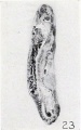
Figure 23
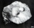
Figure 24

Figure 25
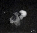
Figure 26
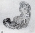
Figure 27
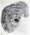
Figure 28


That lysis of the entire conceptus, even including cyemata belonging in this group, may become complete is indicated by such a specimen as No. 2197, shown in figure 29. The cyema of this specimen not only has become markedly modified in external form, but upon microscopic examination is composed of nothing but a web containing remnants of nuclei, the whole forming a perfectly homogeneous mass. In view of this fact, the external form of this specimen really was remarkably preserved.


An interesting specimen belonging in this group is No. 587, represented in figures 30 and 31. While studying this specimen attention was attracted by the small cavity in the wall of the main chorionic vesicle, which contained the cylindrical cyema. Upon further examination it became evident that this smaller cavity was surrounded by a degenerate chorionic membrane which was not a diverticulum of the main vesicle, and that in fact we were dealing with a twin pregnancy similar to No. 1840, to be referred to later. The larger of these two chorionic vesicles, in the villi of which the smaller is contained, measures 58 by 38 by 29 mm., but the smaller measured only 9 by 6.5 mm. in the fixed condition. The latter contained no trace of the cyema at the time of examination, and the very degenerate condition of the chorionic wall, and especially of the amnion, makes it decidedly unlikely that the small though not necessarily rudimentary cyema which it may have contained earlier in its history was lost before it came to my attention. The photograph of the portion of this abortus shown in figure 31 suggests, and an examination of the gross specimen confirms, the surmise that the smaller vesicle was located at the point indicated by the leader in figure 30, the photograph originally taken of the specimen, which shows the presence of the cylindrical cyema. Since a degenerate decidua still separates the villi of the respective vesicles in the area of most intimate contact, it is evident that the larger vesicle must have captured the smaller, as it were, by surrounding it. It thus probably hastened its strangulation and death, which may, however, have been fully assured by the existence of an inflammatory decidua. The only content of the smaller chorionic vesicle is coagulum (magma) with some faint traces of the amnion in the form of coagulum. The villi of the smaller specimen are almost completely degenerate, and the stroma of the villi of the larger, which is non-vascular, has undergone mucoid degeneration.
From what has been said, it is evident that we have here not an evidence of superfetation but double-ovum twins, one of which survived for a considerable period beyond the time of death of the other. Moreover, although both ova may have begun their uterine development equally well, the smaller must soon have been throttled or surrounded by the larger. Or the smaller, perchance, may have become implanted in a more unfavorable portion of the decidua and therefore have succumbed earlier. Since it is wholly unlikely that any portion of the cyema was preserved in the smaller vesicle, this specimen becomes classified in group 3 or 2, accordingly as we do or do not recognize a trace of the amnion in the fine line of coagulum which extends along the interior of the chorionic membrane. The larger, on the other hand, falls into this group. It may be recalled that a similar instance of twins falling into different groups was mentioned in the discussion of group 4, and two more instances will be mentioned in the succeeding group.
The disproportion between the size of the cyemata and that of the chorionic vesicle, though pronounced, is not so great in this as in the preceding group. What was said there regarding the relation of the dimensions of the abortus to those of the chorionic vesicle applies here also. As all the specimens of this group contain cyemata varying from 2.5 to 13 mm., it is evident that the amnion must have developed considerably in some of them. Subsequent retrogression of the cyema or hydramnios, then, left the amnion proportionately too large as compared with the cyema, for (so far as I am able to see) there is little retrogression in the size of the amnion, even when that of the cyema is considerable or total. The amnion generally was relatively too large, although rarely it was folded about the cyema very intimately. When large it frequently was greatly folded, with adjacent folds adherent to each other, and in one instance a small vesicle so constituted could easily have been mistaken for a yolk-sac. The amnion in this case had undergone decided structural changes.
The number of chorionic vesicles wholly encased in blood or in blood and decidua was relatively small. The layer of blood which was interposed between the decidua and chorion varied from 1 to 10 or more millimeters in thickness. Whether the condition of this blood is of any value in determining the time that has elapsed since the detachment of the conceptus and its expulsion from the uterus, I am at present not able to say. The contrast between the states of preservation of the blood and the contained conceptus naturally was sharp in many cases. Often the blood was preserved well, even in instances in which the stroma of the chorionic vesicle was very degenerate. The conceptuses in this group have progressed somewhat in development beyond those in the preceding group, both as to external form and the internal organs, but in general the changes noted in the cyema and its appendages, or in the chorion or amnion, or even in the decidua, did not differ in kind from the changes noted in the previous groups. However, since the specimens in this group are somewhat older, it would seem to follow that the duration of these changes must have been somewhat longer. Nevertheless, the rate of change must have been much slower. Hence a longer duration does not necessarily imply the production of a more pronounced change. It is also not impossible that, with increasing maturity, the tissues may become more capable of resistance and thus affect the rate of change produced by a given cause of constant intensity.
As was true of some of the nodular cyemata, so also several of the cylindrical were sessile, being attached directly to the amnion, sometimes by their caudal extremities, apparently without the instrumentality of an umbilical cord. But, as a rule, a short umbilical cord is present, even if not wholly normal in form, structure, or relations. Indeed, the cord quite frequently is distended greatly, and microscopic examination usually shows that the condition of the umbilical vessels, even when present, is not normal. In many instances they are absent altogether and the tissues of the cord, like those of the cyema itself, have undergone great changes. Rarefaction of the mesenchyme and cavity formations are very common. Portions of the allantoic stalk were frequently preserved, although sometimes in a form so greatly modified as to make their recognition doubtful.
The yolk-sac also frequently was present, though changed so very much that recognition of it could not always be certain. It frequently had a greatly thickened, fibrous wall which was fused completely with the chorion.
The decidua was present in only 31.2 per cent of the 48 specimens in this group and was infiltrated in 80 per cent of these, excluding 2 doubtful cases. This infiltration was marked in 58.3 per cent and slight in 41.7 per cent of the specimens. Abscess formation was relatively rare. The infiltration also was less marked than in the preceding groups, especially in the first three, and cases suggesting interference were rare, although several were noted.
Hydatiform degeneration was present in 38.5 per cent, with a probable additional 6.3 per cent. Infiltration of the decidua was present in only 3 'or 4 of these specimens, but not much reliance can be placed upon this small number.
Since the decidua was absent in 6 of the 10 cases in which it was definitely stated in the history that there was no venereal disease or evidence of it, it is impossible to throw any further light on these specimens; for, as already stated, infiltration does not occur in the chorionic vesicle until the latter is extremely degenerate. Hence, in the absence of the decidua, one can only draw inferences as to its condition from changes in structure of the chorionic vesicle. Since hydatiform degeneration was present in 4 of these 6 cases, the inference that the decidua of these, if present, would also have shown infiltration is justified. In 4 of the 9 cases in which the absence of venereal disease was reported, and in which the decidua was present, the latter undoubtedly was infiltrated. This infiltration was mild in 2 cases and severe in 2. In one of these instances in which it was especially emphasized that the patient stated that she had never had leucorrhea, and in another in which it was stated that leucorrhea was present but that the patient had never had venereal disease, the infiltration was very marked. Further confirmation of the clinical diagnosis was found in three cases of lues and in one of endometritis.
In two cases of this group in which the alleged cause of the abortion was respectively a blow in the abdomen and a fall, severe infection and even abscess formation were present; and in a third, in which an accident was blamed, hydatiform degeneration was recognized, and hence endometritis probably existed. In a fourth case in which abortifacients were supposed to be responsible for the termination of pregnancy, a mild endometritis and changes suggestive of lues were present; and in a fifth instance, in which the abortion was attributed to quinine, the specimen plainly suggested that it came from an induced abortion. The sixth case, which was said to be spontaneous, had the microscopic appearance of an old retained specimen.
The structural changes noticed in the decidua, in the chorionic vesicle, and in the amnion, did not differ in kind from those seen in previous groups. Calcification was present in small areas in only a few specimens, which had the characteristics of old retentions. Coagulation necrosis, or so-called "infarct" formation, so common in the next group, was rare here; but fibrosis and cytolysis of the decidua were as common in this as in the other groups. Rarely, the decidua was so fibrous as to simulate the degenerate chorionic membrane very closely.
B. Tubal
Since 2 of the 5 specimens originally placed in this group were found to contain no embryo, only 3 remain. Of these, No. 697 contains a very small nodule of embryonic tissue, the appearance and structure of which suggest that it very probably belongs in the preceding group of nodular cyemata. However, since the sections of this nodule do not form a complete series, one can not be absolutely certain of this. Another specimen, No. 346, also belongs to the group of nodular cyemata, for neither the caudal nor the cephalic end can be distinguished with the naked eye. This leaves only one specimen, No. 729, in the group of cylindrical tubal cyemata. Since the only remnant of the cyema itself found in this specimen is a small group of cells on the end of the umbilical cord, the existence of a cylindrical cyema in this case can be a matter of surmise only, for the condition of the cord has not been found to be a reliable criterion for distinguishing between the previous existence of a nodular or a cylindrical cyema. Moreover, since Mall thought it probable that the cyema was normal, this specimen can be retained in this group only if we desire to classify portions of the cords as cylindrical cyemata. From these considerations it follows that the group of cylindrical cyemata is left without a true representative from among the tubal pregnancies found in the first 1,200 accessions. This does not mean, however, that cylindrical forms can not or do not occur among tubal pregnancies.
Infiltration of the tube was present in 3 of the 5 specimens originally placed in this group in which it was included in the sections. Hydatiform degeneration was found in 3 specimens, but the tubes of 2 of these have not been examined microscopically. One of these 3 cases was a very fine example of hydatiform degeneration. A second was very clearly an early case, but too few villi were included in the third to make a definite decision possible.
Group 6. Stunted Cyemata
A. Uterine
It was emphasized in Chapter III that stunting, in a physiological sense, can not be limited to cyemata in any particular group; but since the difficulty of recognition increases with the decrease in the size of the specimen, it is only rarely that stunting can be detected by inspection alone in cyemata below the stage of limb formation. Furthermore, if the limbs themselves are to be used as a criterion, limb formation must not merely have begun, but must have proceeded far enough, and normal growth must have been inhibited sufficiently to effect a change in form clearly recognizable with the unaided eye. This, however, does not imply that the effect or the recognition of stunting is confined to the limb-buds. Rarely, it is noticeable quite early in the entire caudal or cephalic extremities, or even in the head itself. This is illustrated by Nos. 2173 and 2233, shown in figures 32, 33, and 34. As is evident from these illustrations, the last cyema borders very closely upon the cylindrical in form.
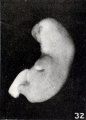
Fig. 32.
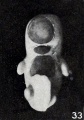
Fig. 33.
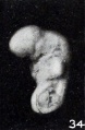
Fig. 34.


The word "stunting," as here used, also implies a disproportion in normal form, not merely a reduction in size of the entire specimen. Hence none of the stunted specimens are merely undersized, normally proportioned fetuses; for although an equal, universal retardation in growth is conceivable, it is unlikely that such a thing really occurs. Such an assumption would imply that all organs and tissues are equally resistant to interference with the blood or nutritive supply, or to toxins or other influences. It may seem strange, indeed, that no fetuses beyond the length of 20 mm. are included in this group. This fact would seem to imply either that the causes productive of stunting which may be operative up to this time cease to be effective later, or that portions of the cyema have become so resistant that they can no longer become affected in such a way as to- produce stunting. It has long been recognized, however, that stunting of an organism can occur as the result of various influences at any time throughout its period of growth in post-natal life. Hence an assumed increase in resistance of the tissues sufficient to withstand all influences affecting such young fetuses as those here concerned can hardly be offered in explanation for the absence of stunted fetuses among those more than 20 mm. long. Nor can one assume that the causes operative until this stage is reached, or at some particular time before this stage of development has been reached, always come into abeyance when a fetal length of 20 mm. has been attained.
If, on the other hand, we assume that the various tissues or organs are affected only during a transitional sensitive period, perhaps in certain formative stages, we are in conflict with the facts; for upon this assumption we should be able to group the stunted fetuses by the effects produced upon their tissues, organs, or systems of organs even, but this does not seem possible. Nor are the organs affected serially in the order of their development; for, entirely aside from certain things to be discussed in a subsequent chapter, some changes present within these stunted cyemata are quite universal and comparable, no matter at what stage complete inhibition of further development has occurred. Moreover, what impresses one most is not the striking modifications of external form, pronounced as these at times are, but the marked structural changes within the organs themselves. To what extent these changes may be attributed to maceration with consequent disintegration, or to dissociated or uncorrelated growth, and whether or not true stunting can be simulated by the effects of maceration alone, will be considered more fully in a succeeding chapter. The explanation for the absence of stunted fetuses beyond a length of 20 mm. is not only the failure to recognize stunting in them, but that maceration changes often become so pronounced that they mask the changes which have been characterized as stunting. When such is the case the specimen is placed in the next group.
Although gradual death of cyemata or of entire conceptuses in consequence of interference with the source of nutrition has not as yet been established, such a phenomenon is not only possible but highly probable. Death of the cyema or of the entire conceptus in consequence of chronic endometritis, or in consequence of other chronic changes, or even perhaps in consequence of toxins, seems likely, however. Hence, although the cyemic circulation be maintained in the main, lack of nutrition nevertheless might occur locally and show itself first in outlying cyemic parts, as in the tips of the extremities or in the facial relief, in which places slight changes in form easily become evident. However, the fact that the present group of stunted cyemata is limited to a comparatively early age very seriously contradicts such an assumption, and the well-known fact that ill-nourished, stunted, viable infants are born with certain constitutional diseases would seem to imply the occurrence of stunted forms in all groups of pathologic cyemata and perhaps also the occurrence of stunted chorionic vesicles as well.
Excellent examples of stunting are No. 2473, a 13 mm. fetus, and No. 675, a 10 mm. fetus, represented in gross and in median section in figures 35 and 36. The head of No. 675 has been so deformed as to look like that of a frog, and the whole outward form has become rounded. Since this specimen was retained long after death, marked maceration changes had occurred, and it is a question to what extent the changes in form here shown can be attributed to maceration alone. The limb-buds in these stunted embryos soon become more rounded, smoother, and shorter, or even somewhat clubbed, losing all details of form as illustrated by No. 2233, represented in figure 34. The head loses its natural curves and becomes atrophic or reduced; the maxilla is often beaked and the mandible depressed greatly, thus producing gaping. In some cases the mandibular region may fuse with the chest, as represented in figure 37 (No. 788a), and with increase in flexure the eyes may come to lie practically opposite the liver. The caudal region looks stubbed or flattened, the caudal process being less evident. Both local and diffuse epidermal thickenings are not uncommon, and sloughing of the epidermis is frequently preceded by bleb formation. The umbilical cord is often distended, especially near its attachment to the abdominal wall, and at this point decided bleb formation is common.
Miiller (1847) also noticed instances of bleb formation on the umbilical cord and stated that Albinus and Sandifort called them "processus infundibuliformis" when they occurred at the point of attachment of the cord to the abdomen. Miiller further stated that Ruysch described a cord so full of "hydatids" that he spoke of it as being a " concatenatio vesiculorum. "
The internal organs of the specimens become distended, lose their clear outlines (as illustrated by No. 983a, shown in figure 38), blood-cells seem to pass out from the vessels in all directions, the normal cell relations are disturbed, cytolysis occurs, nuclei of various cell types become pycnotic and circular in outline, until all areas of the specimen seem to be composed of one type of cell only. This is due to the fact that all nuclei have assumed a similar form and appearance, but in more advanced stages these nuclei, too, disappear and only a homogeneous necrotic mass remains to represent the fetal tissues.
The length of the fetuses in this group varies from 4.5 to 20 mm., and the menstrual age from 30 to 127 days. But neither the former nor the latter can be regarded as more than roughly indicative of their true age; for, although 30 days is approximately the correct age for an embryo of 4.5 mm. long, 127 days is entirely too old for one with a length of 20 mm. Specimens of approximately the same alleged menstrual age are found to differ widely in length, and fetuses of approximately the same length may differ greatly in menstrual age. Nor can these discrepancies be attributed to the unreliable character of the menstrual histories, for specimens with identical menstrual ages often vary considerably in size. This is illustrated splendidly in cases of double-ovum twins. In one of these instances (No. 1840) the fetuses have a length of 15.9 and 38.5 mm. The larger of these twins is normal in every respect and macerated but slightly, but the smaller is greatly macerated and stunted. Since both these specimens are of identical age, they illustrate how misleading even a reliable menstrual history may be.
In a second case, that of No. 788 a, b, one is a nodular cyema described under group 4 and the other a stunted specimen included in this group. The organs of the latter or larger fetus, though dissociated, are all well outlined, and the vertebral disks especially well defined, in spite of the fact that it must have been dead a considerable period of time. The umbilical vessels are degenerate and the limbs atrophic. The menstrual history unfortunately is not known, or it would be possible to make some conjecture at least as to the probable length of retention in utero after fetal death. However, if we assume that the large fetus is smaller than it should be, as undoubtedly is the case, and take its anatomical age to be that of a length of 20 mm., it would have a normal menstrual age of about 7 weeks. The small nodule (7886), which was only 2 mm. long, does not show a development corresponding to that of a normal specimen of this length, and on the basis of size would have an age of less than one month. This, however, does not necessarily imply that it died at that time. Indeed, the relative preservation of the tissues of the two fetuses makes this very unlikely, although we must remember that such a small nodule as 7886 could probably be preserved for a long time in such a large quantity of sterile amniotic fluid as that in which it was contained.
Another instance which shows the misleading character of the menstrual histories, even when known, is that of No. 1914, a case in which a large mass of hydatiform cysts accompanied a normal, living fetus of 7 months. A still better illustration is the case of Slemons (1917 a ), in which a nodular mass, apparently a twin, accompanied a normal viable fetus. Since it is not improbable that such specimens as some of these have been taken to establish the occurrence of superfetation, they will be mentioned in a later chapter.
In the other three cases of twins included in this group the cyemata fall into the same instead of into different groups. In two instances (Nos. 207 a, b, and 341 a, 6) this may be due to the fact that they are single-ovum twins, even if they are not of the same size. In the third case (No. 330 a, b), although from double ova, both fetuses were of the same length. The appearance of these conceptuses indicates that they were retained for a considerable period after fetal death, and the history confirms this. Syphilis was suspected in this case and bleeding began six weeks before the abortion occurred. Furthermore, the sizes of these fetuses, as well as the condition of the tissues themselves, clearly indicate that both had probably been dead some time before hemorrhage began.
Whether or not twin cyemata fall into the same group of the pathologic very evidently depends upon whether they have been subjected to identical influences. That the exact conditions under which they develop may easily vary is evident, and that they may be variously affected previous to implantation is also possible. Nor does the occurrence of twins which fall into different groups necessarily imply that the processes responsible for the physical differences were necessarily or essentially different from those which are responsible for the disproportion in size of normal, full-term twins. It may be simply a matter of degree. In the case of 788 a, b, for example, the deciduse were infiltrated, but not to the same degree.
A few of the specimens in this group are of particular interest because of the great disproportion that exists between the size of the fetus and that of the chorionic vesicle. In No. 135, for example, in which the fetus was 9 mm. long, the dimensions of the chorionic vesicle were 105 by 65 by 65 mm. Since the villi practically were absent and the surrounding layer of decidua was very thin, the above dimensions practically are those of the amnion also, for it had fused with the chorion. Although the wall of this abortus is composed of these three layers, it nevertheless is almost papyraceous. Microscopic examination shows it to be decidedly degenerate and to contain many leucocytes. The amnion, too, is very degenerate, although its epithelium is still present. Even if mechanical distension were the chief factor in the enlargement of this vesicle, it is unlikely that it was solely responsible for the disproportion between the chorionic vesicle and the size of the fetus. Part of this disproportion is attributable to some recession in the size of the embryo, and more to the failure of the latter to develop normally. To what extent the chorionic vesicle grew after the death of the fetus must remain a matter of conjecture; but in view of its very degenerate character, it seems unlikely that it could have grown much. The absence of the menstrual history unfortunately leaves us without data that might be of some assistance in arriving at a conclusion regarding this matter. Although the organs of this fetus have lost their clear definition, and although dissociation and maceration are marked, yet on the whole it is preserved better than one would expect from an examination of the chorionic vesicle. Though epidermal blebs are present, not all the epidermis has sloughed. Indeed, in some places the superficial layer has formed marked local thickenings which now and then are so definitely circumscribed that they could probably be designated as warts or papilla?. Similar, although more diffuse, epidermal thickenings are found also in No. 201, a fetus of 20 mm. The same phenomenon apparently was observed by Rokitansky (1861), who spoke of papillalike growths of the epidermis in a fetus.
The most interesting thing seen in the stroma of the villi of this group was the presence of epithelial vesicles at various depths from the surface in No. 276. Although many of these vesicles which are not well preserved could easily be confused with degenerating vessels, in the case of others this is impossible, because their relations to the epithelium can be established easily. The epithelial lining of many of them is fairly well preserved, and in some instances the vesicles can be shown to communicate with the surface by means of an epithelial invagination. Most of them, however, lie isolated in the stroma.
In looking about for a possible explanation for the presence of these vesicles, it became evident that they had their origin in epithelial invaginations. The opposite walls of these invaginations apparently become fused proximally and their strands of epithelium then connect the vesicles with the surface. Further examination of the sections of the villi in this specimen shows that instead of having a fairly regular border, the epithelium is decidedly crenated or folded, quite in contrast with the usual condition of things. Hence it would seem that proliferation of the epithelium has resulted in foldings into the stroma of the villus, instead of accumulations on the surface. The presence of proliferation of the epithelium in this case seems to lend further confirmation to the presence of hydatiform degeneration.
Out of 73 specimens originally placed in this group, 16 were found to belong elsewhere, and 23 were added, making 80, from which 1 must be subtracted because of twins in one chorionic vesicle. The decidua was present in 45.5 per cent and infiltrated in 88.9 per cent of these, excluding 2 in which infiltration was doubtful. This infiltration was marked, even up to abscess formation, in 56.2 per cent of the specimens and slight in the rest. Hydatiform degeneration was present in 24 per cent, excluding 5 in which its occurrence was probable. Infiltration was present in 90 per cent of the specimens in which the decidua was included and not too necrotic for examination.
In attempting to correlate the clinical histories with the objective examination, it was found that the clinical diagnosis of infection is confirmed in 4 out of 5 cases. In the fifth case the decidua was not included, but since hydatiform degeneration was present, infiltration can be assumed and the clinical diagnosis of infection be regarded as confirmed in this case also. Of the 6 cases which were reported as showing no evidence of venereal disease, the decidua was not included in one and was too degenerate to assist in the diagnosis in a second case. Of the remaining 4 cases, 3 show the presence of a severe and 1 of a mild infiltration.
Although mechanical interference with the course of the gestation seldom was reported in this series, several specimens were noted in which abortion probably was induced. In one case in which the termination of pregnancy was attributed to fright, the decidua, unfortunately, was not included; but since the chorionic vesicle showed hydatiform degeneration, the decidua undoubtedly was infiltrated. In a second case in which fright was given as the alleged cause, Mall noted that the decidua was "markedly inflamed. " In a third case, in which the termination of pregnancy was attributed to work, death of the conceptus must have occurred long before. As stated in the introduction, I fully realize that this contradiction between the alleged cause and the findings may be apparent only, for it is entirely possible that exertion, psychical disturbances, or an accident may precipitate an abortion which is more or less imminent at the time.
The structural changes encountered in this group do not differ in kind from those present in previous groups. However, the incidence of infiltration of the decidua was more than twice as high as in group 5, and was divided equally between the severe and the slight cases. The presence of hydatiform degeneration was somewhat more common, although the percentage of these cases in which the decidua was included was somewhat lower. In 90 per cent of the latter the decidua was infiltrated.
Although they frequently were not especially noted, "appendici durate" seemed somewhat commoner in specimens of this than in those of the previous groups.
B. Tubal
The small number of stunted tubal cyemata among the first 1,200 accessions is especially interesting, because the average length of these specimens is only half as great as the average length of those in the corresponding uterine group. After certain specimens had been excluded only 6 stunted tubal specimens remained. The corresponding uterine group, on the other hand, contained 61 specimens. But in spite- of the small number of stunted embryos in the tubal group, the significance of the fact, nevertheless, is clear. It is only very rarely that a tubal pregnancy lives beyond the earliest stages of development, while in the uterus, on the contrary, this is relatively very common. This conclusion is not based upon a comparison of the relative number of specimens found in these two groups of uterine and tubal pregnancies alone, nor upon the comparative size of the specimens, but upon the respective percentages that the total number of specimens in the uterine and tubal groups of the stunted form among the entire series of specimens in the pathologic division which were examined. Although the stunted embryos form 19 per cent of the total number of uterine specimens, stunted embryos form only 5.7 per cent of the total number of the tubal specimens. This marked difference scarcely can be explained in any other way than as suggested above.
Although 11 specimens originally were placed in this group of stunted tubal cyemata, 5 were found to fall elsewhere. One (No. 338c) was a uterine specimen, being accompanied by considerable decidua. Two others (Nos. 196 and 934) belong in group 4. The former has a length of only 3 mm., and the latter, although 6 mm. long, was recorded as having no extremities, and described as looking like a grain of wheat. Moreover, the anatomy, especially of No. 934, is that of a nodular rather than of a stunted cyema. One specimen (No. 992) belongs in group 3, and another (No. 881) can not properly be classed as stunted, in view of the fact that it was only 3 mm. long.
Of the 6 remaining specimens, 5 lay in tubes the walls of which were infiltrated intensely in the case of 2, and but slightly in 3 specimens. In the sixth case too little of the tube was included to serve as an adequate basis for examination.
Hydatiform degeneration undoubtedly is present in 4 of these 6 cases, or in 66.6 per cent. In another specimen (No. 477) some villi seem to show incipient changes indicative of hydatiform degeneration, but since marked maceration changes also are present, one can not be certain of this case. Too few villi were included with the remaining specimen to make a positive diagnosis justifiable, but the structure of those present did not suggest a hydatiform change.
The cyemata were all macerated and some were fragmented. In two specimens (Nos. 838 and 882), Mall stated that limb-buds were not present. Since the respective specimens were 6 and 8 mm. in length, it is evident that they should have been present, and their absence must be attributed either to inhibition of growth or to disintegration. In the absence of a complete series of sections, this point unfortunately can not be decided. It was possible to recognize outlines of various organs in one specimen only, and since most of the specimens were very young, they had a structure comparable to that of the nodular and cylindrical, rather than of the stunted uterine group. Maceration and dissociation were very marked, as a rule.
Group 7. Fetus Compressus
A. Uterine
The term fetus compressus was used by Mall in a much more general sense than usual. It has been customary to restrict this term almost wholly to one twin which died early and became softened and compressed by the living, welldeveloped companion. Instances of a single fetus similar to a true fetus compressus are recorded in the literature, however. Strahl and Henneberg (1902), for example, stated that they found such a specimen, although the presence of abnormal pressure exerted directly upon it by the uterus could not be established. Hohlweg (1903) also implied that Kiistner thought that cases of fetus compressi could occur in single pregnancies, and I am quite certain that I am representing Mall correctly when I say that his observations were to the effect that quite typical fetus compressi occur not rarely in single pregnancies in the presence of abundant amniotic fluid. Although Schickele (1907) regarded macerated forms from the second months as rare, His (1891) emphasized that the softness of aborted forms easily results in abnormal folds and flexures, and Waldstein (1913) stated that von Winckel found deformities of the extremities extremely common in tubal embryos.
As previously intimated, this group is a very inclusive one. In some respects it really is a group of left-overs not characterized by maceration alone. Specimens in any of the preceding four groups may be, and indeed they nearly all are macerated. However, since many of those in the normal division also are macerated, the presence of maceration can not be regarded as distinguishing any group. Differences in the degree of maceration may and do exist; but since maceration becomes evident externally much more quickly in older fetuses, gross appearances often are a very unreliable guide for the determination of the degree of maceration. One thing, however, is true of all specimens in this group, except such as are composed of material from curettage, and that is that maceration often is present even in specimens composed solely of fragments of wholly normal embryos or fetuses.

Some of the specimens are macerated and swollen and others are shriveled and mummified. The latter Mall distinguished as fetus compressi. The use of this term does not imply, however, that they have necessarily been subjected to pressure, although such usage is at variance with the historical meaning and the usual application of the term. Moreover, very macerated fetuses, the forms of which have not been very materially changed, are not included. These things must be borne in mind in order to avoid misunderstanding. It is true that some of the so-called fetus compressi look as though they had been subjected to pressure, and in some instances, even of single pregnancy, there is no doubt whatever about this. Other fetuses look compressed, although a quantity of liquor amnii seemingly sufficient to prevent direct uterine pressure still surrounds them. The head is collapsed from side to side, the hands and feet are flattened, and the trunk also is collapsed. In most of these cases the appearance of compression is apparent only, and probably resulted from the softening and collapse of the tissues consequent upon maceration and dehydration under aseptic conditions. The skull sometimes is practically devoid of brain tissue and decidedly flattened, all the tissues have become degenerate and necrotic, and those of adjoining parts have often fused more or less, so as to obliterate the natural boundaries.

If, as stated, mummification sometimes may occur while the fetus is contained in a large amount of fluid, it would seem to have to result from chemical changes in the amniotic fluid. Indeed, it not only is conceivable but inevitable that the composition of the amniotic fluid is changed soon after the death of the fetus. As is well known, death of the membranes makes them more permeable, and hence offers greater facility for the absorption of fluid, thus leading to the greater concentration of the liquor amnii, with consequent dehydration of the fetus. It is evident, however, that absorption must be slow, for otherwise the amniotic fluid, although rarely large in quantity, would soon be absorbed and the fetus would subsequently be subjected to direct pressure from the uterine wall. Sometimes, indeed, such is the case, and then the macerated or mummified fetus may be rolled up firmly into a ball, as illustrated by Nos. 921 and 1041, shown in figures 39 and 40. In the last case the decidua, placenta, and membranes were wrapped tightly around the fetus, the whole constituting a firm, rounded mass.

It is difficult to characterize this group as a whole, because it contains such diverse things as portions of a cord, material from curettage, embryonic fragments of mutilated normal specimens, as well as macerated fetuses falling among the groups of the nodular, cylindrical, stunted, or normal. Figures 41 to 50 inclusive afford some idea of the diversity existing among the specimens of this group. This diversity is extreme, not only in form, size, and age, but also in the matter of preservation. No. 1245 (fig. 41) is a good illustration of fragments. Whether they came from specimens which developed under normal or pathologic conditions it was not always possible to state, even upon microscopic examination, for the decidua was not always included. No. 1806 (fig. 43), though classed in this group, was originally described as normal in form and could be placed among grade 3 of the normal division, for some specimens in this grade show even greater maceration.

Specimen No. 651a (fig. 58) and to a lesser degree No. 1301 (fig. 44) illustrate beginning changes in form which, though slight, are quite characteristic of many of the specimens of this group, and when present always exclude them from the division of normals which it should be recalled contains specimens with developmental anomalies. Besides being macerated, both these specimens are characterized by a peculiar sag or droop of the extremities present on the left side only in No. 1301. This is noticeable first in the arms. These distortions, though slight in these fetuses, are shown in a much more advanced stage in No. 1931 (fig. 51), a considerably older fetus which had been retained longer after death. Although this specimen is only 35 mm. long, it unquestionably is anatomically older than No. 1301, which measured 50 mm., a difference largely attributable to the presence of marked maceration. Nos. 1925, 1239, and 1515, shown in figures 48, 49, and 50, are merely older and still more macerated and deformed specimens, No. 1515 being in the early stages of dismemberment.
In order to illustrate the fact that many specimens in this group of the pathologic division are quite comparable to some of those in grade 3 of the normal division, I have selected a number of roughly corresponding stages and placed the photographs parallel, for the sake of comparison, in figures 53 to 63 inclusive. Anyone familiar with the material here concerned will know that photographs are not adequate evidence for illustrating the classification of a specimen; but even an inspection suggests, and the microscopic examination supports, the statement that there is no sharp dividing-line between some of the specimens in the third grade of the normal division and others in this group of the pathologic division. Specimens found in the pathologic division often are better preserved in every way and wholly without developmental anomalies, while some specimens in the normal division are macerated and have pronounced anomalies. Indeed, this is what one should expect, for whatever the cause or causes of the changes represented by specimens which have been classified in group 7, these changes must have a beginning. However, these beginnings undoubtedly would not be clearly recognizable by the unaided eye or perhaps not even with the aid of the microscope. Consequently, the same person may classify one specimen differently from succeeding similar ones, merely because various aspects or characteristics of it impress themselves more upon him at different times. These cases apparently would constitute a border-line group as far as the morphological changes are concerned.
The lengths of the specimens in this group included among the first 1200 accessions range from 1 to 210 mm., and those of all those in the entire collection from 1 to 286 mm. This range is due partly to the fact that the idea of the group developed as the material accumulated. Moreover, a number of different individuals were engaged at widely different times in the classification of the material. For these and for other reasons this group also has been reviewed in accordance with the wishes of Mall. It is interesting that the reclassification has affected almost solely the swollen, macerated specimens and only a few so-called fetus compressi. Hence it is evident that there is practical unanimity of opinion among those concerned as to the gross characteristics of a fetus compressus, even if some of these were placed in the normal division. This unanimity of opinion is explained largely by the fact that the term is applied almost wholly to larger fetuses. The smaller specimens disintegrate completely before the stage of mummification is reached, while in older fetuses the skin, the cartilages, even when still wholly unossified, and the ligaments, as well as the fibrous tissue as a whole, resist disintegration more. The smaller specimens soon become so soft that they literally fall to pieces before they can become dehydrated and mummified. This is illustrated splendidly by Nos. 379 and 717. It is recorded by Mall that the former fell to pieces upon being handled, and of No. 717 it was stated that a slight amount of shaking would cause the entire specimen to disintegrate.
All the small specimens found in this group undoubtedly could be classed among the other groups of the pathologic or normal divisions, for they have been placed in this group merely because they are macerated more completely than the specimens usually classed in the normal divisions. The exclusion of these would limit this group to fetuses beyond the length of 25 to 30 mm. At this time a stage of development much more resistant to disintegration is reached, and the changes in form regarded as peculiar to this group can then arise. As recorded by Mall, small specimens macerated so thoroughly that they almost fall apart, even when handled in fluid, sometimes have undergone very slight changes in form and could be placed in the other groups.
It is an old story that the outward appearances of the specimens in this group often are grotesque. This is true particularly of the so-called fetus compressi. These strange appearances are very largely attributable to the distorted extremities, the gaping mouth, and the pointed features. There is no limit to this distortion, as No. 1925, shown in figure 48, illustrates. Sometimes the greatly and abnormally bent extremities become adherent, as in case of the legs of No. 1859, shown in figure 64, and in other instances, as in Nos. 1860 and 627, bleb formation of considerable size occurs and later the skin may slough or become rolled up and fused so as to form marked welts. Sometimes it may hang in shreds from the fingers and toes or from other parts of the body. These strands of sloughed epidermis may then simulate amniotic bands, and when accumulated about the fingers and toes may hide them completely, giving the appearance of a mittened hand. However, epidermal thickenings, local or diffuse in character, are sometimes present, as shown in figure 65. These give one the impression that they result from epithelial proliferation rather than merely from fusion of accidentally apposed cells. Fusion of adjacent surfaces of the digits is also common, and occasionally the upper or lower extremities may be united quite firmly in places through fused areas of epidermis, as is illustrated by No. 1859.
The contortions of the extremities are due not merely to the relaxation of the joints and consequent abnormal mobility, but result also from a bending of the cartilaginous shafts of the bones, even up to or beyond an angle of 90. The smaller cartilages of the hands and feet may lose their distinct outlines, and under conditions of advanced maceration may fuse somewhat with one another. Rarely, areas of coagulation necrosis are also seen at the distal extremities of the terminal phalanges. Degeneration of the precartilage and cartilage opposite the ossified areas may also be complete, thus presenting the appearances of bones with extremely enlarged cartilaginous extremities. Although no measurements were taken and no exact comparisons made with normal specimens, it is not unlikely that the extremities of some of these developing cartilages are somewhat enlarged, although not necessarily as the result of growth.
A survey of a considerable number of these fetuses shows that the distortion of the extremities, and indeed also those of the trunk, are purely haphazard. They seem to follow no law, except perhaps that of gravity, but often are in diverse directions in the different extremities of the same fetus. Whether or not the initial sag is in the same general direction in all the extremities depends considerably upon the age of the fetus. Below a length of 25 to 30 mm. the limbs still extend stiffly from the body, and in these specimens both pairs of the extremities usually droop, as represented by Nos. 651o and 1710, shown in figures 58 and 66. After the extremities assume their normally flexed posture the abnormal positions which they then take become far more diverse. This is well illustrated by Nos. 1751 and 1931, shown in figures 67 to 68. Similar distortions are present also in the digits, as shown in figure 69. Nor do fetuses of corresponding ages necessarily show the same degree of distortion, or possess distortions in the same general direction, even if they show approximately the same degree of maceration. However, the degree of distortion nevertheless is roughly correlated to the degree of maceration and the duration of the post-mortem intrauterine retention. Fresh and unmacerated specimens never show the characteristic distortion.
The haphazard character of these distortions seems to indicate quite conclusively that they did not result from contractures arising during the gradual death of the fetus. To be sure, the loss of tone accompanying gradual death or following it must finally become extreme, but since the specific gravity of the amniotic fluid, even when altered, is so little if any less than that of the fetus itself, it is difficult indeed to see just why marked changes in the position, even if not in the form, especially of the extremities, can occur without the instrumentality of external pressures.
The umbilical cord of many of these specimens, especially of the fetus compressi, is abnormally and often extremely twisted and thin. As is well known, this twisting is unlike that normally present, though it seems always an accentuation of it. I have never seen it occur in a direction opposite to that normally present in a given cord. That most of the extreme torsion of the cord is purely a post-mortem and not an intra-vitam phenomenon is indicated by the fact that these extremely tortuous cords always can be untwisted easily up to the point of the customary twisting. The excessive tortuosity also varies more in degree, and a single turn does not always involve the same length of cord in fetuses of approximately the same age, even when about the same amount of twisting is present in the entire cord. Indeed, a single turn may occupy different lengths of the cord of the same fetus, and it is not uncommon to find only a portion of the cord twisted and the rest of it wholly untwisted, a fact which probably can be attributed in part to the varying degree of maceration.
While observing the nature of this twisting more closely, it became evident to me that it probably was not produced intra-vitam, but rather resulted from post-mortem passive fetal movements. Had this twisting occurred during the life of the fetus and been the cause of its death, one would expect to find some adjustment to it on the part of the growing tissues. Such an adjustment, however, does not exist. The tissues are necessarily displaced mechanically, but no adhesions between adjacent turns were observed save such as easily could be attributed to maceration changes. Since no external forces save those of the uterine contractions can reach a dead fetus, it seems that this twisting, even if not always present, must result from uterine contractions. Since these contractions in many instances may have been more pronounced and prolonged than usual until abortion finally occurred, the greater twisting present in these cords could easily be explained in this way. If we assume that in a given uterus this peristalsis occurs in a uniform direction, and that in a small percentage of uteri it may occur in an opposite direction, torsion in different directions could be accounted for. I realize fully that direct observations upon the living uterus are necessary to make this supposition valid, but in any case it is difficult to conceive of any other agent than uterine contractions that possibly could rotate a dead fetus. It also is evident that this rotation probably would have to occur very largely before marked absorption of the amniotic fluid had occurred and entirely before the fetus became embedded in a mass of coagulum, such as is illustrated by No. 261, a specimen with a very tortuous cord.
Although it is conceivable that coils of the cord could cause the amputation of an extremity, this probably could occur only in comparatively early stages of development and hardly so late in fetal life as implied in figure 187 of Broman (1911). It is also noteworthy that, although coils of the cord about the extremities were frequently found, not a single instance of partial amputation or of macerated amputated extremities is contained among over 2,000 specimens in the Carnegie Collection.
It does not seem unlikely that the location of the placenta in utero may be instrumental in determining both the amount and the extent of torsion, but until more is known regarding uterine peristalsis all this remains merely a matter of surmise. The tubal and ovarian specimens unfortunately do not throw much light on this question, for they usually are very young and too macerated or damaged. Only one of all the tubal specimens, among both normal and pathologic, contained in the Carnegie Collection, had a cord sufficiently long to be of any value in this matter, and in this specimen definite torsion was present. This fact does not prove, however, that peristalsis is not a factor in torsion, for tubal peristalsis could produce similar results.
Not infrequently the cords show marked bleb formation, as illustrated in Nos. 1475 and 1523 (figs. 54 and 70), but bleb formation, which is present elsewhere in many of these specimens, is not peculiar to the group, for it is common also in younger cords and fetuses, and may also be localized elsewhere on the body, as in Nos. 1523 and 2261 (figs. 71 and 72). In other instances, as in No. 590, the entire cord is decidedly swollen, or alternately swollen and constricted, and in still others it is practically without a turn and wholly without knots, although long and filamentous. Coils of the cord about various portions of the fetus seem to be very common in this group of macerated fetuses, and probably can be attributed to the same factors which produce the twisting. This, however, does not imply that the structural changes in these cords are different from those present in other groups or that torsion was the cause of fetal death. It is largely, if not wholly, a matter of degree of difference, and this degree is determined very largely by the age of the fetus, the length of retention, and perhaps by the irritability of the uterus. The greater tortuosity of these, as compared with normal cords, as stated above, can be explained in the same way.
The decidua was present in 45.3 per cent of the 53 specimens remaining in this group. It was infiltrated in 17, or 70.8 per cent. The infiltration was marked in 29.1 and slight in 70.9 per cent. Although decidual infiltration was present in 88.9 per cent of all cases in group 6 in which the decidua was included, this was the case in only 70.8 per cent of the cases in group 7. The percentage in this group is lower than in any other save group 5, suggesting that the infiltration is not entirely dependent upon the length of retention in utero after fetal death.
Hydatiform degeneration could be recognized in only 10.9 per cent of the cases. The decidua was infiltrated in the 2 cases in which it was present, but since the number of cases concerned is so small, no special importance can be attached to this fact did it not accord with the findings in the other groups. All chorionic vesicles in which this change was noted were in relatively early stages of development.
In trying to correlate the clinical histories of the uterine specimens with the objective examination, nothing especially interesting was revealed. The correspondence between the two is no better in this than in the other groups. In general it may be said, however, that the specimens of this group have been retained longer after death, and that this fact is supported by the great frequency of the presence of calcification and coagulation necroses, or so-called "infarct" formation, which was relatively common in the placenta. The greater maceration of the specimens clearly is evident from inspection alone. Subchorial hematomata were present in several specimens, and chorionic cysts were noticed in one. Changes reported by Welch (1888) as hyaline metamorphosis of the placenta were present in a fair percentage, especially in the older and longer retained placentas, and will be considered further in a subsequent chapter.
The frequency of divergence from the normal form on the part of the fetuses in abortuses also impressed Granville (1834), and Panum (1860) stated that he found the frequency of double malformations among museum collections to be twice that in actual life. Panum found cyclopia in 16 of 618 human deformities, 25 among 143 simple deformities in sheep, and 37 in 91 deformed fetuses. He also believed that various deformities of the jaws in certain quadrupeds probably are due to intrauterine pressure. He came to this conclusion, it seems, because of his experience while incubating eggs. However, according to Panum, spina bifida occurs much more commonly in man than in animals, and he expressed the opinion that the study of comparative teratology may contain the open sesame for a comprehension of many, even if not of all, abnormalities.
His (1882, 1891) found 22 per cent of 62 aborted specimens pathologic, and J. Kollman (1889) stated that it is generally known that one-half of all abortuses are pathologic. Giacomini (1894) claimed that as many as 80 per cent of all abortuses are abnormal, and Phisalix (1890) declared that normal specimens are rare among abortuses; but Mall (1917) found only 39.6 per cent pathologic conceptuses among the first 1,000 accessions in the Carnegie Embryological Collection. However, cyemata classed as pathologic formed only 26.8 per cent among these accessions, the rest being composed merely of vesicles classed as pathologic.
Because of the presence of certain changes in the chorionic vesicle, Crosti (1896) concluded that the cause of abortion is primary in the vesicles. Giacomini (1894) declared, however, that it was impossible to decide whether the change noticed by him was primary or secondary, and emphasized particularly that the decidua should be studied because of its great influence on early development. His (1882) concluded that all abortive forms are primary, but later (1891) decided in favor of a secondary origin, a view held also by Mall (1917 C ). One would hardly seem justified in assuming, however, that all cyemic abnormalities or malformations are secondary in origin, for such an assumption would completely ignore or deny the existence of true hereditary anomalies such as polydactyly, hyperphalangism, etc. However, the mere possibility of the occurrence of anomalies of secondary origin makes the identification of these extremely desirable and important, although some of the achievements of experimental teratology seem to suggest that it may remain impossible to distinguish between anomalies which are primary and those which are secondary in origin.
Since the extension of our knowledge of the subject of human embryology necessarily has been from the older to the younger, it was inevitable that attention should be first directed especially to placental changes. Hence the literature upon the subject of placental pathology is extensive, while that upon the pathology of the cyema and chorion and also upon the decidua is small or even fragmentary. However, now that our knowledge of extremely early conceptuses is being extended much more rapidly, we soon shall be able to recognize the normal with more certainty, which alone will be a great step in advance.
Giacomini (1894) stated that many observers have noticed that abnormal changes arise in the membranes as soon as the development of the embryo is interfered with. He described these changes as the formation of vesicle-like structures in relation to both amnion and chorion.
A very interesting question is that of the invasion of the conceptus by cells of maternal origin. As long as the chorionic epithelium remains intact I never have seen any evidence of it, not even in the stroma of villi which were directly in contact with an abscess. These observations also are in accord with those of Nattan-Larrier and Brindeau (1905 b ). Nor have I ever seen anything suggesting direct invasion of the fetal circulation by cells from the maternal circulation. When one recalls the intimate relationship of the two circulations, it is surprising that such an invasion does not occur, for it would seem to be especially easy, but it is only after the chorionic epithelium is destroyed that maternal leucocytes seem to gain access to the stroma of the villi. Indeed, the only invasion of the stroma which I have ever seen in preserved villi is due to invaginations of and extensions from the Langhans layer, for the presence of large numbers of Hofbauer cells hardly can be regarded as an invasion, even if these cells themselves are not fixed in position.
Besides lytic and degenerate changes due to retention or putrefaction, the occurrence of Hofbauer cells and other appearances suggesting tissue-cultures, thickening of the amnion and chorion was also frequently present. Amniotic villi and fusion of adjacent amniotic folds were seen quite frequently, but the rarest of all changes encountered were such strange appearances on the surface of the amnion as represented in figures 73 and 74. As indicated by the accompanying scales, both of these chorionic vesicles were small. Upon microscopic examination of these amnions it was seen that the hummocky appearance was due to local thickenings formed by increase in the connective tissue of the amnion. The amniotic epithelium was never found to be involved in these thickenings, and since only two specimens were observed I am at a loss for an explanation of this peculiar condition.
If the condition of the uterine mucosa at the time of implantation of the impregnated ovum may show variations in structure at all comparable to those frequently seen in the deciduse accompanying abortuses, then it is easily conceivable that the fate of the conceptus may be determined by the structure of the implantation site. Not infrequently a small area of the decidua surrounding an abortus shows all the transitions represented in figures 75 to 77. Figure 75 shows the fine, large, clear, polygonal decidual cells slightly infiltrated and hence rather homogeneous in appearance. Figure 76 shows considerable infiltration and autolysis, but there are marked changes in the cell-form which remind one of the condition of the post-menstrual endometrium, as described by Hitschmann and Adler (1908). The decidual cells in these areas seem to take on the character of fibroblasts, and this characteristic is brought out still more in figure 77. In some specimens the decidua is composed of a decidedly fibrous mass, which reminds one at once of connective tissue. I do not know how far these changes of fibrosis may be present before implantation occurs or before placental differentiation has taken place; but if at all pronounced they can scarcely fail to affect profoundly the nutrition and growth of the conceptus.
It also may be urged that fibrosis of the decidua is but an effect of the death and retention of the conceptus rather than of pathologic conditions pre-existent to the advent of pregnancy, but the many instances in which the decidua is very degenerate and also infiltrated would seem to argue against such an assumption. Besides, many deciduse surrounding specimens retained for a considerable period of time do not show comparable changes, and (as Orloff, Iwanoff, and L. Fraenkel have shown) restoration of the mucosa actually may begin before the conceptus is expelled from the uterus. In the few cases I have seen of partial regeneration of the mucosa, the decidua was not markedly fibrous. Infiltration of the decidua may possibly arise after the death of a conceptus, but that it is frequently present long before this time would seem to be indicated by the fact that it does not apparently bear any definite relation to the duration of the retention. Moreover, the existence of endometritis is in itself evidence of the latter fact.
B. Tubal
Although one of the six tubal specimens among the first 1,200 accessions was 80 mm. long, the others were relatively small. One (No. 535) was composed of a fragment including only the upper two-thirds of the trunk. The length of No. 685 apparently was estimated from the development shown in the few cross-sections, which showed it to have been macerated and disintegrated. Two specimens were 20 and 22.5 mm. long, respectively, and the other measured 8 mm. Hence it is evident that the average length of the uterine specimens falling in group 7 was very much greater, for their length was almost always above 25 mm., instead of below, as is the case with tubal specimens. This difference between the two groups is attributable, no doubt, verly largely to the different conditions under which development proceeds. As already indicated, mummification probably is impossible below a length of 3 mm. Consequently, such specimens in tubal cases could occur only if the surgeon failed to intervene and if both the patient and the fetus survived the rupture of the tube and its sequelae. Moreover, in the case from which the 80 mm. tubal fetus was obtained, the specimen would never have lived to reach such a length had the patient come into experienced hands earlier, as is evident from the history. Furthermore, since Mall recorded that all the changes in this fetus "can be accounted for by the death of the fetus, which must have occurred at the time of rupture of the tube," it is clear that he regarded this specimen simply as a macerated normal fetus. The placenta which accompanied this fetus is of particular interest, because it illustrates an atypical form of hydatiform degeneration not previously seen. Although cross-sections of many of the villi are enlarged and have the clear stromata characteristic of certain stages of hydatiform degeneration, many of them contain vessels which, though relatively small in caliber, also contain blood-cells. Giant cells are quite common, and epithelial vesicles also are present in the stroma of the villi.
Upon examining the placenta with the unaided eye, and also under low magnification with the binocular, the villi again appear quite atypical and different from any examined so far. Some of them are very fine, long, and filamentous, but others, which are far more numerous, lack the typical hydatids, though bulbous. Many of them look blunt and swollen and possess enlargements lacking the tapering ends so well shown in cases typical of hydatiform degeneration. This modification in form of the villi may have resulted from the crowding to which they were subjected by the surrounding tube.
Regarding No. 478, which is 22.5 mm. long, Mall wrote: "It looks normal but unnatural." This fetus is very dark and hemorrhagic, and evidently is deformed by pressure, either within or without the tube, or probably even after its removal from the amniotic cavity, which the protocol states was contained in the blood-clot. Of another specimen (No. 535) it is stated that it also was apparently normal, and consequently these 3 may be regarded as macerated normal fetuses, which leaves only 3 others. Of these, No. 307 had no head in any true sense, for the place of the head was taken by an empty fold which overhung the chest, and gave to cross-sections of the cephalic extremity of the embryo a form resembling the head of a grasshopper. Marked maceration and dissociation were present throughout the entire body of the specimen. The other two specimens (No. 685 and No. 846) were small, macerated, and dissociated fetuses, No. 685 being also disintegrated. Although No. 846 had not been cut serially, it was very similar in structure to No. 685, and it is not unlikely that both of these specimens also could be regarded as macerated normal cyemata; or, if we emphasize the hydatiform degeneration, we may prefer to group them as macerated, stunted, or cylindrical. Hydatiform degeneration was present undoubtedly in two specimens, and probably in four. In a fifth (No. 846) the appearance was that of a very 'degenerate and macerated hydatiform change. This does not necessarily imply, however, that the cyemata contained in all these chorionic vesicles were abnormal, for I have found hydatiform degeneration in a number of chorionic vesicles, the cyemata in which appeared perfectly normal and which had been regarded and classified as such. Moreover, since relatively few villi were contained in any of these specimens, it is possible that the hydatiform degeneration, especially in these, although also in some of the specimens in other groups, may have been purely local instead of general in character. Furthermore, the villi contained in the clots which surrounded the specimens in this group usually were very macerated and necrotic even, so that the true picture was often decidedly masked. Marked infiltration of the wall of the tube was present in 2, or 40 per cent, of the 5 specimens in which it was included in the section. In the other 3, or 60 per cent, the infiltration was slight.
Summary
Uterine. As shown in table 4 (Chapter III), the number of specimens in the different groups varied from 17 in the first to 74 in the sixth. The other groups contained almost an equal number of specimens. The percentage of instances in which the decidua was included in the specimens varied from approximately 44 to 76.8 per cent, the first and last groups being comparable in this respect. The high percentage of presence of the decidua in the first group is due to the fact that implanted villi remain attached to the decidua until the latter is expelled, but this explanation manifestly can not hold for the high percentage of its presence in the specimens in group 7. Here it probably is due to the fact that a sluggish but contracting uterus often molds into a ball the secundines, the chorionic vesicle, and the contained fetus, together with the decidua and blood-clot, the ball being finally expelled as a unit. In the younger specimens, and especially in undelayed abortion, this is less frequently the case, and decidua and conceptus are more frequently expelled separately. In some instances, too, the decidua is purposely removed by the practitioner in order to expose the contained conceptus and free it from the surrounding clot.
The variation in the percentages showing infiltration of the decidua is considerable, ranging from 45.5 to 100 per cent. In the first three and in the sixth group the percentage ranges from 90 to 96. It is 100 per cent in the fourth group and 46 and 50 per cent in the fifth and seventh groups, respectively. There is also considerable variation in the incidence of slight as compared with intense infiltration. The former ranges from 11 to 86 per cent, and its incidence is approximately comparable in the first, second, and third groups, in which it ranges from 11 to 19 per cent. Slight infiltration was most frequent in group 5, that of cylindrical embryos, where it was present in 86 per cent. Its incidence was quite comparable in the stunted fetuses (group 6), but very low in group 7. The relatively high incidence of intense infiltration in this group, unless largely secondary, is surprising, in view of the fact that many of these specimens have been retained so long and are so degenerate. That intense infiltration is so frequent in group 1 is not surprising, and its presence likely leads to an early termination of the gestation, no matter whether the endometritis existed previously to the gestation or was incidental to or consequent upon interference.
The occurrence of such a decided variation in the presence of infiltration of the decidua would seem to be the best possible evidence that this infiltration has a pathologic significance. Were the leucocytes, upon the presence of which the opinion regarding infiltration was mainly based, a constant constituent of the normal decidua, they should have been present in all specimens. This could fail to be true only if this infiltration occurred in the earlier months of pregnancy alone.
A slight increase in the frequency of the occurrence of hydatiform degeneration is seen in the first three groups, in which it rises from 69 to 85 per cent, being lowest in group 5 and next lowest in the last group. The gradual increase in its presence in the first three groups is easily explained by the fact that some time is necessary for its development, and that it is difficult to recognize it in its early stages or in macerated fragmented villi. The decline in its occurrence in the last groups may be due to abortion of the conceptus soon after the development of the degeneration. Its relatively extreme rarity in full-term births can be explained similarly. The most interesting thing in this connection is the fact that the decidua was infiltrated in every instance in which it was included in the cases of hydatiform degeneration occurring in the first four groups. Since the last three groups contained so few specimens, the percentages for each group are of very little value, but the presence of infiltration seems to be far less common in the specimens in these groups. This may be due to the fact that some low-grade infections spontaneously disappear, but not before they have seriously damaged the conceptus, which may then be retained for some time.
The occurrence of changes suggestive of lues rises gradually from zero in the first group to 10 per cent in groups 3 and 7, the incidence being roughly comparable in groups 4, 6, and 7, although group 5 contains only about half as many as these three groups.
Tubal. Approximately 73 per cent of all the tubal specimens classed as pathologic in the first 1,200 accessions fall into the first two groups, which are composed of villi only and of empty whole or of fragmented chorionic vesicles. This leaves only 27 per cent to be distributed among the other five groups of the pathologic division. Furthermore, the preponderance of the specimens falling into the first two groups actually is even more pronounced than this, for not a single tubal specimen formed of villi only is included among the accessions below No. 415. This is due mainly to the fact that Fallopian tubes containing what appeared to be blood-clot only were not retained in the earlier days of the collection.
If we assume that the incidence of specimens containing villi only was the same among the first 400 as in the last 800 of the first 1,200 accessions, then the total number of specimens composed of villi only rises from 39 to 78, and the percentage of those in the first two groups from 73 to 78. These percentages stand in very marked contrast to those in uterine abortuses, in which specimens composing the corresponding groups form only 18.4 per cent of all the. pathologic. Since all the specimens contained in these two groups necessarily are young, and hence also relatively small, it is evident that it probably would be incorrect to attribute to the unusual conditions within the tube this difference between the relative number of the uterine and tubal specimens contained in the first two groups. It would seem that the tube, although small, is sufficiently large and sufficiently distensible to permit growth to proceed sufficiently actively to assure development beyond the stage represented by the specimens composing these two groups. Consequently, it seems that the death of the embryo, with subsequent disintegration of it and of the surrounding amnion, and in almost half of the cases also of the chorionic vesicle itself, must be due to other things than a mere lack of space within the tube. Nor can the large percentage of young tubal specimens be attributed solely to the diagnostic skill of the surgeon, for in this case the chorionic vesicles falling into group 2 not only should contain an amnion and an embryo, but the whole conceptus should still be in a splendid state of preservation. This, however, is not true of any of these specimens, not even in the instance of twin tubal pregnancy already referred to, although in this case both chorionic vesicles are still implanted within the tube-wall throughout the whole area of contact.
The high incidence of hydatiform degeneration, especially in these groups, as well as the presence of infiltration of the wall of the tube, probably points directly to the real cause for this difference, which very likely is to be found in the absence of a nidus sufficiently favorable for implantation and growth. Nevertheless, development within the tube must proceed under more unfavorable mechanical conditions than within the uterus, for the latter probably undergoes expansion more spontaneously, pari passu with the growth of the conceptus. The tube, on the other hand, undoubtedly does not do so to the same degree, but becomes distended while its walls are being eroded by the trophoblast. Consequently (as so well illustrated in the case of No. 825, a twin tubal pregnancy), the tube, by virtue of its elasticity alone, must subject the contained, growing conceptus to considerable pressure. Nor is it only the elastic recoil of the tube-wall that the growing conceptus must overcome, for the latter is periodically, or at least intermittently, subjected to the force exerted by tubal peristalsis whenever the tube attempts to rid itself of its unusual guest.
It is interesting that there does not seem to be the least evidence that the tubal musculature regularly undergoes a marked hypertrophy in these cases. Although I have examined carefully a series of specimens with regard to this point, I have never been able to find any indication of a protective reaction on the part of the tubal musculature or of the other tissues which were being invaded and destroyed by the proliferating trophoblast. Aside from a certain amount of hyperemia and of infiltration, which may be provoked by the presence of the conceptus within the tube, the latter seems to be largely passive, except for the intermittent attempts at tubal abortion. These may be provoked very largely by the destructive and irritative effects of the trophoblast, and only to a minor degree through mechanical distention incident to the increase in size of the conceptus, for this distention necessarily is an extremely gradual one. This, however, does not affect the fact that if more or less sudden hemorrhage rapidly distends the tube, the latter probably may be thrown into violent peristalsis through this agency alone and also undergo marked hypertrophy, under conditions of gradual distention by clot alone.
The tissue changes observed in the tubes were far less varied than those seen in the uterus. This was due partly to the absence of anything comparable to the decidua, and also to the absence of specimens that had been retained an unusually long time. Densely fibrous villi were not common; hyalin degeneration was rare, and calcification, except perhaps in its incipient stages, was not observed. Since the villi were scattered about in the blood-clot, coagulation necroses, or matting, were practically absent. Marked maceration changes were exceedingly common, however, and most evident where the surrounding blood was least well preserved. Almost all save a few specimens were contained in much clot, but since nothing comparable to a detachable decidua intervened between the implanted villi and the tube-wall, such villi frequently had been left in loco, even when they had undergone marked degeneration. In several specimens the degenerate muscle-cells, together with the degenerate trophoblast overlying them, from which they could not be distinguished, formed a considerable layer between the tips of the villi and the tube-wall. In others this layer appeared roughly like a decidua, although a layer of fibrinoid was never seen.
Aside from the many instances of splendid hydatiform degenerations, no other change than a fibrosis of the stroma of the villi was encountered. This fibrosis seemed to be more common in cases of severe infection, especially if evident also in the contained clot. Hofbauer cells, when present at all, were few, and instances of villi which were filled with them were not found. Aside from maceration, the changes in the stroma and in the vessels of the villi were limited to hydatiform degeneration and fibrosis. The syncytium and trophoblast often gave evidence of having been very active, however, a fact which may be due largely to the recurring accessions of fresh blood which bathed the chorionic vesicle, and which not only nourished but probably also stimulated both villi and syncytium to proliferation. The tendency to decidual separation, with the onset of hemorrhage, would seem to make the conditions of the uterus much less favorable in this regard. Since most of the tubal specimens were very young, changes suggestive of lues were seen but once.
A comparison of the summaries of the findings in uterine and tubal abortions shows that a marked parallelism exists between them. The incidence of infiltration, of hydatiform degeneration, and of the presence of infiltration in cases of the latter, is remarkably similar. In 315 uterine specimens the incidence of infiltration of the decidua was 79.9 per cent, while infiltration of the tube wall was present in 78.8 per cent of the tubal specimens. In spite of the remarkable agreement between them, these percentages tell only a part of the truth, for they do not express the differences in intensity of infiltration or the presence of abscess formation. My impression is very definitely to the effect that, as a whole, the uterine infiltrations were far more severe than those of the tube. Many of the latter were so slight that one possibly might regard them as due to the pregnancy itself; not that normal pregnancy is necessarily accompanied by infiltration, but this might especially be the case if pregnancy occurred in abnormal surroundings, as it does in the tube.
Hydatiform degeneration was present in 31.2 per cent of the uterine and in 51.6 per cent of the tubal specimens. The higher incidence of hydatiform degeneration in the tubes may be due to the fact that three-fourths of all the tubal specimens fall into the first two groups, while only 57.1 per cent of the uterine do so. It is, of course, in the early stages of development that hydatiform degeneration is particularly frequent. Moreover, in comparing the incidence of hydatiform degeneration in the first four groups of the tubal and uterine specimens, we find that they are practically identical. That for the tubal is 41.4 per cent and that for the uterine specimens actually somewhat higher, or 42.1 per cent. Furthermore, the incidence of infiltration in all cases of hydatiform degeneration in the tubal specimens is 78.8 per cent and in the uterine 79.9 per cent. This shows not only that a parallelism between the two groups exists in regard to these changes, but that there is also a parallelism between the incidence of hydatiform degeneration and infiltration in the specimens in each group. It will be seen that in the first four groups the incidence of infiltration of the decidua in the uterine specimens is just about as much higher than infiltration of the tube in the tubal specimens as the incidence of hydatiform degeneration in the uterine specimens is higher than that in the tubal. This is not what one would expect a priori, but it is interesting that the greater incidence of hydatiform degeneration in the first four groups of the uterine specimens also is in harmony with the slightly greater incidence of the presence of infiltration in these specimens.
A similar parallelism is found to exist also between the incidence of infiltration in all the tubes, portions of which were examined, and its incidence in the decidua. It was 88.5 per cent in the former and 81.5 per cent in the latter. Hence, infiltration was found somewhat more commonly present in the tubes than in the decidua. This fact might seem to stand in contradiction to the relationship between infiltration and hydatiform degeneration just referred to, but such is not necessarily the case. It is accounted for, in a large measure, by the greater frequency of very poorly preserved deciduae, which often make the determination of infiltration exceedingly difficult. The tubes, on the other hand, always are sufficiently well preserved, even if not perfectly so, to make a decision in this matter comparatively easy.
Although the series of cases showing hydatiform degeneration is unprecedentedly large, only 315 uterine and 104 tubal specimens were included in this study. This, of course, is too small a series for statistical purposes, but the relatively small size of these groups is compensated for to a remarkable degree by the very striking parallelism existing between them. This parallelism was found to exist not only between the incidence of hydatiform degeneration and of infiltration in the first few groups of the tubal and uterine specimens, but also between these two, and seems to indicate an extremely close relationship between infiltration and hydatiform degeneration. It is not unlikely, however, that it is not so much the presence of an infection itself as the changes induced by it, especially in the decidua, which may be the real cause for the advent of hydatiform degeneration.
Description of Specimens
Readers will notice direct contradictions between the first and last portions of some of the following descriptions. This is due to the fact that the first portion of the description is based upon the gross appearance alone and the second upon histologic examination in addition. Things taken for embryonic rudiments were not always found to be such. Moreover, since Mall wrote the original protocols at widely different periods, some of the descriptive terms, among which are fibrous, hyaline, and mucoid, are not always used in exactly the same sense. With changing conceptions old terms also took on a new meaning. In the earlier protocols the term dissociation is used largely, if not wholly, in the sense of disintegration, but later it takes on the further idea of an uncorrelated or helter-skelter growth. Hence, in the earlier protocols a nodular embryo is said to be completely dissociated when it shows practically no differentiation whatever, having a homogeneous structure. The boundaries of any organs which may have been there have become effaced so completely that the individual organs can no longer be recognized.
The chief items in each protocol have been numbered for the sake of convenience. The figures mark (1) the name and address of the physician who was the donor, (2A) the dimensions of the abortus or chorionic vesicle, (2B) the length of the embryo or fetus, (3) the relevant clinical data, (4) notes on the gross specimen, (5) notes from the microscopical examination of a portion of the specimen, and (6) comments or some significant facts bearing upon the abortus. In some instances the dimensions of the abortus also represent those of the chorionic vesicle, both with free and matted villi. It was not the purpose to make a final diagnosis, but merely to call attention to facts which might throw an interesting sidelight upon the condition of the conceptus.
These protocols are not presumed to be complete descriptions of the specimens. To have made them such would have required considerably more revision and would often have been superfluous for the present purpose. In many cases the decidua and the chorionic vesicles really require further description, but since my attention was directed especially to them, and since their condition was particularly noted in the discussion, this deficiency in the protocols in no way prejudices the discussion.
The clinical histories we owe to the donors of the specimens; the original descriptions of the first 827 specimens were made by Dr. Mall himself, and the later gross descriptions are based on original notes by various associates of Dr. Mall, expecially Drs. Evans and Streeter. A few protocols were entirely rewritten by Dr. Wheeler at Dr. Mall's request.
Group 1
No. 223
(1) Max Brodel, Baltimore, Maryland.
(2) A 40X18X15 mm.
(4) At the point of attachment to the uterus the " fibroid mass" is very rich in villi. At its rounded end it is composed wholly of blood.
(5) The entire mass is surrounded by a layer of pus and necrotic decidua. In certain portions the intcrvillous spaces are filled with degenerating trophoblast. Where the trophoblast cells are far removed from the blood they are often necrotic. Some villi contain numerous Hofbauer cells and the stroma of many suggests a rapid degeneration of a rather fibrous non-vascular stroma.
(6) Marked infiltration of the decidua.
No. 290
(1) S. P. Warren, Portland, Maine.
(2) A 50x15x10 mm.
(3) The specimen is said to be from a six weeks' gestation and the abortion is believed to have been induced by some emmenagogue.
(5) Sections were cut from different portions of this irregular mass and the remnants of a few villi, more or less infiltrated with leucocytes, were found. The bulk of the specimen is composed of decidua, mucous membrane, blood, fibrin, pus, and a few degenerate, necrotic villi.
(6) Marked infiltration of the decidua.
No. 323
(1) V. Van Williams, Baltimore, Maryland.
(2) A 120x90x65 mm.
(4) The specimen was brought fresh to the laboratory and was found to be composed of enlarged villi and vesicles, most of which measure 5 mm. and a few fully 20 mm. in diameter. On one end the specimen is fibrous, and from there the villi extend into a bloody mass.
(5) The latter villi are very irregular in form, the mesqderm being hyaline, with numerous spindle-shaped nuclei. Between the villi there are great masses of necrotic trophoblast, some blood, and occasionally small masses of leucocytes. A few of them contain irregular clefts with cells, a clear fluid, and some coagulum.
(6) Slight infiltration and decided hydatiform degeneration.
No. 395
(1) R. M. Pearce, Albany, New York.
(2) A 17X10X7 mm.
(3) Dr. Pearce writes: "I am sending you today a small encapsulated mass, found among curettage material, which appears to be a young ovum. I have refrained from attempting to determine definitely whether or not it contains an embryo, for fear of injuring a specimen which might be of value to you. The specimen was removed six weeks after the last menstruation. The uterus was emptied because the patient had eclampsia three years ago, and since then has had premature delivery of two dead children. The specimen is preserved in'lO per cent formalin."
(4) The whole mass was cut into serial sections, but no embryo was found.
(5) The sections show it to be composed of a few very degenerate villi and inflammatory decidua. Most of the villi are also fibrous and degenerate. A few, however, contain blood-vessels filled with blood; others contain obliterating vessels. The fragmentary wall of the chorion is very fibrous and the growth of the syncytium is very irregular. Undoubtedly the ovum "collapsed" some days before the uterus was scraped. The whole specimen is buried more or less in a slimy mass rich in leucocytes, which indicates that the uterine mucosa was markedly inflamed.
(6) Marked infiltration.
No. 565
(1) Dr. W. A. Duvall, Baltimore, Maryland.
(2) A SOX 20X20 mm.
(4) The solid mole appeared to be composed of mucous membrane of the uterus, fibrin, and blood. Its macroscopic appearance was much like that of the decidua.
(5) Sections show that it is composed almost entirely of decidua containing spaces lined with fibrinoid substance. These seem to be degenerating uterine glands. There are isolated villi which have undergone marked degeneration, and also a few buds of syncytium. Portions of the mass are very markedly degenerated, there being large groups of pycnotic nuclei in certain necrotic areas.
No. 644
(1) Edwin B. Fenby, Baltimore, Maryland.
(2) A 45X29X35 mm.
(3) Patient a rather frail, nervous woman, 35 years old, who, since January, had been nursing a sick child. Last regular period December 15. February 15 she felt badly: menses appeared and continued with intermissions until March 9, when her physician curetted.
(4) The specimen consists of a pear-shaped mass, the small end of which includes long, delicate, bulbous villi, some of which had the appearance of blood-clot.
(5) Transverse section of the block shows that its interior is filled entirely with blood. Around the periphery are villi and remnants of a degenerate decidua showing very extensive inflammatory reaction. Most of the villi are necrotic, but a few of them still seem to be preserved. These have undergone fibrous and mucoid degeneration, and occasionally are capped by trophoblast which has undergone almost complete fibrinoid changes. Extremely degenerate portions of the chorionic wall are present throughout the clot.
(6) Marked infiltration and hydatiform degeneration.
No. 698
(1) N. E. B. Iglehart, Baltimore, Maryland.
(2) A 50X20X13 mm. Decidual cast with some syncytium and trophoblast.
(3) Patient 38 years of age, mother of three children, this being her first abortion. Last period April 11 to 14; abortion June 6. No history of infection.
(4) The specimen looks like a solid irregular mole and measures 50X20X13 mm. On sectioning, it is found filled with blood, with a space between the inner wall and the decidua.
(5) Transverse sections of the same suggest that it is composed entirely of decidua and clot. What appears to be the chorionic wall is the decidua capsularis, the interior of which is filled entirely with blood. Between the slight traces of the chorionic wall and the blood-clot containing them a few small masses of syncytium and trophoblast are found. The ovum apparently has been destroyed almost entirely, so that the sections remind one very much of the type of pregnancy found in the uterine tube.
(6) Marked infiltration.
No. 749
(1) Dr. G. C. McCormick, Sparrows Point, Maryland.
(2) A 100X100X100 mm.
(3) Patient aged 19 years, married five months and thought she was pregnant four months. Symptoms resemble placenta prsevia. Irregular. Hemorrhage for two months.
(4) The specimen is a hydatiform mole in several pieces, which, when placed together, form a large mass 100 mm. in diameter. The exterior is covered with numerous wartlike prominences from 1 to 3 mm. in diameter, and with hypertrophic yilli, some of which are 40 mm. in length and over 10 mm. in diameter. Most of these are attached to the main wall of the chorion by thread-like processes. No embryo was found.
(5) The villi are very large and most of them have undergone mueoid degeneration, the interior not staining well by any of the methods used. The trophoblast is mostly nccrotic, but at points it seems to be active. The mesenchyme of some of the villi appears to be more or less fibrous and contains large nests or groups of cells appearing to come from degenerating blood-vessels. There are also masses of Hofbauer cells in these villi, which are covered by a fairly active trophoblast.
(6) Hydatiform degeneration; decidua absent.
No. 861
(1) C. W. R. Crum, Brunswick, Maryland.
(2) A 35X25X15 mm.
(3) Woman, aged 40, married 16 years. Eleven pregnancies: eight full-term children; abortion between first and second child; two abortions since last child, now 2% years old. Last menstrual period February 14 to 18 and abortion March 29 following. No infection apparent in condition of uterus. No venereal diseases. Family fertile.
(4) The specimen consists of two irregular masses which measure 11X10X5 mm. and 35X17X11 mm., respectively. The large mass is firm and at several points villi, which attain a length of 7 mm., protrude from it.
(5) These have undergone fibrous as well as mueoid degeneration and are matted together by an inflammatory exudate which contains great masses of leucocytes. The trophoblast is scanty.
(6) .Severe infection; some hydatiform degeneration.
No. 866
(1) Thomas S. Cullen, Baltimore, Maryland.
(2) A 26X20X9 mm.
(3) Patient aged 36, married 17 years. Seven pregnancies: children 16, 14, 10, and 7 years; miscarriages 1910, 1913, and this one, March 16, 1914. Patient missed no menstrual period. In July 1913 there was flooding and afterwards a little continuous bleeding until the abortion. Last periods March 6 and one month before abortion. Uterus slightly enlarged. No venereal diseases.
(4) The specimen measures 26X20X9 mm. and is covered with villi, which are so flattened over the greater part of the surface as to form an almost smooth exterior, but a portion of the circumference of the specimen supports thin, shaggy villi from 5 to 7 mm. in length. The ovum is solid, and on being cut open no chorionic cavity it apparent.
(5) The completely degenerated villi which are outlined by fibrinoid only are matted together with fibrin. The extensive trophoblast is necrotic. It appears as though the specimen had been completely detached for some time before the abortion, and that it must be far older than the history suggests.
(6) Necrotic early confluent hydatiform degeneration.
No. 914
(1) Benjamin O. McCIeary, Baltimore, Maryland.
(2) A 40X10X10.
(3) First pregnancy of a woman aged 24 years, married in 1910. Last menstrual period May 12 to 16, 1914, and abortion June 17 following. Condition of uterus negative. Venereal diseases negative. Fertility of family good.
(4) The specimen is a firm elongated abortus 40 X 10 X 10 mm.
(5) It is composed of a large piece of hemorrhagic and inflamed decidua, scattered through which are large strands of fibrinoid substance and a few yilli which have undergone mueoid degeneration. There is some trophoblast.
(6) Marked infiltration.
No. 920
(1) Hiram Fried, Baltimore, Maryland.
(2) A 50X50X50 mm.
(4) The specimen consists of large hemorrhagic fragments which together constitute a mass about 100 mm. in diameter. One of these fragments forms a shell 50 mm. in diameter, which contains a cavity about 30 mm. in diameter.
(5) Sections through its wall show it to be composed of blood-clot, throughout which are scattered occasional villi more or less completely dissociated. In some of them the mesenchyme is fibrous. Some of the remnants of the villi are surrounded by two zones of nuclei the remnants of the two epithelial layers and lie in masses of leucocytes, thus giving an appearance such as is frequently seen in tubal pregnancy.
(6) Severe infection.
No. 929
(1) R. A. Hammack, Manila, Philippine Islands.
(3) Patient aged 22 years. Two pregnancies, both ending in abortion, this being the second. Normal period was delayed about one month before abortion. No history of infectious diseases.
(4) The specimen consists of numerous small fragments of blood, measuring together about 30 mm. in diameter,
(5) Sections through a piece of what appears to be chorionic tissue proved it to be a scrap of uterine mucous membrane and a large clot of blood, a portion of which ia fairly rich in leucocytes. A few very small fibrous villi are found, and attached to them are very small, irregular clumps of trophoblast.
No. 941
(1) John Wade, Baltimore, Maryland.
(2) A 30X20X10 mm.
(4) The embryonic mass measures 60X40X20 mm., and sections, as well as careful inspection, show it to be composed mostly of decidua. On one side of it an ovum, measuring 30X20X10 mm., was found, completely filled with dense reticular magna, and a small vesicle 3 mm. in diameter was closely adherent to the chorion. On opening, it appears to contain granular debris. In general its walls are tough, and at its attachment to the chorion there is a small nodule, probably the umbilical vesicle. The villi of the ovum are well formed, but long and slender.
(5) Sections show that they have undergone mucoid degeneration and are matted together with a great mass of inflamed decidua.
(6) Marked infiltration.
No. 994
(1) A. F. Ries, Baltimore, Maryland.
(2) A 68X26X24 mm.
(3) Patient aged 25 years, married about 7 years. Four pregnancies: normal birth five years ago, another three years ago, an abortion in May 1913, at four months, and this abortion. Beginning of last period, September 5, 1914. Abortus protruding from mouth of cervix November 25; curetted November 28. Fertility of family good.
(4) The abortus measures 68X26X24 mm. and consists of villi and a pointed hemorrhagic mass. Probably this lay within the cervix. The villi are of very irregular shape, some of them long and shaggy, and have attached to them numerous opaque nodules which, in some cases, are more than a millimeter in diameter the trophoblastic nodules or appendici durate.
(5) Sections of the mole show the villi to be matted together with a large mass of fibrinoid substance, to contain considerable numbers of leucocytes, and to have undergone mucoid degeneration in many cases. Between the villi there is more or less fresh blood, within which are buds of syncytium showing a quite active growth.
(6) Marked infiltration and early confluent hydatiform degeneration.
No. 1015
(1) A. F. Ries, Baltimore, Maryland.
(2) A 41X37X31 mm.
(3) The patient had a miscarriage 10 years ago, this being the third one. Menses appeared "on and off" from July to November 26, with alternate weekly flowing. On the latter date the bleeding was profuse and abortion occurred on December 7.
(4) Specimen consists of "a hard, firm mass, which on section gives no evidence of pregnancy. Apparently a tumor."
(5) Microscopic examination reveals a mass of necrotic dicidua and villi.
Group 2
No. 20
(1) J. W. Williams, Baltimore, Maryland.
(2) A 20X14.6 mm.
(4) From the exterior the ovum appears to be quite normal, with well-developed villi, which are represented better near one end and look somewhat swollen and matted. Within the ccelom, however, there is a great quantity of magma, within which were buried several nodules. These were removed and sectioned.
(5) Sections revealed no amnion lining the chorion, and the small nodules are only masses of magma which contain no cells. The villi, the stroma of which is clear, are covered by the usual quantity of trophoblast. They are non-vascular and slightly macerated. At isolated points between the villi are small masses of a granular substance which present the appearance of coagulated albumin.
(6) Probably early hydatiform degeneration. Decidua not included.
No. 21
(1) T. S. Cullen, London, Canada.
(2) Chorion 12X9X5 mm.; vesicle within, 5.5X3.5 mm. (4) From external appearances, the ovum is apparently normal, with well-developed villi branching a number of times. Upon opening the chorion it was found that the coelom was filled with a quantity of magma re'ticulS, within which was embedded a very large transparent vesicle.
(5) The main vesicle is brought into contact with the chorion by means of a small secondary vesicle; both are inclosed by a layer of mesoderm within which are numerous blood-islands. The smaller vesicle is lined with a layer of large, spindle-shaped cells. The cavities of both vesicles contain cells which are scattered throughout the magma. There are no blood-vessels in the chorion. The syncytial layer is diminished but well formed upon the tips of the villi. Here in many places it accumulates in layers, forming masses. Both chorion and yolk-sac are very degenerate. The amnion is absent. The yolk-sac has fingerlike extensions, some of which may represent cyemic remnants, but this is unlikely.
(6) Decidua not included.
No. 29
(1) W. D. Booker, Baltimore, Maryland.
(2) A 30X30X30 mm.
(4) The ovum was covered by a few atrophic villi, and within no trace of an embryo could be found. The coslom was filled with a cheesy mass of granular magma as usual. After the magma had been searched through most completely, the portions of the chorion which might have a remnant of the embryo attached were stained and cut into serial sections, but nothing whatever could be found.
(5) Sections of the chorion show that its walls and villi are fibrous and thickened. The amnion is absent. A few villi show mucoid degeneration, but the stroma of most of them is fibrous and macerated. Some few show very intense so-called granular hypertrophy or, better, hyperplasia. Many are non-vascular. Appendici durate are numerous, and groups of degenerate villi are contained within degenerate masses of trophoblast. The latter and fusion of the villi are most numerous opposite areas of greatly thickened chorion. Leucocytic infiltration in wholly absent.
(6) Decidua absent.
No. 55
(1) W. T. Watson, Baltimore, Maryland.
(2) A 35X20X14 mm.
(3) Last period, January 18 to 22; abortion March 13.
(4) The specimen is a very fleshy mass containing a sharply defined, spherical cavity, 15 mm. in diameter, with smooth walls, but no trace of an embryo.
(5) Sections showed that the sharply defined cavity was the ccelom, as its walls were formed by the chorion. The thick, fleshy mass is composed of villi, syncytium, blood, fibrin, and pus. The fibrous walls of the chorion, which contain remnants of blood-vessels and decidua, are invaded by leucocytes and partly disintegrated. The main bulk of the villi and syncytium stains poorly and appears necrotic. The stroma is fibrous, more or less degenerate, invaded by leucocytes, and covered in part by very active syncytium. The cavity of the coslom is partly filled with a granular magma, in which are embedded some cells. The whole picture is that of severe infection. Very little decidua is present, and this is fibrous, infiltrated, and necrotic.
(6) Marked infiltration of the decidua; severe infection.
No. 70
(1) C. M. Ellis, Elkton, Maryland..
(2) A 45X30X28 mm.
(3) Patient had regular periods until July 28, 1896, when she missed one. On October 20 she had a profuse hemorrhage; after this there was no flow until February 4, when this specimen passed.
(4) The gross specimen is very solid because of the large amount of clot at one end, at which groups of characteristic hydatids are found.
(5) Sections show it to be composed of a mass of distended and cystic chorionic villi undergoing mucoid degeneration characteristic of hydatiform mole. Between the villi there is a large quantity of blood, with an excessive amount of trophoblast, which on one side forms a large mass. Within the center of the specimen there is a small collapsed chorion with poorly defined walls. The specimen was not cut into serial sections, so it is impossible to state whether or not the embryo has been entirely destroyed, but in all probability this was the case. (6) Marked infiltration; hydatiform degeneration.
No. 71
(1) G. H. Whitcomb, Greenwich, New York.
(2) A 10X9X5 mm.
(3) Dr. Whitcomb writes: "The specimen is from a woman 23 years old, who had been married three months before the abortion occurred. She had been troubled with chronic cystitis and endometritis, but menstruated regularly. After marriage she had two menstrual periods, but the third failing to appear, she concluded she was pregnant. Seven days after the lapsed period she slipped while descending the stairs, and this was followed by some tenesmus. Four days later I examined her and found a free flow of unstained mucus from the uterus, with tenderness, hyperjemia of the pelvic organs, and irregular pains. An examination of the urine on the following day showed it to be loaded with pus and blood, and it contained also the ovum. Two days later the decidua was discharged. The specimen was preserved in 50 per cent alcohol. Shortly after this the woman became pregnant again and went to full term." From the above data the abortion occurred 40 days after the beginning of the last menstrual period.
(4) When the ovum came to the laboratory three years later it was well preserved and had not been opened. The villi were even but slightly deficient on one side. Within there was a small amount of magma r6ticu!6, and at the bottom of one of the halves of the chorionic vesicle there was found a very small nodule.
(5) The nodule, which was embedded and cut into sections 20 microns thick, appears to be of foreign material. The syncytium of the chorion is normal, except for maceration changes. There are no blood-vessels.
(6) Decidua absent.
No. 82
(1) H. F. Cassidy, Baltimore, Maryland.
(2) A 75X60X40 mm.
(3) "Last period began June 3, 1896, and the tumor was passed March 8, 1897, 40 weeks later."
(4) The specimen was brought to the laboratory fresh. It was pear-shaped, purulent on the pointed end, and the interior appeared to be composed of fresh blood-clots.
(5) Sections of the large, solid mass, mainly composed of clot, show that it contains a collapsed ovum with folds of the fibrous chorion extending throughout the specimen. On one side of the specimen there are a few slender villi. Most of the folds of the chorion are composed of double walls, usually in apposition and occasionally completely blended. All of the chorionic membrane and some of the few remaining villi are composed of relatively well preserved, dense, fibrous connective tissue. There is no amnion. Along the main central body of the chorion large quantities of fresh blood are found. The rest of the tumor is composed of blood-clots and nests of leucocytes and of syncytium. The syncytial nests, located in great part along the chorion, are preserved better where they come in contact with fresh blood, but are necrotic elsewhere. At no point does the syncytium invade the chorionic membrane. It is impossible to interpret this specimen without assuming that the chorion continued to grow long after the death of the embryo.
(6) Infection. Decidua absent.
No. 93
(1) H. F. Cassidy, Baltimore, Maryland.
(2) A 40X20 mm.
(4) The specimen contained a cavity into which projected a large tongue of fleshy tissue. Within the latter there is a blood-clot, as well as a sharply defined cavity.
(5) Sections through different portions of the specimen show the outer sac to be the decidua and the tongue of tissue, the chorion. Within the central cavity of the tongue (ccelom) lies the greatly macerated amnion. It can not be stated definitely whether or not remnants of the embryo are present, as the specimen was not cut into serial sections. The walls of the chorion are thickened and irregular, and around it are packed hypertrophied villi, with great quantities of blood between them. Covering the villi is a layer of blood and fibrin separating them all from the decidua. The latter is fibrous and shows decided general infiltration.
(6) Marked infiltration.
No. 123
(1) H. J. Boldt, New York.
(2) Ovum 17X14 mm., with vesicle 1.8X1.5X1 mm.
(3) "The last menstrual period prior to the abortion occurred August 14 or 15. Abortion September 10. The whole ovum was placed in 95 per cent alcohol within 10 minutes after abortion."
(4) The entire ovum was covered with villi, apparently normal, but surrounded by a layer of pus and blooa. After opening it, the ccelom was found filled with a mass of coagulated fibrinous albumin, the magma reticu!6, within which no embryo could be seen. The two halves of the ovum were then stained, which brought out prominently a small vesicle embedded in the magma. This vesicle had a rounded opening upon one side, with a long pedicle upon the other, which extended towards, but waa not attached to, a small mound on the inside of the chorion. Vesicle and chorion were both cut into serial sections.
(5) The sections of the vesicle appear to be those of the normal umbilical vesicle. The opening on the side is undoubtedly due to a tear, judging by its broken edges.
(6) Marked infiltration of the decidua.
No. 147
(1) A. C. Pole, Baltimore, Maryland.
(2) Ovum 30X27X20 mm.
(3) "Last period began January 1, and the specimen was discharged March 23."
(4) The ovum is only in part covered with villi, the remaining portion of the chorion being clear and transparent. The ccelom is completely filled with magma, which has turned very white hi the alcohol in which the specimen was preserved. On one side of the choripn and closely attached to it, there is a small vesicle and an irregular mass which may represent the remnants of the embryo. The magma contains some degenerating erythroblasts.
(5) Sections of the chorion show that the mesqderm is very fibrous and rich in cells. The vesicle within is about a millimeter in diameter, and is located 2 mm. from the chorion, but not attached to it. Its walls are composed of only one layer of cells on one side of the vesicle, while on the opposite side there is a second layer of mesoderm 0.5 mm. thick, in which are embedded numerous bloodvessels filled with blood. There are also a few bloodvessels in the chorion in the immediate neighborhood of the vesicle, which are likewise filled with blood.
(6) Decidua not included.
No. 153
(1) E. W. Stick, Glenville, Pennsylvania.
(2) A 50X20X20 mm.
(3) Last period began April 30; abortion July 15.
(4) The mass is pear-shaped and proves to be a ruptured chorion partly inverted and embedded in an organized clot of blood and fibrin. The chorion is, of course, ruptured, and at the point of rupture there is a mass of blood which forms the large end of the pear-shaped mass. There is no amnion within the degenerate chorion, nor could the embryo be found. A portion of mucous membrane of the uterus is attached to the chorion. This shows marked inflammatory infiltrations and fibrosis.
(5) The villi of the chorion are swollen, show marked mucoid degeneration, and the stroma of many of them has undergone a kind of coagulation necrosis. The stroma is non-vascular and Hofbauer cells are common. The trophoblast cells are generally normal in appearance. There are many leucocytes, especially within the decidua, considerable areas of which are purulent. Except for relatively small regions, the chorionic membrane, although devoid of endothelium and disintegrated in places, is but slightly invaded, in spite of the fact that it is surrounded in part by a narrow zone of leucocytes.
(6) Marked infiltration and hydatiform degeneration.
No. 173
(1) L. R. Jump, Tesla, California.
(2) A 25X15X10 mm.
(3) Marriage December 15, 1899; last menstrual period December 20; abortion February 12. Several other masses about the same size, which appeared to be composed of blood-clots, were passed at this time.
(5) Upon section the specimen was found to be composed of a collapsed ovum well covered with villi. The chorionic membrane is almost wholly destroyed. Within the cavity of the cffilom there is an irregular, ill-defined mass of blood-clot. The small sclerotic fragments of accompanying decidua are markedly infiltrated.
(6) Severe infiltration; early hydatiform degeneration.
No. 181
(1) D. S. Lamb, Washington, District of Columbia.
(2) A 18X18X10 mm.
(4) The ovum is filled with reticular and granular magma, and no remnants of an embryo could be found, although every particle which might contain it, with the adjoining chorion, was cut into serial sections.
(5) The mesoderm of the chorion and villi is edematous and shows mucoid degeneration. The epithelial covering is poorly developed, often being composed of but one layer of cells.
(6) Decidua absent.
No. 185
(1) F. R. Sabin, Baltimore, Maryland.
(2) A 40X25X15 mm.
(3) The abortion occurred seven weeks after the beginning of the last menstrual period.
(4) The specimen was brought to the laboratory in formalin. Upon opening, it was found that the ccelom was filled with reticular and granular magma. No trace of an embryo could be found, although the entire ovum was cut into serial sections.
(5) The main wall of the chorion is decidedly thickened and completely filled with leucocytes with nuclei in all stages of fragmentation. They form a fairly sharp border on the ccelom side, making the chorion appear as the wall of an abscess. The leucocytic invasion must have been merely from the ccelom side, for the villi are not affected to any extent. Some of them are edematous; others show marked mucoid degeneration.
(6) Severe infiltration of the decidua and infection of the chorionic vesicle.
No. 190
(1) C. M. Ellis, Elkton, Maryland.
(2) A 25X22X12 mm.
(4) The ovum is filled with magma, within which no trace of an embryo can be found, although the entire specimen was stained and cut into serial sections.
(5) The chorion and villi are apparently normal, except for slight maceration and hydropic degeneration. Some villi contain blood-vessels.
(6) Decidua absent.
No. 191
(1) C. M. Ellis, Elkton, Maryland.
(2) A 16X11X11 mm.
(3) The specimen is from a supposedly induced abortion, and had been in Dr. Ellis's collection for 10 years before it was sent to us.
(4) The cavity of the spceimen is filled with a small amount of granular magma, and the villi are matted. No. embryo was found.
(5) The chorionic wall is somewhat fibrous, as are also the cores of the villi. The trophoblast may be normal, and there is no indication of surrounding inflammation.
(6) Probably macerated, induced. Decidua absent.
No. 195
(1) D. S. Lamb, Washington, District of Columbia.
(2) A 30X30X30 mm.
(4) The specimen was well covered with villi and contained some reticular magma. No embryo could be found, although the entire ovum was cut into sections. The mesoderm of the chorion appears normal and is rich in blood-vessels filled with blood, but that of the villi is non-vascular and rather clear.
(6) Dedicua not included.
No. 204
(1) D. S. Lamb, Washington, District of Columbia.
(2) A 14X12X8 mm.
(4) The specimen, said to be three weeks old, was found filled with a mass of granular magma.
(5) The whole ovum was stained and cut, but no trace of an embryo could be found. The chorion and villi appear normal.
(6) Decidua not included.
No. 233
(1) W. P. Miller, Hagerstown, Maryland.
(2) A 70X45X40 mm.
(4) The irregular mass appears to be an ovum filled with blood.
(5) Sections, however, show that there is a mixture of distorted villi, blood, syncytium, decidua, and pus. Most of the villi are completely degenerate, their place being occupied by detritus mixed with leucocytes. The decidua shows marked infiltration and fibrosis.
(6) Severe infiltration.
No. 243
(1) Max Brodel, Baltimore, Maryland.
(2) A 30X20X10 mm.
(4) The specimen is pear-shaped, with smooth, thin walls, over which are scattered a few thin villi.
(5) The latter and the chorionic membranes show maceration changes.
(6) Normal macerated. Decidua absent.
No. 255
(1) Max Brodel, Baltimore, Maryland.
(2) A 20X20X10 mm.
(5) The villi are matted; some are fibrous, but others mucoid. At points the syncytial layer is well mixed with leucocytes, which also have invaded some of the villi as well as the mesoderm of the non-vascular chorion. The whole chorion was cut into serial sections, but no trace of an embryo or remnants of amnion were found.
(6) Infected. Decidua not included.
No. 278
(1) E. M. Stanton, Albany, New York.
(2) A 6X4 mm.
(3) This specimen was found accidentaljy in curettings from a woman supposed to have chronic endometritis following pregnancy. There is nothing in the history by which the age of the specimen could be estimated. Before the specimen was sent, part of it had been cut into sections, and the accompanying record stated that no embryo had been found.
(4) The part received at the laboratory contained a chorionic vesicle 3X2.5 mm., filled with magma, in which was a cavity about 1.5 X 1 mm.
(5) Sections showed that the cavity was natural, but not sharply denned, with nothing to indicate that an embryo had been in it. On the contrary, it was found that the magma r<Sticul6 was filled with a loose network of mesoderm cells, which unite one side of the chorion with the other. These cells are directly continuous with those of the mesoderm and resemble them in every particular. At one point there is a small group of epithelial cells, which may represent what was originally the embryo. Otherwise the chorion and villi are normal in appearance, being encapsulated in decidua which has in it some uterine glands. All in all, this specimen reminds one very much of Peters's ovum. There is some general infiltration in the decidua, and also a few accumulations of leucocytes are present in the mucosa. The specimen shows some maceration. We consider this specimen one in which the embryo has been destroyed, leaving a normal chorion.
(6) Slight infiltration.
No. 280
(1) Thomas H. Magness, Baltimore, Maryland.
(2) A 40X25X25 mm.
(4) Within the mole, which is said to be five or six weeks old, there is an irregular cavity with smooth walls measuring 10X5X5 mm.
(5) Sections were cut of the thick hemorrhagic walls. These showed the walls of the chorion to be thin, with considerable retieular magma attached to them on the inside. No amnion was found. The villi, though not very large, are well developed, contain remnants of bloodvessels, and are covered with a mass of necrotic syncytium. The blood and mucus over the syncytium is filled with leucocytes which invade the mesoderm of many of the villi. The latter are very degenerate and are surrounded by severe leucocytic accumulations. The exterior of the clot which surrounds the villi is formed by a thick wall of densely packed leucocytes, which replaces the decidua almost completely. It is probable that the whole ovum had been dead for several weeks, the embryo and amnion having been destroyed entirely.
(6) Severe infection and rapid degeneration.
No. 299
(1) W. B. Burns, Memphis, Tennessee.
(2) A 16X12X10 mm.
(4) The specimen, which apparently is normal, is filled with a mass of dense magma rfiticule'.
(5) Serial sections failed to show even a remnant of an embryo. The structure of the chorion and villi is normal, possibly a little edematous from maceration. No bloodvessels are present.
(6) Possibly a very early hydatiform degeneration. No decidua.
No. 310
(1) W. T. Watson, Baltimore, Maryland.
(2) A 18X14X14 mm.
(4) The specimen is covered with villi, which on section proved to be markedly changed.
(5) The mesoderm has undergone mucoid degeneration and contains vacuoles in which there are free nuclei. The epithelial layer is irregular. The villi are yacuolated, contain some blood-vessels, and are covered with a fairly active and preserved trophoblast. The interior of the ovum is filled with magma reticule 1 , at the periphery of which degenerated erythroblasts Hofbauer cells of Minot are found. Many of the villi also contain numerous Hofbauer cells. There is no trace of embryo or of the amnion. The degenerate decidua is decidedly infiltrated.
(6) Marked infiltration; hydatiform degeneration.
No. 358
(1) C. M. Swett, Bangor, Maine.
(2) A 30X16X10 mm.
(3) Pregnancy of six weeks' duration.
(4) The outer surface of the ovum is smooth, and the specimen runs out into a pedicle which undoubtedly was attached to the uterus.
(5) Sections show that the villi are matted together, and that much blood and syncytium is between them. Around this there is a fibrous decidua contain ng many leucocytes and necrotic areas. The mesoderm of the chorion is swollen from maceration, and some of the villi are rather fibrous. No blood-vessels are present in the L atter 'i TIle coelom measures 8X6X6 mm. and is lined by a layer of reticular magma, but contains no trace of amnion or embryo.
(6) Marked infiltration.
No. 435a
(1) F. A. Conradi, Baltimore, Maryland.
(2) A SOX 20X15 mm.
(4) The ovum is filled with a dense reticulated magma and contains a cavity measuring 20X15X10 mm.
(5) The ovum, which was cut into serial sections, contains no embryo or amnion. The villi are non-vascular and atrophic, having undergone degeneration, partly mucoid and partly fibrous. They are covered with very little trophoblast. The chorionic wall is fibrous, and numerous cells are scattered throughout the dense magma. The degenerate remnant of decidua which is present suggests the presence of an infection.
(6) Suggestive of hydatiform degeneration; too little decidua.
No. 593
(1) Abraham Poska, Hobson, Montana.
(2) Ovum 30X25X20 mm.
(3) Patient said she did not know she was pregnant having had her last menstrual period four weeks before the abortion.
(4) The ovum, which is covered with large villi, has a pedicle. The entire specimen measures 45X25X20 mm. The interior apparently is solid, and upon cutting it open no cavity is found, nor can a remnant of the embryo be seen.
(5) Sections of the pedicle show it to be markedly infiltrated, much blood and pus being scattered through the tissue. Several villi present here are degenerate, but those on the main body of the specimen are large and beautifully branching. Sections through these include their points of attachment, which consist of chorionic membrane practically destroyed by an invasion of leucocytes. Apparently an abscess destroyed the main body of the ovum almost entirely. The villi are disintegrating, for the mesenchyme is breaking down. The spaces are becoming larger and the nuclei fewer. The stems of the villi are being invaded by leucocytes, but there are small remnants of embryonic blood-vessels. The trophoblast is scanty.
(6) Marked infiltration and early hydatiform degeneration.
No. 594
(1) Abraham Poska, Hobson, Montana.
(2) A 47X40X30 mm.
(3) There were several severe hemorrhages previous to the abortion, which were interpreted by the patient as frequent menstrual periods. She passed much clotted blood.
(4) The specimen, which is solid, is composed mostly of blood containing a solid ovum with a long, narrow, slitlike ccelom. No remnant of the embryo could be found.
(5) The folds of the swollen, hemorrhagic chorion have coalesced in certain areas, and at points numerous large ridges of mesenchyme have grown into the ccelom. These folds contain a great many Hofbauer cells. The chorionic wall is thickened, edematous, and also contains some Hofbauer cells and blood-vessels filled with blood. A very few villi are present, and these are undergoing mucoid degeneration, but most of them are fibrous. The degenerate, transformed remnants of the decidua show extensive leucocytic infiltration, but in general the leucocytes do not reach to the chorionic wall. There is some coagulation necrosis. (6) Marked infiltration.
No. 596
(1) T. C. Smith, Washington, District of Columbia.
(2) A 25X20X15 mm.
(3) Last menstruation February 21, this being her first pregnancy. Patient claimed at the time of her abortion that this was her regular menstrual period.
(4) The ovum is covered almost entirely with villi which branch three or four times, and which on one side are much less numerous and atrophic. Upon opening the ovum it was found to be lined with a smooth surface, but although examined under the most favorable conditions in direct sunlight, no trace of an embryo could be found. On one side there appeared to be a small mass of reticular magma, and it was thought possible that the embryo might be within it. Since the specimen was perfectly white and transparent, however, it would have been almost impossible to miss the embryo had it been there.
(5) Upon section, the wall of the chorion appears to be normal in structure, but the villi have undergone partial mucoid degeneration. The trophoblast is not plentiful, but there are numerous nodules of it and tufts of syncytium. Within the nodules of trophoblast there is some fibrinoid and, often, scattered groups of vacuolated syncytium. Although the entire ovum was embedded in paraffin and cut into serial sections, no embryo was found. Fine examples of vessels in the last stages of degeneration are found in some of the villi. No decidua is included in the sections.
(6) Early hydatiform degeneration. Decidua absent.
No. 606
(1) C. S. Parker, Baltimore, Maryland.
(2) A 18X13X8 mm.
(4) Part of the ovum is covered with branching villi, quite uniform in size and 2.5 mm. long. The entire specimen projected would measure about 18X13X8 mm. Viewed in direct sunlight, no trace of an embryo could be seen, but it may have been lost with the missing portion of the specimen.
(5) The entire specimen was cut into serial sections, which do not show any trace of an embryo. However, the chorion and villi are so badly macerated that it is difficult to make out any structure whatever. In fact, even the nuclei of the chorionic membrane have disappeared entirely, leaving only a fine reticulated structure.
(6) Almost complete lysis. Decidua not included.
No. 661
(1) G. L. Wilkins, Baltimore, Maryland.
(2) A 45X25X25 mm.
(3) Patient is a recently married woman who had missed one period. No history of uterine or venereal disease. Patient decidedly anemic.
(4) The specimen is a smooth, pear-shaped mass, with a stem 25 mm. long. The walls are thick and spongy, and within there is an irregular cavity about 8 mm. in diameter, which is lined with a smooth membrane and filled with delicate reticular magma. It was thought that the embryo mighl be present.
(5) Sections show that the very thickened fibrous chorion is encircled by a large hemorrhagic mass containing many villi and buds of syncytium, surrounded by a thin layer of markedly inflamed decidua. The chorionic wall, which is markedly fibrous, contains numerous blood vessels filled with bood. The magma is very dense and directly in contact with the chorionic wall. In the center of the ccelom the magma is partly granular and contains a few Hofbauer cells. No trace of an amnion could be found. Most of the villi are decidedly fibrous, but a few show mucoid degeneration, with fenestration of the stroma. The former retain some vessels. (6) Marked infiltration.
No. 663
(1) Lindsey Peters, Columbia, South Carolina.
(2) A SOX 15X10 mm.
(3) Patient 30 years of age. Five pregnancies: first, term, child still living; second, twins at term, one child living; third, stillbirth at term; fourth, term, child living; fifth resulted in this abortion. Last menstrual period, January 18 to 22; abortion, March 15. No infection of uterus; position normal.
(4) The specimen is a smooth, almond-shaped body containing a small cavity filled with reticular magma. No remnants of an embryo could be seen. The specimen had been opened when received.
(5) The chorion and some of the villi are fibrous; others are mucoid and are attached to the inflamed decidua by means of an extensive blood-clot. There is very little fibrinoid substance or trophoblast, but numerous buds of syncytium intermingled with fibrin and many leucocytes. No amnion could be found.
(6) Marked infiltration and some hydatiform degeneration.
No. 702
(1) George H. Hocking, Govans, Maryland.
(2) A 50X20X20 mm.
(3) Patient is 42 years old and has two children, the younger aged 6 years. Last menstrual period April 16, but on May 20 there was a slight flow of a dark color. On June 3 the mass was expelled without pain or special discomfort.
(4) The specimen apparently is solid, measuring SOX 20X20 mm. It appeared to be composed of numerous clots and had a smooth external surface. Within it was composed mostly of blood and contained a cavity a centimeter in diameter, filled with reticular magma. In the center of the magma there was an opaque body, 5 mm. in diameter.
(5) Sections of the specimen do not show any remnants of an embryo or amnion. The chorionic wall is loosened up and destroyed almost completely for a considerable extent. Large quantities of blood are present between the villi. The entire mass is encircled by an intensely inflammatory necrotic decidua. The non-vascular villi mostly have undergone mucoid degeneration. The interyillous spaces are filled with blood and the trophoblast is necrotic.
(6) Intense infiltration.
No. 7236
(1) L. L. Iseman, Chicago, Illinois.
(2) A 45X25X25 mm.
(4) Chorionic wall smooth and thin.
(5) Sections show it to be composed of necrotic membrane and a few necrotic villi. The intervillous spaces are engorged with fresh blood. A necrotic, somewhat infiltrated decidua encircles the chorion, marked off on the inner side by a layer of fibrinoid substance. No trophoblast.
(6) Marked lysis of the decidua and slight infiltration.
No. 736
(1) K. B. Varden, Mercersburg, Pennsylvania.
(2) A 35X16X16 mm.
(4) Almond-shaped hard mass measuring 35X16X16 mm., and entirely filled with hard, coagulated blood. Towards one end there is a cavity 7X4 mm., lined with smooth membrane.
(5) This cavity is in a collapsed ovum filled with reticular magma and some blood-vessels in the chorion which do not stain with the Heidenhain method. The chorionic wall and most of the villi are fibrous and locally thickened. The latter are degenerate, surrounded by necrotic zones, and anastomose freely. A strand of reflected mesenchyme bridges one end of the ccelom. A few buds of syncytium protrude into the extensive mass of surrounding blood. The entire specimen is encircled by considerable fibrous substance, as well as by an outer, markedly inflammatory, and in part necrotic decidual zone. (6) Marked infiltration.
No. 750
(1) J. M. Jackson, Pittsburgh, Pennsylvania.
(2) A 20X15X15 mm.
(3) Patient aged 23 years; married March 25, 1913; no previous pregnancies; last menstrual period June 6 to 10, 1913, and abortion August 19 following. First intercourse two weeks after June 10 (Jewish law). Conditions of uterus normal; no veneral diseases. Family fertile.
(4) The entire mass consists of a blood-clot, decidua, and a large piece of an ovum, which together measure 45X30X25 mm. The ovum alone measures 20X15X15 mm., and is covered with numerous knob-like villi. The main wall of chorion is very thin and transparent, and when opened was found to be lined with a delicate membrane. One half of the specimen is filled with an extremely delicate reticular magma, and although the inspection was made under favorable conditions, no trace of an embryo was found. After the specimen was thoroughly examined, it became more and more doubtful whether an amnion was present.
(5) Sections later showed that it was absent. Small fragments of decidua which were in the specimen were decidedly fibrous. The chorionic membrane and villi have undergone fibrous and mucoid degeneration. The trophoblast is scanty, and there are a few buds of syncytium, surrounded by a small amount of mucoid substance, between the largely non-vascular villi.
(6) Some hydatiform degeneration and probably chronic endometritis. Decidua fibrous.
No. 753
(1) L. A. Peek, West Palm Beach, Florida.
(2) A 45X30X25 mm.
(3) Patient 23 years old; married about a year; first pregnancy. Usually about 32 days between menses. First day of last period, May 19; abortion August 1 following. No infection of the uterus; no venereal diseases.
(4) The specimen was found to consist of blood-clot containing a cavity about 12 mm. in diameter, lined with smooth membrane. No trace of embryo or amnion could be found.
(5) The sharply defined cavity of the ccelom is surrounded by a thin, somewhat macerated chorionic membrane. The magma is granular and contains disintegrating cells. In the fresh blood a few fibrous and mucoid villi are contained. Outside there is a decidedly hemorrhagic and inflamed, partly necrotic decidua. In many respects the chorionic wall resembles that of No. 752, group 6.
(6) Marked endometritis; lysis in decidua.
No. 7656
(1) J. B. Harvie, Troy, New York.
(2) A 55X35X20 mm.
(3) Patient unmarried; one miscarriage 18 months before the present one.
(4) The specimen, which was expelled spontaneously while the patient was being etherized in the hospital, measures 55X30X20 mm. and appears to be composed of a collapsed, very hemorrhagic chorion. No trace of an embryo could be found.
(5) The cavity of the ovum, which is small (about 10 mm. in diameter), is infected, and the chorionic membrane is practically destroyed by a severe inflammation. The villi, which are packed together, show mainly fibrous and some mucoid degeneration. A great quantity of mucoid substance is found between them. In this numerous buds of syncytium and some small abscesses are contained. The surrounding trophoblast and decidua are markedly inflamed and the latter necrotic in several portions. (6) Marked infiltration; hydatiform degeneration.
No. 770
(1) C. S. Parker, Baltimore, Maryland.
(2) A 12X9X6 mm.
(3) Negro patient, aged 30 years, married 12 years, bix births at terms and three or four abortions. Copulation September 22. Menstrual period due September 28, appeared September 30, lasting 9 days. Abortion October 5, probably induced. Vaginal discharges not foulsmelling. No venereal diseases. Husband's family fertile.
(4) Specimen consists of chorion with a few blood clots attached. It is covered uniformly with villi, all of which are equally developed. The ovum measures 12X9X6 mm. It was opened with great care and found to be well filled with granular and reticular magma. The magma was removed in a single mass, and on one side of the chorion an irregular body about 2 mm. long was found. This was at first thought to represent the embryo, but when cut into serial sections was found not to be such.
(5) The chorionic wall and villi are quite normal in form and the trophoblast is plentiful. The specimen is somewhat macerated, but it appears as though there is some mucoid substance, in addition to the trophoblast, between the villi. The nodular mass looks much like an extension of mesenchyme into the crelom, with strands of empty blood-vessels on either side of it. There is an epithelial tube-like structure, which might pass for the allantois or for the central nervous system. Adjacent to this structure is an umbilical vesicle, the blood-vessels of which communicate freely with those of the chorion.
(6) A normal, slightly macerated specimen. Decidua not included.
No. 791
(1) Ira L. Fetterhof, Baltimore, Maryland.
(2) A 30X20X20 mm.
(3) The specimen is said to be two months old.
(4) The ovum, which measures 30X20X20 mm., is entirely covered with villi about 4 mm. long, and from one end arises a pedicle about 15 mm. long. The interior is filled with a delicate reticular magma, and on one side is an irregular body, 3 mm. long, which is adherent to the chorion.
(5) This small nodular mass is not well formed, and appears to be largely disintegrated. Apparently it is composed of two bodies, between which there is a clear body, no doubt representing the yolk-sac. Sections of the chorion show that the wall is somewhat thickened. The villi are mostly mucoid and filled with a clear mesenchyme without any blood-vessels. The trophoblast is composed mostly of small patches arising directly from the surface of the villi, and at points it forms rounded nodules, the centers of which are sometimes necrotic. There is a small mass of mucoid substance between the villi.
(6) Hydatiform degeneration. Decidua absent.
No. 829
(1) Austin Miller, Portersville, California.
(2) A 38X38X30 mm.
(3) Woman about 30 years of age, married 5 years. Two previous pregnancies at term. Last menstruation about November; abortion January 20. Uterus normal. No venereal diseases. Family fertile.
(4) This specimen consists of a white, shaggy ovum, measuring 38X38X30 mm. About one-half of it is devoid of villi, and in the denuded area a perfectly transparent chorion can be seen. On the opposite side the shaggy villi reach probably 10 mm. in length. They are extremely complex, branching many times, and almost all of the stems have a clear, bulbous appearance. The ovum, which was opened through a careful slit in the denuded area, contained a voluminous cavity filled with a fluid, and was crossed by extremely delicate strands of reticular magma. An embryonic remnant was not found. In the part of the wall where the most luxuriant villi occur, many circular, opaque white nodules, about 1 mm. in diameter, could be seen.
(5) The villi seem to have undergone mucoid degeneration. The trophoblast is scanty and mostly takes the form of buds of syncytium and nodules which have undergone partial fibrinoid degeneration. These nodules of trophoblast are frequently vacuolated.
(6) Hydatiform degeneration. Decidua absent.
No. 865
(1) Walter Tobie, Portland, Maine.
(2) A 50X20 mm.
(3) Woman aged 35 years, married 10 years. Six pregnancies: five abortions and one birth at 7 months, dead. Beginning of last menstrual period uncertain; said to have been about \Yi to 2 months pregnant. At operation amputation of uterus at cervix; double salpingo-oopherectomy and appendectomy. No definite history of venereal disease. Dilatation and curettage, with ventral suspension, 4 years before. Said to have had tubal pregnancy one year ago. Family fertile on both sides and easily pregnant.
(4) The specimen consists of the uterus with both tubes and ovaries. The uterus measures 65X58X58 mm. It was cut six times in the antero-posterior plane, disclosing an implanted ovum 55X20 mm., apparently beginning to dislodge, the portion pointing toward the cervix being free. The small slit in the chorion measures about 26X3 mm.
(5) The right ovary measures 32X18 mm. and contains six old copora and a small carcinoma measuring 10X12 mm. in cross-section. This shows to the right in figure 78, and upon higher magnification is seen to be studded with numerous small cell-nests which are barely visible in the mounted section with the unaided eye. Practically all of these nodules are discrete, as shown in figure 79, and all look decidedly quiescent, for very few nuclear figures suggesting mitosis were seen. All these cell-nests are well defined and none are edematous in character. Some of them contain a few small areas of calcification, and some of the latter are contained also in the ovarian stroma of the carcinomatous nodule.
The left ovary, shown in section in figure 80, which contains several old corpora and a recent one, measures 35X19 mm. and is cystic. The largest of these cysts, which by reference to the original specimen is found to be approximately spherical, measures 11.5X9 mm. on section. It is devoid of contents and immediately outside of the lining epithelium a small teratoma is found. Examination of the original specimen shows that this teratoma extended somewhat farther into the lateral half, and that the greater part of it still is contained in the more medial portion of the ovary (which is embedded in celloidin) and lies in the dorso-medial area of this portion. As shown in figure 81, it is represented by a grayish-white cartilaginous plaque, 8 mm. long by 1.5 mm. thick, with a markedly triangular process extending outward from its medial side. An examination of the stained sections shows this teratomatous mass to be composed of cartilage, at one extremity of which small plaques of bone are found. Surrounding this there is some adipose tissue which contains a number of tubules cut mainly in cross-section which from their appearance suggest those of suderiparous glands, but which probably are sebaceous. At the opposite extremity a considerably larger lobulated mass of glandular tissue is found, which seems to be sebaceous in nature. A few small tubules also are found near these lobules, which are partly separated from each other by trabeculae of fibrous tissue. No remnants of epidermis, but some of a few hairs were found. A few degenerate hair-follicles also are present, and considerable accumulations of round cells are found near some portions of the teratoma.
After cutting the uterus into blocks, it was found that part of the ovum is detached and protrudes from the cervix. A portion of the chorion is well implanted and to the naked eye shows mottling. These spots, when cut into sections, are shown to be composed partly of hypertrophic degenerated villi, mostly with a mucoid stroma, some of which are very rich in Hofbauer cells. The villi, which are of all shapes and sizes, are matted together with fibrinoid substance and a great quantity of leucocytes and pus. At points the leucocytes burrow into the trophoblast and also enter the mesenchyme of the villi. Most of the trophoblast is necrotic and contains a great many plaques of nuclear dust. The leucocytes have entered the cavity of the ovum and line the inside of the chorion, which is undergoing fibrous degeneration. Neither .'iininon nor embryo was found.
(6) Severe infiltration; hydatiform degeneration.
No. 876
(1) C. W. R. Crum, Brunswick, Maryland.
(2) A 46X30X23 mm.
(3) Patient aged 25, married in 1909. Five pregnancies: two children at term, followed by three abortions; August 1913, January 1914, and this one, April 19, 1914. Last menstrual period February 3 to 8. On February 15 patient was nauseated as in early pregnancy. No infection of uterus. Temperature normal. Retroversio uteri. No venereal diseases. Family fertile.
(4) The specimen consists of an irregularly shaped, firm abortion mass which measures 46X30X23 mm. Villi protrude at one end, but most of the surface seems to be firm decidual tissue. The specimen contains a perfectly smooth-walled cavity measuring about 32X22 mm. in diameter, with no trace of an embryonic rudiment.
(5) Sections of the wall show that the chorionic membrane is of normal texture, but thickened. Some of the villi are somewhat fibrous, but most of them show marked mucoid degeneration. Some are intermingled with active trophoblast, which in some instances contains fibrinoid substance and coagulated blood. The surrounding decidua is markedly inflamed and contains abscesses.
(6) Marked infiltration; hydatiform degeneration.
No. 883
(1) G. H. Hocking, Govans, Maryland.
(2) A 37X26X20 mm.
(3) Patient aged 34 years, married May 4, 1905. Two pregnancies; birth at term P'ebruary 2, 1906, and abortion April 21, 1914. Slight flow which continued until abortion on April 21. Condition of uterus negative. No venereal diseases. Family small.
(4) The specimen consists of a small, pear-shaped, mass measuring 37X26X20 mm. It is friable, and on being opened, a smooth- walled, oval chorionic cavity 20X9 mm. is disclosed. This is filled with a coagulated albuminous material containing cellular elements. The remains of the embryo could be seen.
(5) Sections show that the cavity of the ccelom is almost obliterated. On one side there are curious globules of degenerated pus which stain with eosin. The chorionic membrane and villi have undergone mucoid degeneration, and the latter are embedded in a clot of blood. There is a great deal of leucocytic infiltration, especially in the degenerated inflamed decidua. Leucocytes have invaded the mesenchyme of the chorionic membrane as well as that of the villi, and numerous small pus cavities are found between the chorionic epithelium and the stroma of this membrane.
(6) Severe endometritis and intra-chorionic infection; some hydatiform degeneration.
No. 968o
(1) D. P. Quezon, Manila, Philippine Islands.
(2) A 60X45X25 mm.
(3) Specimen from young Filipino woman.
(4) The chorionic sac, which measures 60X40X25 mm., had been cut into irregular pieces, and was found intermingled with blood clot. In addition, about 50 c.c. of free masses of blood were separated from the chorion.
(5) The chorionic membrane appears normal, but the villi are almost wholly non-vascular and many show mucoid degeneration. The trophoblast is active and the tips of the villi are well implanted in the large piece of decidua. Where the two come into contact the fibrinoid substance is very evident. The chorionic vesicle contains no blood-vessels, but a great many Hofbauer cells. In the decidual fragments which have undergone fibrinoid changes there is considerable infiltration.
(6) Slight infiltration; hydatiform degeneration.
No. 970
(1) R. W. Hammack, Manila, Philippine Islands.
(2) Chorion 3X5 mm. with ccelom. In situ.
(3) The specimen came from a Filipino girl, aged 16 years, who had taken hydrochloric acid with suicidal intent four days before her death. At the autopsy acute broncho-pneumonia, congestion of the kidneys, and hyperplasia of the endometrium were diagnosed, which led the physicians to suspect early pregnancy.
(4) The description of the uterus at the time of autopsy is as follows: "Uterus large and soft and the serosa red. The folds of the vaginal mucosa are present. The entire vagina is purplish red; cervix slightly enlarged and soft. In the fundus the mucosa is red, greatly thickened, soft and irregular. On the posterior wall beneath the mucosa is a firm, slightly movable nodule, a little less than 10 mm. in diameter. This was not opened. The cavity of the uterus is enlarged; the musculature is very slightly thickened and pink. The Fallopian tubes are of normal size, moderately hyperemic. The right ovary contains a large corpus luteum. The left ovary contains a small cyst, 20 mm. in diameter, containing reddish fluid."
The specimen received at the laboratory consists of vagina and uterus, with both tubes and ovaries, all in one mass. The uterus had been opened, and adherent to the mucosa were masses of blood. The entire uterine mucosa is covered with hemorrhagic nodules measuring in general about 10 mm. in diameter. One of these, located medianly, is larger than the rest, and a narrow block of tissue cut out of this and sectioned was found to contain part of the ovum. All of these sections were saved, and an adjoining block was taken and cut into serial sections in celloidin. Even this did not contain the entire ovum, but careful examination of the next block revealed the remainder.
(5) The ovum with its villi measures 3X5 mm. The coelom is filled with a homogeneous substance, through which are scattered individual cells and also some strands of tissue from the chorionic membrane. The villi are about 0.5 mm. in length and covered with an active trophoblast. This layer of trophoblast, which ramifies into the adjacent tissue, is intermingled with a great deal of fibrinoid substance and cells, and penetrates the bloodsinuses. There are many buds of syncytium and considerable inflammatory reaction in the surrounding tissues. Towards the lumen the ovum is covered with decidua reflexa, marked off with a layer of fibrinoid substance. The sections examined show no trace of an embryo. On account of the swollen condition of the chorionic membrane and the lack of sharpness of the mesenchyme, the specimen does not appear normal.
Sections through the corpus luteum show it to be lined by a layer of lutein cells about 2 mm. in thickness. There is no central cavity, although the surrounding ovarian tissue is very hyperemic. There is no blood within the central cavity, but regarding this point it is impossible to make a definite statement, as both ovaries had been cut into before the specimen reached the laboratory. (6) Slight infiltration; intra-uterine absorption.
No. 978
(1) G. C. McCormick, Sparrows Point, Maryland.
(2) A 31X16X15 mm.
(3) Patient aged 39 years; married Otcober 8, 1902. Six pregnancies: five at full term and this abortiot, November 13, 1914. Last menstrual period September 19 to 23. Condition of uterus good. No venereal diseases. Family fertile.
(4) The specimen consists of a shrunken chorionic sac, partly embedded in blood-clot. The whole mass measures 31X16X15 mm. On opening the sac the interior was found to be filled with caseous magma. No embryo could be seen.
(5) The chorion is composed partly of well-formed villi and partly of a hemorrhagic mass. Sections through the latter show that the blood is penetrated by villi which have undergone fibrous and mucoid degeneration. There are also numerous buds of syncytium in addition to masses of syncytium which have undergone necrosis. The decidua is thin, necrotic, and somewhat inflamed. Clumps of erythroblasts lie along the inner surface of the chorionic membrane. Neither the latter nor the villi contain preserved blood-vessels.
(6) Marked infiltration; some hydatiform degeneration.
No. 986
(1) W. C. Stick, Hanover, Pennsylvania.
(2) 45X28X17 mm.
(3) Patient aged 24 years; married in 1912. Two pregnancies, both ending in abortion, May 27, 1914 (specimen No. 905), and this one, November 19, 1914. Last menstrual period began about September and lasted for six weeks. No infection of uterus. Venereal diseases negative. Family fertile.
(4) The specimen consists of an irregularly flattened sac 45X28X17 mm., to which is attached a blood-clot. On one end there is an oval area measuring 27X27X22 mm., which is covered with chorionic villi. On opening the chorion it was found to be filled with a pronounced layer of thick, creamy material which could be pulled off only with difficulty. The particles thus detached proved to be very tough.
(5) Sections of the chorion show that it is covered with villi which have largely undergone mucoid degeneration. They are largely non-vascular and some of them are fibrous. Between the villi are numerous islands of trophoblast and also considerable stringy matter in which masses of syncytium are buried. The overlying decidua is marked by a zone of fibrinoid substance and contains numerous small abscesses.
(6) Severe endometritis with abscess formation; probably very early hydatiform degeneration.
Group 3
No. 77
(1) A. Horn, Baltimore, Maryland.
(2) A 70X50X30 mm.
(4) When the specimen was cut in half there was found within it a spherical cavity 20 mm. in diameter, lined with a smooth, fibrous membrane and filled with a clear fluid which permitted a careful inspection of its interior. On one side of the cavity was a small elevation 1 mm. in diameter and 0.25 mm. high.
(5) Sections are made of the walls of the specimen through the elevation, which proved to be a fibrous thickening of the amnion at its junction with the chorion. There are no blood-vessels in any portion of the chorion which have not undergone mucoid and fibroid degeneration and contain some Hofbauer cells. Except for a new nuclear remnants, the stroma of some villi is completely degenerated. Between the villi there is a great quantity of trophoblast, fresh blood, and fragmented leucocytes. When fresh blood and syncytium come in contact there are many fragmented leucocytes. Slight infiltration of the decidua is present. The latter is also fibrous. (6) Mild infiltration; hydatiform degeneration.
No. 130
(1) P. G. de Saussure, Charleston, South Carolina.
(2) Ovum 15X10X6 mm., with vesicle 4X3X1.5 mm.
(3) "The specimen was passed by the patient while urinating, 14 days after the beginning of the last menstrual period. She had no idea that she was pregnant, and thought that the specimen was a piece of mucous membrane from the bladder. It was hardened entirely in 50 per cent alcohol."
(4) When received the specimen was only half covered with villi, the other half haying apparently been stripped off. There was also a tear in the chorion through which a vesicle was protruding. Upon lifting the ovum this vesicle fell out. The ovum was then carefully cut open and was found to contain a considerable quantity of magma reticule 1 . Within this there was a long pedicle, measuring 7X2 mm. There was also a space in the magma large enough to hold the vesicle which had escaped. Both ovum and vesicle were cut into serial sections.
(5) The serial sections of the ovum show that the amnion is still unbroken. Its greatest measurements are 10X4 mm., into which extends the umbilical cord. At the end of the cord there is a mass of tissue, mostly broken down, the remains of the embryo. This mass is ragged, without any form corresponding to an embryo, and had the amnion been torn no doubt it would have fallen out. The blood-vessels of the cord are gorged with nucleated blood-cells, but they do not extend into the embryo. The chorion is normal in appearance. The umbilical vesicle is pear-shaped and completely closed. At no place is there a break to show its attachment to the cord. Although considerably macerated, the sections show the characteristic structure of an umbilical vesicle.
No. 143
(1) W. C. Stick, Glenville, Pennsylvania.
(2) Large double sac, 15X10 mm., attached to the wall of the chorion.
(4) The chorion appears normal. The double, cystlike body has thin walls and is filled with a clear fluid. The specimen has been in strong alcohol for nearly 20 years.
(5) Serial sections show a very degenerate chorion to which the double vesicle is attached. The structure of the walls of the two sacs is identical with that of the mesoderm of the chorion with all of the epithelial cells fallen off. The two sacs do not communicate; the larger has smooth walls; the smaller has numerous small vesicles, about 1 mm. in diameter, opening into it, and the cluster of vesicles are directly blended with the mesoderm of the chorion. The specimen undoubtedly belongs to the vesicular forms, peculiar only on account of its size.
No. 159
(1) W. W. Golden, Elkins, West Virginia.
(2) Fragments of chorion with amnion.
(3) "From a woman in good health who had aborted about a year before during the third month. During the second month of the pregnancy from which the present specimen was obtained there was a slight flow of blood without any pain. It continued for 2 days; 10 days later it recurred and continued for 24 hours. Three days later it recurred again, became profuse, and the abortion followed. The supposed duration of pregnancy is 10 weeks. No indication whatever of endometritis. Both father and mother are perfectly healthy and are very anxious to have children."
(4) The specimen consists of portions of the mucous membrane of the uterus, large portions of the chorion, the amnion, but no embryo.
(5) The decidua is markedly infiltrated, and leucocytes have invaded portions of the chorion. The syncytium is very active, and at numerous points the syncytium and leucocytes have invaded the mesoderm of the chorion. Some villi show a g9od deal of "granular hyperplasia" and are being fused into a solid mass. The membranes are greatly thickened and composed of a dense connective tissue. The vessels are largely obliterated. The amnion is curled up and thickened, and its walls have undergone hyaline degeneration. The amniotic epithelium has proliferated, forming islands at many points.
(6) Mild infiltration.
No. 180
(1) C. W. Dodge, Rochester, New York.
(2) Ovum 20X15X10 mm.; vesicle 2 mm.
(3) The woman was a patient of Dr. Edward Mott Moore, of Rochester. On March 28 her right ovary was removed. She left the hospital on April 15 and coitus occurred on May 13. On June 19 menstruatipn appeared and this ovum was expelled.
(5) Sections of the chorion show that its mesoderm is of normal thickness, but that it is fibrous and rich in nuclei. Throughout the main wall of the chorion, but not in the villi, there are numerous blood-vessels filled with blood, showing that at one time an embryo may have existed. The villi are normal in form, with a very extensive syncytial layer of cells over them. At points this forms large islands which can easily be seen with the naked eye. . Within, immediately over the vesicle, an island of this kind, a millimeter in diameter, arises from the main wall of the chorion and sends processes up between the villi. The mesoderm just below this island is thinner than the rest, making it appear as if the violent growth of the syncytium took everything before it, but that in the attempt to produce new villi the fibrous mesoderm of the chorion would not follow. At many points between the villi there is a slimy mass of albumen, well infiltrated with leucocytes and numerous small islands of syncytium, some of which can be followed back to their origin from the villi. The vesicle is composed of but one layer of cells, that of the mesoderm with blood-islands embedded within it. No trace of an entoderm can be made out, although the lumen of the vesicle extends into a pedicle which, as a single strand of cells, attaches itself to the chorion.
No. 257
(1) A. W. Lankford, Baltimore, Maryland.
(2) Ovum 55X40X40 mm., containing a pedicle 14X2 mm., to which is attached a nodule 4X0.5 mm.
(4) A large portion of the chorion is covered with wellformed and apparently normal villi. One portion is hemorrhagic and another fibrous, appearing as though it had protruded through the os.
(5) Sections through this portion show that the villi are atrophic and have undergone fibrous degeneration. The chorion is thickened and the decidua infiltrated with leucocytes. The inside of the thickened chorion is lined with epithelial cells which are continuous with those over the cord; it appears as if the amnion had become completely blended with the chorion. The cord is also fibrous with some spots which have undergone mucoid degeneration. It contains three large blood-vessels a vein and two arteries which show perivascular infiltration and fibrosis. The body at the end of the cord is simply its continuation, with the umbilical vein running through its entire length.
(6) Marked endometritis.
No. 279
(1) Dr. Kemp, Baltimore, Maryland.
(2) A 100X60X60 mm.
(4) Part of the ehorion is hemorrhagic; the rest appears normal. Sections show that some of the villi are nearly normal, but with a deficient amount of syneytium, even where they are well-embedded in blood. The amnion and a worm-like process (30 X 5 mm.), which proves to be the umbilical cord with its three blood-vessels, are present. The latter are well-developed and fully 1 mm. in diameter. There are also numerous vessels in the villi of the ehorion. The tissue of the chorion is hyaline and contains a diminished number of nuclei.
(6) Marked infiltration and early hydatiforin degeneration.
No. 408
(1) J. Park West, Bellaire, Ohio.
(2) A 50X30 mm.
(3) The specimen came from a delicate, anemic woman who had been sick for a week with peritonitis. She menstruated normally June 10 to 14, then slightly for a day and a half, and considerably for three days before the specimen was expelled August 13.
(4) The solid specimen was cut through lengthwise and found to be composed mostly of a hemorrhagic mass containing a collapsed cavity lined with an amnion and measuring 12X4 mm. The chorionic membrane is greatly thickened and very necrotic.
(5) 'The solid mass is filled with necrotic and fibrous villi matted together in an irregular manner, and of many nests of trophoblast which in places are reduced to masses of nuclear dust. Beginning calcification is present. At numerous points the villi are invaded by trophoblast, showing that the latter continued to grow long after the death of the villi (?). At the periphery of the mole there is an extensive infiltration with leucocytes, indicating an inflammatory process of the uterus.
(6) Marked infiltration.
No. 429
(1) Elizabeth Dunn, Chicago, Illinois.
(2) A 45X25X10 mm.
(4) The cavity of the vesicle is filled with a smooth membrane the amnion. The chorion measures 45 X 25 X 10 mm. and the amniotic cavity 35X15 mm. Sections of the chorion, as well as two small nodules which accompanied it, were cut, but no remnants of the embryo could be found.
(5) The villi are matted together and degenerate, and many are irregular and atrophic. The amnion is in apposition with the chorionic membrane. Apparently we have here a specimen from which the embryo has escaped after it had been well developed, and which, on account of the detachment of the chorion, became necrotic. There is but little decidua, and this shows inflammatory reaction.
(6) Decidua infiltrated.
No. 468
(1) J. M. Jackson, Pittsburgh, Pennsylvania.
(2) A 14X11X5 mm.
(3) The specimen was discharged spontaneously a week after the cessation of one menstrual period. The mother has the following history: At about the age of 18 she acquired a gonorrheal infection which was not treated, and about a year later she was treated for an infection of one tube and ovary, at which time the vaginal and cervical discharges were examined, but no gonococci were found. After a later exacerbation of the tubal trouble, one ovary and tube were removed by the abdominal route. Three years later she married, and after two years of .sterility became pregnant. Pregnancy normal, and fetal life was determined up to within three days of delivery. No fetal heart-beat could be heard thereafter, and after an easy labor a slightly macerated dead child was born. The placenta was adherent and the microscopic appearance presented a gritty, granular area at the adherent portion. Specimen No. 4(38 was discharged one year later. She is now pregnant for the third time at six months.
(4) The specimen is evidently a collapsed ovum, measuring 14X11X5 mm. Its external surface is smooth and on it blood-vessels are coursing. Along the edge of one surface club-shaped villi (?) are seen. The specimen is very hard from the alcohol. On opening, a fine moldlike magma fills the cavity, from which it can be extracted, hanging together so as to form a cast of the cavity. The cavity is smoothly lined. No embryo could be seen. The fragments were then stained in cochineal, and the specimen afterwards cut ;n serial sections, but still no trace of an embryo was found.
(5) Sections of the chorion show that the ovum is lined by an amnion which is more or less adherent to the chorionic wall. The latter and the villi are very fibrous and matted together with blood and pus, indicating that there had been a very extensive inflammatory reaction within the uterus. The decidua is decidedly fibrous in places. Practically no trophoblast is attached to the decidua.
(6) Severe infiltration.
No. 469
(1) Joseph M. Jackson, Pittsburgh, Pennsylvania.
(2) A 27X30X10 mm.
(3) Father had several attacks of gonorrhea before marriage, at which time he was supposed to be well. Never had syphilis. Mother is a sound, healthy, robust woman, age 26 years, married 6 years. Her family history is bad; many cases of tuberculosis on both sides of her line. She has one healthy child 4 years old. One miscarriage almost 3 years ago at 2 months; no ascertainable cause. The present specimen was discharged spontaneously 3 weeks after skipping one regular period. She is now pregnant for the fourth time and has gone 2 weeks over one period. She has no symptoms of adnexal and no evidence of tubercular trouble. Menstruation normal, but of unusually foul odor.
(4) Collapsed ovum 27X30X10 mm., covered completely on one side and partially on the other by branching villi about 3.5 mm. long, although some along one lateral margin are fully 6 mm. long. A large, relatively bare area, 20X15 mm., exists on one side, where the small scattered villi are seldom branched and not over 1.5 mm. long. On opening the ovum, no distinct cavity ia met, as many white "cheesy" trabeculse obscure it. No evidence of an embryo is found. The magma is very extensive, completely filling the exocoelom, and may be described as reticular with much white granular or cheesy substance in its meshes.
(5) On one side of the chorion a shaggy body, about 3 mm. long, was found embedded in the magma. This, with the adjacent chorion, was cut into serial sections. These show that the body seen before cutting was the umbilical vesicle, all of which is filled with round cells. No embryo could be found. The chorionic wall and villi are somewhat fibrous, and the trophoblast is scanty. At points it seems to be invading the mesoderm of the chorion. Some of the villi contain blood-vessels. No amnion, or only a remnant of it, is present. The magma, which forms a thick layer within the chorion, is composed of fine granules, through which are scattered numerous .small round cells.
No. 483
(1) Charles A. Lamont, Canton, Ohio.
(2) A 20X12X6 mm.; B 3 mm.
(3) The patient is a primipara, age 33 years. Last menstruation October 23; previously had been invariably regular. On January 3 she noticed a slight flow which increased at times, but had little pain until the night of January 5. The following morning the ovum was expelled.
(4) The specimen consists of a smooth sac which proved to be the amnion, and upon which the embryo is sessile. This sac contained a granular deposit and a cylindrical embryo 3 mm. long, the external form of which is irregular.
(5) Sections show extensive changes within. The central nervous system is irregular and the otic vesicles are lacking. The eye-vesicle can still be made out and the heart can be outlined. Within the body the spinal cord is irregular and contains a lumen which at points appears to communicate with the pharynx. In other portions of the body the cord is absent. The amnion is attached along the entire length of the body.
No. 498
(1) Guy L. Hunner, Baltimore, Maryland.
(2) A 40X40X40 mm.
(3) "Patient aged 30 years, with one child 6 years old and no miscarriages. Physician first saw patient June 10, 1909, because of backache, dragging feeling, and slight piles. Menstruates every 5 or 6 weeks, stops 1 day, then a scant flow for 3 or 4 days. I found uterus far back in pelvis and containing a small round fibroid fundus. Correction of position and introduction of pessary. January 28 period began with terrible bearing down in lower abdomen. No flow until February 1. A large clot and free flow on February 2; no more until Monday, February 6. Another terrible cramp and some blood. This period was 3 weeks late; is often 1 week over time. Pessary removed about 1 week before. Examination: Uterus far back in pelvis, easily brought forward. Size of 2 months' pregnancy, but rather too firm feeling. Cervix soft, enlarged. Breasts enlarged, firm colostrum. Diagnosis: Probable pregnancy; fibroid. Patient was advised to keep quiet and report in 1 month. On March 1, after pains during the night, passed the specimen."
(4) The chorion is partly covered with villi and entirely encircled by the decidua. In the portion of the chorion which is devoid of villi there is a large space between the chorionic membrane and decidua. The interior of the ovum is lined by a smooth membrane the amnion and between it and the chorion there is a gelatinous mass of reticular magma about 1 mm. in thickness and distributed equally around the whole circumference of the ovum. No remnant of the embryo could be found.
(5) Sections through the wall include the amnion, the chorion, a thick layer of fibrinoid substance with cells, and a layer of decidua. The latter is infiltrated with leucocytes. The chorion is thin and the villi are atrophic and well separated, having undergone fibrous and hyahne degeneration. There is considerable trophoblast, some large masses of which are degenerate. There are also small nodules of necrotic trophoblast scattered between the villi and fusing with the chorionic epithelium. There is a great deal of blood in the magma which lies between the amnion and chorion, into which numerous strands of mesoderm cells from the chorion radiate. Between the blood and the chorionic membrane there are some Hofbauer cells, which also are found within the mesenchyme of the chorion and its villi, which also contain some bloodvessels.
(6) Mild infiltration; early hydatiform degeneration.
No. 505
(1) Thomas S. Cullen, Baltimore, Maryland.
(2) A 70X35X25 mm.
(4) The mole has a thick wall and contains a smooth cavity measuring 50X20X10 mm. A small nodule was found attached to the lining membrane, but serial sections show that it contains no embryonic remnant.
(5) Sections of the chorion and amnion show that both are fibrous. The wall is degenerate, many of the villi having undergone fibrous degeneration, while a few are mucoid. Others show "granular hyperplasia." Some of the villi contain large central cavities. There is considerable fresh blood between the villi which seems to nourish them. The main wall of the chorion is necrotic in places and has been invaded by leucocytes. There are many islands composed of degenerate trophoblast and areas of villi are matted and fused.
(6) Severe endometritis.
No. 518
(1) Thomas C. Smith, Washington, District of Columbia.
(2) A SOX 45X40 mm.
(3) The specimen came from a patient whose period was two weeks overdue.
(4) The ovum is covered with well-formed yilli which appear to be normal in shape as well as in their mode of branching.
(5) Sections show masses of trophoblast between the villi, many of which have undergone necrosis. The villi and chorionic wall contain numerous large blood-vessels, showing that at one time a normal embryo must have been present. The cord-like structure seen within is 10 mm. long and 1.5 mm. in diameter. This proves to be the umbilical cord and contains a very large cavity, the tissue of which is more or less dissociated. Scattered through this tissue are a few very pronounced groupings of cells which appear much like syncytium or trophoblast. It is remarkable that this large, cord-like structure contains no blood-vessels.
(6) Hydatiform degeneration. Decidua absent.
No. 531
(1) C. A. Rhodes, Atlanta, Georgia.
(2) A 19X19X19 mm.
(3) The specimen is from a multipara. This was her sixth pregnancy, menses having been 17 days overdue.
(4) One side of the spherical specimen is covered with a crescent-shaped group of villi, and the rest of it is nearly naked, only a few very small villi being present. Within the chorionic vesicle there is a delicate reticular magma and a detached umbilical vesicle suspended in the center of the crelom. There are also very opaque particles present.
(5) The sections stained by the Mallory method show beautifully the continuity of reticular magma with the mesenchyme. The chorionic villi are covered with a relatively small amount of trophoblast and contain remnants of many blood-vessels. The walls of the free umbilical vesicle are dissociated, and numerous strands of cells radiate from it into the adjacent magma. In addition to this, another vesicle was found closely attached to and directly continuous with the chorionic wall. This probably is a rudimentary amnion.
No. 533
(1) J. L. Fewsmith, Newark, New Jersey.
(2) A 35X30X30 mm.
(3) The patient missed one period which should have occurred on May 17. She aborted June 15.
(4) The ovum is pear-shaped and well covered with villi and some decidua. In general its external surface is irregular and ragged. The ccelom is well filled with a dense reticular magma containing two irregular bodies, one 6 mm., the other 5 mm. long. The latter lies in the amnion (?), the former in the middle of the exocoelom.
(5) Sections were cut of these bodies and of the chorion, but they did not stain well. The chorionic wall is macerated and nonvascular and some of the delicate villi arising from it are macerated and coalesce; others are extremely degenerate. The trophoblast is scanty. The round body, which appeared to be abnormal, was cut into sections and found to be inclosed by a tough membrane which does not stain. Its interior is filled with a granular mass uniform throughout. Apparently we have here a portion of the amnion with remnants of the yolk-sac, but without any remnants of the embryo. The decidua is very degenerate.
(6) Very early hydatiform degeneration.
No. 555
(1) G. C. McCormick, Sparrows Point, Maryland.
(2) A 30X20X20 mm.
(3) Specimen said to be of a few weeks' gestation.
(4) The entire mass measures 45X30X30 mm., and contains a cavity lined with a smooth membrane. This cavity is separated from the chorion by a delicate reticular magma.
(5) The chorion and villi are fibrous, and many of the latter are embedded in blood and disintegrating. The trophoblast is necrotic, and its nuclei are turning into dust, while the stroma is being invaded by leucocytes. The decidua shows marked general infiltration and some necrosis.
(6) Marked infiltration.
No. 556
(1) E. J. O'Shaughnessy, New Canaan, Connecticut.
(2) A 30X30X30 mm.
(3) Patient has one child, 13 months old. Since birth of child menstruated only once, September 3. Coitus only once, about October 15. Abortion November 28. Patient induced, or tried to induce, abortion by taking 12 gr. calomel and 20 gr. quinine about a week before.
(4) The mole is the usual fleshy variety, with a cavity lined by the amnion. The whole mass is 50X30X30 mm., within which is a cavity 30 mm. in diameter. In this hangs a peculiar mass about 20 mm. wide, with a ragged ending, which is probably the cord. It appears as if the ovum had been injured mechanically in some way, and that the embryo had been broken off.
(5) Sections of the chorionic wall show it to be fibrous, and many of the villi are also undergoing mucoid degeneration. The trophoblast is scanty and mostly necrotic, the greater portion containing nuclear dust. Where it comes into contact with the decidua, however, it appears to have been growing with some activity. The fibrinoid substance over the villi is very extensive, and this in turn is partly covered with a degenerate decidua infiltrated with leucocytes.
(6) Slight infiltration of the decidua.
No. 564
(1) Josiah S. Bowen, Mount Washington, Maryland.
(2) A 35X25X25 mm.
(3) The ovum was found protruding from the uterus and was removed with the curette. This is the second time the patient has been curetted.
(4) The specimen was perfectly smooth, but with spotted surface. The larger part of it is solid. In the smaller end there is a cavity measuring 20X10 mm., which is filled entirely with granular magma. Deep down on one side there is a smooth, pear-shaped opaque body, about 1.5 mm. in diameter and about 4 mm. long, which appeared to represent the head of the embryo. The solid part of the ovum is composed of an extensive mass of villi.
(5) Part of the chorion containing the suspicious mass shows no trace of an embryo in serial sections. There are, however, numerous other large cheesy masses within the amnion which probably represent the disintegrating embryo, but which are without structure. The chorionic wall and villi are very fibrous, and some villi show granular infiltration. There is considerable fresh blood within the intervillous spaces. The trophoblast is quite abundant and active. The decidua has a great deal of fibrinoid substance upon its chorionic side, and is extensively infiltrated with leucocytes, showing that the uterus was markedly inflamed. There is a degenerate, thickened amnion.
(6) Marked infiltration.
No. 5846
(1) Henry Rohlfing, St. Louis, Missouri.
(2) A 37X31X21 mm.
(4) About two-thirds of the ovum is coyered with long, irregular villi, branching two or three times, while the remaining part is denuded. Within there is an extensive mass of reticular magma. By varying the illumination, the faint outline of a sac with a transparent body within was suspected.
(5) Sections show the chorion and villi to be somewhat macerated and hyaline. There is no trophoblast, but a few buds of syncytium. The ccelom is filled with a dense reticular and granular magma, and its serial sections reveal the stub end of the umbilical cord about 2 mm. in diameter, around the edges of which are attached large folds of collapsed amnion. The outline of several bloodvessels within the cord can be followed, and within the amniotic cavity there is a debris of cells, showing that the embryo has disintegrated.
(6) Decidua not included.
No. 605
(1) Charles S. Parker, Baltimore, Maryland.
(2) A 45X50X25 mm.
(3) The specimen was aborted after uninterrupted menstruation; an unsuspected case of pregnancy, believed to be illegitimate.
(4) The ovum is covered by a uniform layer of villi which branch two or three times, and on one side is a small patch of decidua. The interior is partly filled with coarse strands of reticular magma, to which are attached numerous granules. On one side of the specimen is an umbilical cord surrounded by ragged amnion. The tip of the cord has what looks like a piece of intestine and stomach attached to it. The larger granules attached to the reticular magma would seem to be remnants of the embryo, parts of which appear to be normal.
(5) Sections include part of the amnion and the attachment of the cord to the chorion. The amnion is disintegrating, macerated, and the tissues of the chorion are dissociated and also macerated. The chorionic membrane is somewhat hyaline and contains a few blood-vessles, around which there is an invasion of round cells. Otherwise it appears to be normal. The villi are hyaline and practically non-vascular, with degenerate stroma and epithelium. Between them there is a great quantity of necrotic trophoblast. A fibrous, degenerate yolk-sac and a stump of the cord with the degenerate vessels are the only embryonic remnants found.
(6) Early hydatiform degeneration. Decidua absent.
No. 611
(1) V. N. Leonard, Baltimore, Maryland.
(2) A 45X24X20 mm.
(4) The specimen is smooth, has thick walls, and contains a well-defined cavity 25X8 mm. There is considerable reticular magma within this cavity, and a loose membrane, probably the amnion, is attached to one side of the chorion. Adherent to this membrane is an irregular fold, 5 mm. long, wlu'ch attaches it to the chorion. On the opposite side a small nodule of irregular shape is closely attached to the walls of the specimen.
(5) Sections show that the thick-walled mole consists of a thin chorion encircled by blood and a very hemorrhagic decidua, the deeper portions of which show marked general infiltration. The amnion was not included in the sections, but can be seen as a crumpled membrane within the chorion among the blocks of tissue which were not cut. The chorionic membrane is hyaline, and through it are scattered numerous nuclei. It is surrounded by villi of the same structure, radiating through much necrotic trophoblast, with quite a good deal of fresh blood between them. The trophoblast shows a little activity where it comes in contact with the fresh blood. The fibrinoid shows a curious stratification in some areas not seen in other specimens. The very degenerate villi are encircled by a very thick layer of fibrinoid substance, following which is the very hemorrhagic and infiltrated decidua.
(6) Marked infiltration.
No. 645
(1) C. A. Bentz, Buffalo, New York.
(2) Ovum 30X30X20 mm.
(4) One end of the ovum is covered with ragged villi. The chorion is lined with a delicate amnion, and at one end is a small pointed body, 3 mm. long, thought to be the remnant of the umbilical cord. This is attached to a part of the chorion which is markedly thickened.
(5) Sections through the chorion include a hemorrhagic mass about 5 mm. in diameter, and show the amnion greatly hypertrophied, much folded upon itself, and more or less adherent to the chorion. What appeared to be the umbilical cord in the gross specimen is probably only a fold in the amnion. There are few blood-vessels in the chorion and one large epithelium-lined cavity. The villi have undergone mucoid degeneration and in some of them there are numerous Hofbauer cells. Where they come in contact with the blood-clot they are covered with active trophoblast, which not only forms islands, but also streams through the blood-clot.
(6) Typical hydatiform degeneration.
No. 651d
(1) G. C. McCormick, Sparrows Point, Maryland.
(2) A 45X25X25 mm.
(4) The ovum is covered with long, tortuous villi. Within there is much reticular magma and the remnants of a disintegrated embryo.
(5) The chorionic wall has lost its sharp contour, and there are irregular strands of mesenchyme reaching from it to the coelom. This tissue comes in contact with a thin membrane which seems to be the disintegrating amnion. The villi have mostly undergone mucoid degeneration, and there are a few clumps of trophoblast. There are also buds of syncytium scattered quite equally throughout the chorion. The villi and exocoelom contain many Hofbauer cells, which are very large, many of them containing several nuclei.
(6) Early hydatiform degeneration.
No. 651e
(1) G. C. McCormick, Sparrows Point, Maryland.
(2) A 25X25X20 mm.
(4) The ovum is covered with regular villi which divide two or three times and measure from 2 to 3 mm. There is much reticular magma within the exoccelom, and within the amnion a layer of granular magma.
(5) The chorion is fibrous and the villi show mucoid degeneration. The trophoblast is scanty, but proliferating in places, with a stringy mass between the villi, invaded more or less by buds of syncytium. A remnant of the yolk-sac is present.
(6) Early hydatiform degeneration.
No. 658
(1) Thomas C. Smith, Washington, District of Columbia.
(2) A 30X20X20 mm.
(3) Patient aged 41 years, married twice; three children by first marriage, none by second. In this case the patient says her menses came on time on March 12. On March 24 she had a sharp pain and the specimen dropped from her.
(4) The smooth vesicle is filled with a jelly-like whitish mass. On one side of the interior there is a transparent mound about 3 mm. in diameter. The chorionic wall is fibrous, covered with a few villi and an inflammatory, rather fibrous decidua. The villi are mostly fibrous, nonvascular, and the trophoblast scanty. Within there is a vesicle embedded in the fibrous amnion.
(6) Mild infiltration.
No. 668
(1) O. S. Lowsley, Bellevue Hospital, New York.
(2) A 40X30X20 mm.
(3) Last period December 26; abortion in March following.
(4) Ragged specimen, to which is attached a transparent vesicle 10 mm. in diameter. The vesicle is attached to a hollow stem which can be traced into the mass, and there communicates freely with the amniotic cavity. Within this cavity is an opaque cylindrical body about 3 mm. long, possibly the remnant of an embryo. No doubt the ovum was injured some time before the abortion, causing the amnion to protrude and leaving it attached to the chorion. More careful examination of the specimen did not reveal the presence of an embryo.
(5) Sections were cut through the chorion, but did not include the amnion. The chorion is degenerate and the villi large and swollen, most of them having undergone mucoid degeneration, which is very complete in some cases. They are matted together by blood, inflammatory exudate, trophoblast, and many buds of syncytium, and contain large Hofbauer cells. Marked leucoeytic accumulations are found at the periphery. Decidua is not included in the sections.
(6) Hydatiform degeneration. Decidua absent.
No. 682
(1) G. H. Hocking, Govans, Maryland.
(2) Chorion with amnion.
(3) The woman had been married 18 years and had eight consecutive births at term, followed by this abortion. Syphilis suspected, but a Wassermann test gives a negative reaction.
(4) The specimen consists of large pieces of decidua and an ovum with a layer of ragged vilG which are more or less bound together by blood-clots and fibrinoid. The mass measured in formalin 45X30X30 mm. Upon further dissection it proved to be a collapsed ovum lined with smooth membrane, which does not contain an embryo. The amnion is easily detached from the chorion, and the remnants of the umbilical cord can be seen. However, the specimen appears to be an ovum from which the embryo has escaped.
(5) The chorionic wall is somewhat fibrous, and over it there are degenerating matted yilli. The encircling layer is composed mostly of fibrinoid substance and a small amount of decidua, which shows some general and considerable local infiltration. There is some active trophoblast where the villi come into contact with fresh blood.
(6) Mild infiltration.
No. 689
(1) Robert B. Slocum, Wilmington, North Carolina.
(2) A 32X28X20 mm.
(3) Patient aged 22 years; married slightly less than a year. The last menstrual period was March 12 to 18, abortion following on May 22. Said to be caused by an automobile accident 2 days before.
(4) The ovum is spherical, measuring 32X28X20 mm. It is well covered with villi, some of which are larger than others, showing marked swellings at their tips. At one pole over 1 cm. square, the villi are scanty and poorly developed. Judging externally, the specimen appears to be abnormal. The clear area of the chorion was then removed, and it was found that the coelom was filled with a very dense reticular magma. Within are opaque granules, one of which is about 1 mm. in diameter. Upon removing most of the magma a nodule, about 2 mm. long and evidently representing the embryo, was found near that portion of the wall of the chorion where the villi were best developed. On examination of the specimen in this way it was found that a portion of the magma was glassy in appearance and fell out easily, while the remaining part was fibrillar, the fibrils radiating toward the embryo.
(5) The entire chorion was cut into serial sections, those containing the nodule being mounted. There are no blood-vessels in the chorionic wall, and the villi appear edematous, with a scanty amount of trophoblast. There are numerous buds of syncytium. The amnion is sharply denned, well folded upon itself, and attached to the chorion at one point, but contains no embryo. There is, however, an umbilical vesicle the nodule which is nearly disintegrated. Its blood-islands are small, but can still be outlined, and its interior is filled with round cells.
(6) Hydatiform degeneration. Decidua absent.
No. 701
(1) George H. Hocking, Govans, Maryland.
(2) A 70X50X30 mm.
(3) Patient 22 years old. Married one year and the mother of one child. Last period March 8; abortion June 7, after a slight bloody discharge which had continued for 2 weeks.
(4) The chorionic mass, which measures 70X50X30 mm., contains a cavity 30X20 mm., lined by the amnion, in which is a cylindrical mass 2 mm. in diameter and 20 mm. long. No remnants of an embryo were found.
(5) The specimen consists of a chorion with many welldeveloped villi covered with a great quantity of necrotic trophoblast, blood, and fibrin. In this irregular mass are growing many buds of syncytium. The mesenchyme of the villi is somewhat fibrous, and scattered through it are many Hofbauer cells. Sections through the umbilical cord show the remains of large blood-vessels, indicating that the embryo was well developed shortly before the time of the abortion.
(6) Marked infiltration; early hydatiform degeneration.
No. 7236 1
(1) Lawrence L. Iseman, Chicago, Illinois.
(2) A 60X40X40 mm.
(4) Ovum filled with magma. Umbilical cord is normal in appearance.
(5) The structure of the villi and of the chorionic membrane is quite normal. The mesenchyme of the villi contains great quantities of Hofbauer cells, and is largely non-vascular. Between the villi there is a great deal of fresh blood, and into this are growing numerous buds of syncytium. The surrounding tissue is markedly inflamed. Remnants of the amnion are attached to one end of the cord.
(6) Marked infiltration.
No. 72362
(1) L. L. Iseman, Chicago, Illinois.
(2) A 50X35X30 mm.
(4) Ovum with smooth, fleshy wall, which is thickened and lined by the amnion, which has fused with the chorion.
(5) The villi, which are necrotic, have undergone partial mucoid degeneration and are surrounded by a degenerated, fibrous decidua. The spaces between the villi are filled entirely with fibroid which apparently has cut off the nutrition. There is very little trophoblast.
No. 757
(1) A. C. Smink, Baltimore, Maryland.
(2) A 50X50X25 mm.
(3) Patient aged 30 years; married in 1909. Two pregnancies. Last menstrual period June 20 to 24, 1913, and abortion September 18 following. Condition of uterus normal; no venereal diseases. Patient belongs to a prolific familiy.
(4) The specimen is disk-shaped and measures 50X50X25 mm. It is covered by a delicate membrane, which suggests that the ovum has been turned inside out. Sections of this mass show the interior to be composed mostly of a stratified blood-clot, intermixed with numerous irregular villi.
(5) Microscopic examination shows that the outer layer above noted is composed of the inverted membranes which surround a blood-clot containing some villi. The stroma of the latter is decidedly fibrous, non-vascular, and degenerate, suggesting rapid destruction. The epithelium is quite well preserved, but the "appendici durate" are necrotic. Hofbauer cells are quite common, and the stroma in many places, and even whole villi, have undergone coagulation necrosis. In other villi it is fenestrated. The blood-cells are well preserved in many portions of the clot, but the stroma of both membranes has undergone hyaline degeneration.
(6) Decidua not included.
No. 771a
(1) B. T. Terry, Brooklyn, New York.
(3) Patient had a miscarriage in 1900. From that time until June 28, 1913, she menstruated regularly, then the periods became irregular, until she was operated upon September 5, 1913. At operation it was found that the ovaries and left tube were normal, but that there was a chronic endometritis. Venereal diseases denied.
(4) The specimen is formed of blood-clot containing a cavity 13 mm. in diameter.
(5) The sections show very degenerate fibrous villi which are matted together in hyaline material and necrotic decidua. Between the villi and the decidua there is fibrinoid substance. The chorionic membrane is thickened, macerated, and degenerated, and neither it nor the villi contain any blood-vessels. The amnion, which is decidedly macerated and degenerate, is folded. No evidence of inflammation.
No. 803
(1) W. J. Weese, Ontario, Oregon.
(2) A 43X26X23 mm.
(3) First pregnancy of a woman aged 21 years, married March 23, 1913. Last menstrual period October 15 to 19, 1913, and abortion November 26 following. No infection of uterus. No venereal diseases. Mother of patient had one abortion.
(4) The chorion has a very thin, hemorrhagic wall lined with a smooth membrane. On one end is a small tuft of villi. There is a considerable reticular magma, and on one side a nodular embryo 3 mm. long.
(5) The chorionic membrane, which is somewhat fibrous, is covered with matted villi which have undergone fibrous degeneration. There is a great deal of fibrinoid and large hemorrhages. On the outside is a layer of unorganized clot. The nodule consists of what appears to be a collapsed amnion. As the sections were not serial, it is impossible to determine the exact relation of this membrane to the ehorion. The decidua is decidedly necrotic, fibrous, slightly infiltrated, and hemorrhagic.
(6) Slight infiltration.
No. 813
(1) H. D. Taylor, Baltimore, Maryland.
(2) A 80X50"X25 mm.
(3) First pregnancy of a woman aged 24 years, married August 10. Last menstrual period June 13 to 17, same year; abortion December 30 following. No venereal diseases. Family fertile.
(4) The specimen appears ulcerated and is composed of a flattened oval mass measuring SOX SOX 25 mm. After opening it, the chorionic wall was found to be sharply defined, with numerous hemorrhages between the villi. The cavity measures 70X35 mm. and is filled with a tough, clear jelly.
(5) Sections of the chorion show that the wall is very thick and fibrous, and that at many points it appears to be composed of two layers. The amnion is closely attached to the chorion. Some of the villi are completely degenerated, and others are fibrous and matted together by a large amount of fibrinoid substance. There is a neerotic decidua, the cells of which are arranged in whorls in some places, and which is markedly infiltrated in some areas.
(6) Marked infiltration.
No. 814
(1) J. M. Melton, Crozet, Virginia.
(2) A 55X25X25 mm.
(3) The sixth pregnancy of a woman aged 43 years, married 18 years. Fourth pregnancy ended in abortion 7 years before. November and December periods missed before this abortion, December 26. No venereal diseases. Family very fertile.
(4) The mole is solid, stratified, and hemorrhagic, measuring 55X25X25 mm. Upon opening, it was found to be composed of a slit-like cavity running into the larger part of the specimen, which upon sectioning proves to be the amniotic cavity.
(5) Into this there extends the stub of an umbilical cord containing several very large blood vessels, and also a large cavity partially filled by a reticular mass. The chorionic wall and villi are very fibrous, and are matted together into a great mass by blood and quite active trophoblast. On the outside are remnants of decidua which are markedly inflamed.
(6) Severe infiltration.
No. 922
(1) H. F. Cassidy, Roland Park, Maryland.
(2) A 60X45X26 mm.
(3) Negro, age 24 years, married in 1908. Two children,
5 and 2 years. This is the only abortion. Last menstrual period May 20 to 22 and abortion June 24 following (1914). Four or five days before abortion just a show, then about a normal flow. Lifted a heavy child and felt some pain then. No history of infection. Pregnancies not attended by fever.
(4) The specimen is a pear-shaped abortion mass measuring 60X45X26 mm., and consists chiefly of thick, blood-infiltrated chorionic walls which inclose a smoothlined cavity 20X9 mm. No trace of an embryo can be seen.
(5) The specimen is very hemorrhagic, with a very thin chorionic membrane and an amnion which is much folded upon itself. The decidua is ill-preserved, markedly infiltrated and hemorrhagic, and near it lie a very few fibrous villi. The trophoblast is mostly necrotic. The wide space (5 mm.) between the decidua and the chorion is filled with fresh blood.
(6) Marked infiltration; some hydatiform degeneration.
No. 960a
(1) Marcus Ostro, Baltimore, Maryland.
(2) A 90X70X25 mm.
(3) Patient aged 32 years, married in April 1909. Three pregnancies, one abortion. Last menstrual period October 3 to 8, 1914; abortion October 16 following. Patient discharged from hospital March 29, 1914, after having had a post-partum hemorrhage. Menstruated regularly every month thereafter until abortion, each period lasting 4 to
6 days. Flow free, no pain. Two days after the last period ceased, bleeding began again and continued up until the time of entrance to the hospital, October 15. The hemorrhage was very profuse during the two days preceding the abortion, and the patient had become very weak and anemic. On October 16 the specimen was passed. Husband has triple-plus Wassermann on blood and spinal fluid.
(4) The specimen consists of a mass of clotted blood 90X70X25 mm., but the greater part is separated easily, leaving a placental mass with torn chorionic sac. No embryo was found. What was apparently the stump of the umbilical cord could be recognized.
(5) Sections of the chorion show a leucocytic infiltration within its cavity which involves the amnion. The villi are well-developed, largely non-vascular, and have undergone a peculiar degeneration which is neither mucoid nor fibrous; they are matted together with inflamed exudate, mucoid substance, and more or less active trophoblast. There are also numerous plaques of nuclear dust. A layer of fibrinoid substance covers the chorionic membrane as well as the tips of the villi. There are nests of trophoblast cells within the inflammatory mass, probably within the bloodvessels. It would appear as though the embryo escaped some time before the abortion, leaving a normal chorion which underwent degeneration.
(6) Intense infiltration.
No. 962o
(1) J. M. Jackson, Pittsburgh, Pennsylvania.
(2) A 35X35X35 mm.
(4) The specimen is covered with decidua and has a smooth inner surface, to which is attached a delicate cord about 15 mm. long. This ends in a collapsed bag, measuring 30X5X8 mm., apparently in the amnion.
(5) Sections of the chorion show it to be thin and lined with a great deal of reticular magma. The villi on the outside are delicate, the mesenchyme having undergone mucoid degeneration, and there is some mucoid substance between them. The individual villi are well covered with trophoblast, all of which is vascular and very much more extensive than usual.
(6) Early hydatiform degeneration.
No. 976
(1) J. C. Bloodgood, Baltimore, Maryland.
(3) Patient aged 38 years; married. One pregnancy 2 years before, ending in miscarriage at second month. Family and past history unimportant. Present trouble began with nausea about 4 months before curettage. At first this was slight and intermittent, but for the last
2 months it had been continuous and severe. Patient noticed no irregularities or abnormalities about her periods until September, when, although appearing on time, the flow was very scanty and lasted only 3 days instead of a full week as previously. After that she became worse, and there was bleeding for 2 weeks before operation. The hemorrhage was slight, and at no time did she pass any clots. During that time the patient was constipated and constantly nauseated, vomiting everything she took. At no time did she have any pain except a dull backache. This had been present for 4 months and was worse at her periods. No leucorrhea.
(4) Specimen consists of about 40 c.c. of uterine curettings, a considerable portion of which is chorionic membrane. Attached to one piece of this membrane is a cylindrical embryo 5 mm. long. Part of the chorion appears to have normal villi, while sections of another show its main wall to be very fibrous and the villi quite edematous, containing many Hofbauer cells. The entire ovum is encircled by a mass of leucocytes and shows an extensive inflammatory reaction in the uterus. The part of the chorion containing the embryo does not seem to have this inflammatory reaction.
(5) The piece of the chorion to which the assumed embryonic mass was attached was cut into serial sections, but upon examination it was found not to be such. The overlying trophoblast at this point and the decidua are very markedly inflamed.
(6) Severe infiltration.
No. 985
(1) A. F. Ries, Baltimore, Maryland.
(2) A 70X50X35 mm.
(3) Patient aged 34 years; married about 14 years. Seven previous pregnancies. This abortion November 19, 1914. Endometritis, cystocele, rectocele, and lacerated perineum. Family fertile. Specimen was passed in two parts, with 12 hours interval between; first the ovum, then the decidua.
(4) Ovum 70X50X35 mm., with thick irregular walls. Apparently it is inverted and contains a fibroid nodule.
3 mm. in length, which, when sectioned, seemed to be the umbilical cord.
(5) Sections show the chorion with many villi, with degenerate nuclear mesenchyme containing some bloodvessels. There are several enormous Hofbauer cells scattered through the villi; in fact, they might be called giant cells. There is a great deal of blood between the villi, and the trophoblast is fairly active. The overlying decidua is very hemorrhagic and infiltrated. The embryonic nodule was cut into serial sections and proved to be the umbilical cord, which has undergone fibrous degeneration. Within the cord there is an epithelial vesicle, possibly the remnant of the allantois. Besides the cord there is the amnion, which is thrown into many folds and which has also become very fibrous and degenerate. (6) Marked infiltration.
No. 1001
(1) Geo. Heller, Baltimore, Maryland.
(2) A 65X43X25 mm. (abortus); 42X30X20 mm. (chorion).
(4) The specimen is composed of a partially torn decidua containing a chorionic sac partly filled with clear fluid, and a small oval mass 1 mm. long which may be the remains of an embryo. The amnion, which is partially fused with the chorion, is approximately two-thirds its size. Where it is separated from the chorion reticular magma is present. When examined under the binocular microscope the presence of hydatiform degeneration easily is revealed. The decidua is only slightly hemorrhagic.
(5) The best examples of hydatiform villi which are already vesicular in form are found near the basalis, which, like all the rest of the decidua, has undergone autolysis and shows a few small areas in which there is an increase in round cells. Otherwise no evidence of infiltration was noticed. Some of the villi and membranes contain small numbers of atypical Hofbauer cells, and the stroma of all the villi is degenerate. The epithelium is not well differentiated and shows no evidence of unusual growth. The trophoblastic nodules also are degenerate and no remnants of an embryo were seen. A fair quantity of illpreserved blood is found between the villi.
(6) Hydatiform degeneration with slight infiltration and autolysis of the decidua.
No. 1005
(1) H. B. Titlow, Baltimore, Maryland.
(2) A 48X36X35. mm.
(4) On cutting the clot, "caseous magma" welled out, clotting immediately in the bichloride in which the clot had been placed. No embryo was found in the necrotic contents, which apparently represents the degenerate conceptus.
(5) Microscopic examination reveals nothing but the very degenerate vesicles with villi and decidua.
Group 4
No. 11
(1) F. E. Kittridge, Nashua, New Hampshire.
(2) A 10X7X7 mm.; embryo 0.8 mm.; umbilical vesicle 1.5X1 mm.
(3) Patient aged 25; menses regular every 4 weeks, periods lasting from 4 to 5 days. She gave birth to a child Spetember 19 and had the first recurrence of menstruation December 19. The second period followed on January 25, was very profuse, and lasted until February 1. She missed her next period, concluded she was pregnant, and called upon the physician a few days later. March 1 she fell and hurt herself, and during the night had a scanty flow. This recurred each day, and on March 7 she passed the ovum.
(4) The ovum is very large for its age, having a long diameter of 10 mm. and a short diameter of 7 mm. It is covered with villi only around its greatest circumference, having two spots without villi, as was the case with Reichert's ovum. The villi of the chorion are from 0.5 to 0.7 mm. long, branched and somewhat fibrous in structure. Upon opening the chorion it was found that the embryonic vesicle was situated just opposite the edge of the zone of villi. About it there was a considerable quantity of magma reticule which was not removed. The portion of the chorion to which the vesicle was attached was cut and from the sections a reconstruction made in wax.
(5) The sections and reconstruction show that the embryonic vesicle is attached to the chorion by means of a stem. The greater part of the vesicle itself is composed of two layers, ectoderm and mesoderm. In the neighborhood of the embryonic stem there is a third outer layer which shows all of the characteristics of the ectoderm. Just beside the attachment of the vesicle to the stem there is a sharp, deep, and narrow invagination of all three embryonic membranes. Within the stem a sharply defined allantois communicates with the cavity of the vesicle just below the cavity of the ectoderin. The ectodermal plate of the evagination is very broad, but not of equal thickness throughout. It extends to the outside of the vesicle and ends quite abruptly in the neighborhood of the stem. The blood-vessels of the mesodermal layer extend to the stem, but do not enter it, nor are there any blood-vessels in the chorion. Since the first publication of this specimen our studies, both of normal and pathological material, have been greatly extended, and seem to prove even more clearly that this specimen must belong to the pathological class. The other pathological specimens in our collection, as well as the perfect, normal specimen described by Peters, all speak for this conclusion. Yet the presence of all three blastodermic membranes, with blood-islands in the mesoderm and an allantois in the embryonic stem, indicate that the specimen can not be far from the normal, but represents the earliest changes in the blastodermic membranes of an ovum of the Peters stage under pathological conditions. The villi coalesce and are matted somewhat. The chorion is macerated.
No. 14
(1) J. Friedenwald, Baltimore, Maryland.
(2) A 30X30X30 mm.; within is a small double vesicle with a short pedicle 1.5 mm. in diameter.
(5) The mesodermal layer of the chorion is thin and decidedly fibrous, with but few cells scattered through it. There are groups of cells in the chorion at the base of the vesicular embryo. The walls of the vesicle are thick, without blood-islands, and covered with a single layer of epithelial cells which have fallen off at points. Scattered throughout the mesoderm are numerous cells. At the base of the vesicle there are a few blood-spaces containing blood-cells. At the base of the larger vesicle is a large closed space. A similar space lies immediately below the smaller vesicle. The small embryonic nodule shows but slight differentiation, and the whole specimen is considerably macerated.
No. 24
(1) C. O. Miller, Baltimore, Maryland.
(2) Chorion 21X16X5 mm., with vesicular nodule. (4) The ovum was completely covered with villi which
branch a number of times. Upon opening the specimen, it was found that the coslom was filled with magma reticule of moderate density; no trace of an embryo could be found. When placed in direct sunlight a small nodule became visible. The walls of the vesicle are composed of three layers, the outer being greatly thickened at points, but retaining sharp borders. The mesoderm is hypertrophic. The inner layer is irregular thick and thin with a tubular branching process which extends to the stem of the vesicle. There are blood-islands in the vesicle and stalk, and the vessels extend to the villi of the chorion. The trophoblast is very extensive, forming large buds upon the chorion as well as upon the villi. At points these buds coalesce to form islands, the centers of which are composed of a necrotic mass filled with fragmented nuclei. The amnion is absent, but a remnant of the allantoic stalk is found in the chorionic membrane. A small mass of tissue near the yolk-sac, and continuous with it, may be a remnant of the embryo. This shows but little differentiation. The villi show typical hydatiform degeneration. (6) Hydatiform degeneration.
No. 25
(1) J. W. Lord, Baltimore, Maryland.
(2) A 25X25X25 mm., with pedicle 6 mm.
(4) The ovum is covered entirely with long villi, and one side of it is hemorrhagic. The pedicle within shows all the characteristics of the umbilical cord of an embryo 5 weeks old. No trace of an embryo, however, could be found, but at the free end of the pedicle, which has very ragged edges, are a number of cells. The amnion lines the entire ccelom and is reflected over the pedicle, just as it would be over a normal cord.
(5) Sections show that the club-shaped, cylindrical body is in fact the cord with its blood-vessels and amnion The free end of the cord is rich in round cells, appearing much like the granulation tissue of healing wounds. At this point the end of the cord is infiltrated with cells, in addition to the nucleated cells of the cord. It appears as if the embryo had gradually fallen off, piece by piece, leaving the ragged stump of a cord, the blood-vessels of which are but sparsely filled with blood. At the base of the cord there is a remnant of the umbilical duct. The chorion apparently is normal, but macerated.
No. 32
(1) W. D. Booker, Baltimore, Maryland.
(2) Ovum 30X30X30 mm.; within is a pedicle 9X2 mm.
(3) Patient is a colored woman. Last menstruation began December 26 and lasted 4 days, the usual duration being from 4 to 5 days. Cohabitation with husband December 12 and January 9. Hemorrhage began March 14 and continued until tlie 18th, when the abortion took place. Time between beginning of last period and the abortion, 82 days.
(4) Within the ovum was a large pedicle 9X2 mm., which had every appearance of the normal umbilical cord of an embryo 25 mm. long. The age of the ovum, as estimated by the menstrual history, calls for a cord of this size, but the chorion is undersized.
(5) There was no embryo, but at the point where the cord should be attached to the body is a mass of cells, making it appear as though the embryo had sloughed away. At this point the blood-vessels are greatly distended with blood, which permeates also into the surrounding tissues. Within the cord is a large as well as a smaller space. The mesoderm of the chorion and villi is fibrous and the chorion thickened. The stroma of many of the villi is decidedly dense, suggesting "granular hyperplasia. " The vessels are degenerate or altogether absent.
No. 37
(1) G. M. Gould, Philadelphia, Pennsylvania.
(2) A 25X18X15 mm.
(4) The entire ovum is covered with villi which appear normal in form, both to the naked eye and under the microscope.
(5) The specimen was macerated considerably, but the thick sections made of it are extremely instructive. The embryonic mass within proved to be an atrophic cord, embryo and umbilical vesicle. The cord with its bloodvessels passes directly over into the head end of the embryo, which contains but a rudimentary nervous system. The mesodermal tissues are characteristic and the form of the pharynx and the lower jaw is recognizable. From this region two branchial arteries pass into the cord. A single vein, however, extends from the cord directly into the center of the body and ends just below the lower jaw. There is no heart, liver, myotomes, or lower end of the body, these being replaced by the cord. The arteries are empty and the vein is distended with blood.
No. 58
(1) W. Howard, Cleveland, Ohio.
(2) A 20X15X12 mm.
(3) The specimen was from the first pregnancy of a woman who has been married one year. The duration of the menstrual period was usually 3 to 4 days, the last one having ended July 25. The August and September periods were passed, and September 30 she had a hemorrhage which she believed to be the usual menstruation; this ended October 1 with abortion of the ovum. The time between the beginning of the last period and the abortion is 71 days. Cohabitation July 25 to August 5, and again on August 15, or several days before the first period lapsed.
(4) The ovum was only partly covered with villi and filled with a jelly-like mass of magma. Floating within this mass was a large vesicle, 6 mm. in diameter, with transparent walls. This vesicle in turn was partly filled with granular magma.
(5) The trophoblast is excessive. The mesoderm of the chorion and inclosed vesicle is very fibrous. There are blood-islands and a cavity lined with epithelium in the stem of the vesicle. The main portion of the vesicle is composed of two layers, but near the stem three layers are present. The mesoderm of the villi is hyaline and edematous, and between them is a stringy mass of fibrin, rich in leucocytes. Some of the villi have coalesced and the stroma of others suggests "granular hyperplasia." The amnion is very much larger than the embryo. Both chorion and amnion show some maceration. The embryonic mass at the base of the amnion is well preserved and contains blood-cells and a small cavity.
No. 78
(1) A. P. Stoner, Harlan, Iowa.
(2) A 36X33X13 mm.; B 1 mm.
(3) Last menstrual period December 1; abortion took place February 26 following. The sac was perfectly smooth when passed. After the abortion two or three pieces of decidua and placenta were passed, weighing together about 30 grams, the right quantity, it seemed, for a 10-weeks' ovum. The woman's husband had been absent for over 10 weeks, making the specimen at least that old. It appears as if there had been an arrest of development of the embryo and that the membranes continued to grow.
(4) When the specimen came to the laboratory the walls were perfectly smooth, without any villi whatever. The cavity was filled with a clear fluid, and within this was attached a small double vesicle, measuring 1X0.6 mm. This was embedded and cut in serial sections.
(5) The nodule within is covered with a single layer of epithelial cells which become thickened over the pedicle. At one point the thickening is greatly increased, and immediately below it there are two small vesicles lined with epithelial cells. The main cavity of the vesicle is lined with a layer of cubical cells, and is filled with a considerable quantity of round cells. This cavity is hourglass in shape and extends to the walls of the chorion. The mesoderm of the vesicle is increased in quantity. At the base are several blood-spaces filled with blood. The chorionic membrane is absent, which accounts for the smooth nature of the specimen; but a few detached, degenerated villi are present.
No. 111
(1) Dr. Gray, Washington, District of Columbia.
(2) B 3 mm. long.
(5) Specimen is a greatly dissociated embryo, in which there are two very large cavities and a remnant of the central nervous system, entirely filled with cells. The amnion is present in some of the sections, but in all of them the chorion is absent. No decidua accompanies the embryo.
No. 134
(1) G. N. Sommer, Trenton, New Jersey.
(2) Ovum 17X11 mm., with compressed vesicle measuring in sections 9x3 mm.
(3) A number of sections of this unique specimen were received from Dr. Sommer with the statement that the ovum had been passed by a young multipara, after considerable pain and hemorrhage due to the introduction of a bougie to produce abortion. The monthly period had been five days overdue when the abortion occurred. The bougie had been introduced several days earlier.
(5) In stirring up the ovum the woman punctured it, and it then became filled with maternal blood, which formed a clot around the embryo. The leucocytes invaded the walls of the ovum, the stem of the yolk-sac, and even the blood-vessels of the embryo, and show all stages of fragmentation within the embryonic tissue. The yolk-sac itself is most interesting, as it shows the effect of an infraction upon a very young normal embryo. The stem of the vesicle is quite extensive, and in it are embryonic blood-vessels filled with blood. Many of them extend into the chorion and some into the yilli. The walls of the yolk-sac are composed of three distinct layers. The inner is composed throughout of a single layer of sharply defined cubical epithelium the entoderm. Immediately next to this is an extensive mesoderm which continues into the mesodermal layer of the stem to the chorion. Near the attachment of the vesicle to the chorion there is a sharp evagination of the vesicle, which is lined with a thick layer of epithelial cells the ectoderm. This layer lines only the evagination and does not extend over the rest of the vesicle. Beyond and on the distal side of the evagination the mesoderm is arranged in five groups of cells which in every way suggest myotomes. In this region there are embryonic blood-vessels filled with blood. The syncytium is very extensive.
The blood-clot from the mother's blood, within the coelom, is recent, as is shown by the fact that many red blood-corpuscles are present. In the periphery of the clot, next to the chorion, the red corpuscles are partly broken down and appear as an imperfect granular detritus containing a network of fibrin. There are as yet no pigmentary changes in the tissues adjacent to the clot. The latter extends through a tear in the chorion into the ccelom, and as this portion is approached it is noticed that its characters change. The red blood-corpuscles diminish in number, and the main coagulum consists of leucocytes which extend through the surrounding tissues. This mass of leucocytes also extends along the border of the red clot into the cavity and walls of the vesicle. The blastoderm cells are intact on one side of the vesicle, whereas on the other they have suffered desquamation and have retracted from its walls. A part of the leucocytes composing this part of the clot are in a very imperfect state of preservation. They show great irregularities in the forms of their nuclei and are in a state of fragmentation. Fragmented leucocytes extend throughout the clot, a great portion of the chorion, and through the walls of the embryonic vesicle. The tissue elements of the embryo are for the most part well preserved. There is no evidence of extensive necrosis. Occasionally where the clot of red and white corpuscles and fibrin becomes clearly intermingled with the villi of the chorion the syncytial cells stain imperfectly. The bloodvessels of the chorion contain numerous leucocytes, constituting in some instances what appear to be leucocytic thrombi. One section was stained for bacteria, but none were found. The process as a whole is to be interpreted as an acute hemorrhagic inflammation of the embryonic structures. The large number of leucocytes undergoing fragmentation indicates that the inflammatory irritant was of a severe nature and had acted with a considerable degree of intensity, as is shown not only by the rich immigration of leucocytes, but by the severe retrogressive changes which they have undergone.
No. 161
(1) H. F. Cassidy, Baltimore, Maryland.
(2) A 50X25X25 mm.; B 10mm.
(3) "Last period at the end of August. Abortion November 17. After missing the next period patient took medicine and had a rubber tube introduced into the uterus. Purulent leucorrhea during the past six months.
(4) The entire ovum was covered with hard clots of blood, but on one side the villi appear to be normal. Upon opening the ovum a mass measuring 10X5X5 mm. was found attached to its walls;
(5) Upon sectioning, this proved to be a strangulated embryo of the fifth week, filled and covered with round cells. These cells have obliterated the structure of the head entirely, but as the tail end of the body is approached the outline of the organs still can be followed. The villi of the chorion are enveloped in a great mass of blood and pus, and the syncytium is excessive. Within the stroma of the villi at many points are numerous round cells which appear to be migrating cells from the embryo. The vessels of some of the villi and of the chorionic membrane appear to be normal.
(6) Decidua necrotic and infiltrated.
No. 162
(1) A. Wanstall, Baltimore, Maryland.
(2) A 70X30X30 mm.: B 1 mm.'
(3) "Last period from September 2 to 7, five days being the usual length of periods. The woman began bleeding November 9 and passed the specimen on November 22. She is the mother of five children and states that this is the only time she has aborted. There is not the slightest indication of uterine disease."
(4) Within the specimen is a cavity measuring 35 X 12 X 12 mm., lined with a smooth wall and filled with a jelly-like substance, within which is a very small embryo.
(5) The sections show a remarkable atrophy of the embryo and umbilical vesicle. The chorion is very thin and composed of mesoderm only. The villi and epithelial cells are lacking, but in their place is a thick layer of maternal blood. The entire chorion is lined with an amnion, and into its cavity the nodule-like embryo projects. Its tissues are not uniform, being thickened at some points, necrotic at others, and mucoid at others. Throughout the center of the nodule are some capillaries filled with blood. At the point of juncture between the amnion and chorion there are three projections from the embryo into the ccelom: (1) the umbilical vesicle; (2) the allantois; (3) the heart. That the second is the allantois is indicated by its cavity which is multiple at points. The heart is within a pocket of the ccelom and has an irregular lumen which is well filled with blood. At the base of the nodule there is a short tube which communicates with the allantois the intestine.
No. 166
(1) H. F. Cassidy, Baltimore, Maryland.
(2) A 40X40X40 mm.; B 2.5 mm.
(3) Last period October 18; on December 29 there was a discharge of blood which continued until the 31st, when the mole was expelled.
(4) The mole is composed of very thick, fleshy walls, within which there is a cavity with a smooth wall, measuring 30X20X20 mm. On one side there is a small atrophic embryo 2.5 mm. long.
(5) The sections of the chorion show that its villi are well formed and are embedded in a mass of blood. Possibly the syncytial layer of epithelium is increased. The ccelom side of the chorion is mooth and in contact with the amnion. Attached to the latter is the embryonic mass or remnant which does not reach to the chorion. No umbilical vesicle is to be found. The amnion and embryo are completely separated from the chorion, which is nonvascular. The embryo is nodular in form, being attached throughout half its length to the amnion, and passes through the latter. In the center of the embryo there is a solid column of cells quite sharply defined the remnants of the central nervous system. At the tail end of the embryo there is a blind tube the allantois. The coelom of the embryo, which lies as a pocket on its ventral side, contains an irregular sac which may be either the heart or the umbilical vesicle, probably the former.
No. 189
(1) T. E. Oertel, Augusta, Georgia.
(2) A 28X25X15 mm.; B 4 mm.
(4) The ovum filled with granular and reticular magma, contains a deformed embryo lying within a distended amnion, 8 mm. in diameter.
(5) The umbilical vesicle and amnion appear to be normal for an embryo of this size, but the body is greatly deformed, the central nervous system is open throughout its extent and partly encircles the dwarfed embryo like a broad hoop around a ball. A number of the motor roots of the spinal nerves are developed, more in the region ot the tail than elsewhere. There are no cranial nerves. The heart is a vesicle filled with blood, hanging into the ccelom and slightly attached to the body-wall. Its vascular connection with the body is entirely cut off. The bloodvessels of the body are irregular in shape and entirely changed from the normal type. 1 hey are filled with blood which extends through their walls into the surrounding tissues. The branchial arches correspond to an embryo of this size. There are still traces of optic vesicles, chorda, and possibly allantois, the liver, stomach, and intestines having degenerated.
No. 198
(1) R. E. Larsen, Chicago, Illinois.
(2) A 25X25X25 mm.
(4) The interior is filled with considerable reticular, and large clumps of granular magma. Embedded in this is a large cylindrical pedicle, 7 mm. long, bent upon itself. Sections of the specimen show the pedicle to be the umbilical cord rounded off at its former juncture with the embryo.
(5) The mesoderm of the cord, thickened cnorion, and villi is fibrous, having also an excess of spindleshaped cells. The blood-vessels are all very large, those of the villi as well as most of those of the main wall being gorged with blood. The large blood-vessels of the cord are empty. Within the cavity of the amnion, scattered throughout the magma, are numerous flakes of tissue of the embryo and a great many free cells.
No. 244
(1) M. Brodel, Baltimore, Maryland. (From Dr. Kelly's sanitarium.)
(2) B 4 mm.
(4) The specimen is inclosed in the amnion, which measures 25X15X15 mm., and is surrounded by a mass of granular magma.
(5) The sections show the amnion attached along most of the ventral side of the embryo, somewhat as it is in the normal specimen at the end of the second week. The central nervous system is still quite sharply defined, being more characteristic in the head than in the trunk. The heart is composed of a solid mass of cells in the front of the embryo, and extends as a horn-like process to the head. Between the heart and the body there is a large group of epithelial cells, in which are scattered some small round cells, probably the remnant of the liver. Otherwise the tissue of the embryo is of even structure, with an occasional necrotic area. Most of the epidermis is missing. Neither umbilical cord nor umbilical vesicle is present, the free embryo being attached to the amnion only.
No. 247
(1) W. S. Seymour, Trappe, Maryland.
(2) A 40X40X17 mm.; B 2.5 mm.
(4) The ovum was found filled with granular magma, and in the center, far away from the chorion, there is a free body.
(5) Sections of the chorion show that it is slightly macerated, and that the stroma of the villi has undergone hydatiform degeneration. The villi are without capillaries and the amnion is missing. At points between the villi the trophoblast cells form mounds below the epithelium, which have a tendency to penetrate the mesoderm of the chorion. The pear-shaped body is probably the embryo. It contains cavities lined with epithelium and surrounded by a considerable amount of mesoderm which contains numerous blood-vessels filled with blood. There are some accessory vesicles in this layer similar to those found in No. 78, described above. (6) Hydatiform degeneration.
No. 253
(1) M. Brodel, Baltimore, Maryland.
(2) A 35X30X15 mm.; B 4 mm.
(5) Chorion and villi are somewhat hyaline, with only indications of blood-vessels within them. The amnion, which measures 19X13X13 mm., is attached at one point, has hyaline walls, and does not contain the embryo. The latter is a swollen infiltrated specimen of the third week, with no brain and little of its spinal cord left. The rest of the structures (heart, ccelom, and Wolffian body) are quite sharply defined, but are well infiltrated with round cells. Most of the epidermis is intact. The armbuds are well denned.
No. 264
(1) Wm. S. Gardner, Baltimore, Maryland.
(2) A 25X20X15 mm.; B 2 mm.
(3) Last period occurred August 12, but menses had been irregular for three months before.
(5) The ccelom is filled with a hard hyaline magma, rich in round cells, in which is embedded the umbilical vesicle, measuring 2.5 mm. in diameter. The chorion is thickened, fibrous, and covered with some villi. These also are fibrous and most of them are non-vascular.
(6) Decidua infiltrated.
No. 275
(1) W. Tobie, Portland, Maine.
(2) A 40X30X25 mm.; B 8 mm. (straightened).
(4) The chorion of this specimen, which was thought to be 2 months old, is thin and covered with some villi embedded in much blood. In structure it is fibrous, with a diminished amount of epithelium, and contains no blood-vessels. Within is an amniotic cavity, filled with a clear fluid, into which the deformed embryo projects. The exoccelom is from 2 to 3 mm. wide and is filled with typical magma reticule.
(5) The structures of the embryo form almost a continuous mass of tissues, in which the irregular central nervous system can still be outlined. Most of the epidermis is still intact. The lenses of the eyes form small pearls inclosed in capsules lying beneath the skin. In front of them are two small bodies connected with the epidermis, which might pass for lenses, but which are probably olfactory pits. In a number of places the tissues are fibrous. The decidua shows some infiltration and the villi are decidedly fibrous, non-vascular, and glued.
(6) Mild infiltration.
No. 291
(1) A. Wegefarth, Baltimore, Maryland. (Brodel collection.)
(2) B 5 mm.
(4) The membranes are devoid of villi and very thin. The umbilicial vesicle is necrotic and filled with an irregular mass.
(5) Sagittal sections of the embryo show that the specimen is pathological, its head being rounded and the epidermis haying fallen off. The spinal cord is distended and the brain is solid. Veins and arteries are greatly distended with blood. The eye-vesicles are atrophic and the lenses dissociated, but encircled by a sharply defined capsule
No. 292a
(1) J. P. West, Bellaire, Ohio.
(2) A 50X30X30 mm.; B 3.5 mm.
(3) "Patient 31 years old, married 10 years, but has never been pregnant before. Last period November 10, and on December 24, after a hard day's work, she had a sudden gush of blood, and since then has been wasting at times until abortion, February 4."
(4) Chorion is partly covered with long villi, which are fibrous in some places and edematous in others. The amnion within, which occupies the entire cavity of the chorionic vesicle, is partly filled with granular magma through which can be seen the outlines of an atrophic embryo.
(5) Sections show that the brain and most of the spinal cord have been destroyed. In the middle of the embryo the aorta and ccelom are sharply defined, but elsewhere the tissues are entirely obscured by numerous round cells. The epidermis is intact. The decidua is decidedly infiltrated and also contains a few large local accumulations of leucocytes.
(6) Marked infiltration.
No. 304
(1) G. L. Hunner, Baltimore, Maryland. (Brodel collection.)
(2) A 15X7X6 mm.; B 4 mm.
(4) The specimen is surrounded by much decidua, which is infiltrated with leucocytes.
(5) The strorna of the villi and chorion are decidedly macerated and degenerate, with remnants of blood-vessels within them, and are covered with an active trophoblast. The decidua is encircled with pus and fragments of uterine mucous membrane. The ovum is partly filled with magma rticul6, in which is embedded an umbilical vesicle 2 mm. in diameter, attached to the remnants of an embryo without myotomes. The neural canal is present and the body runs out into a stem containing a tube (allantois) which does not attach itself to the chorion. Remnants of an amnion are present. All in all, the embryo appears to be much like Graf Spee's specimen, which is 1.54 mm. long. No trace of a heart could be found, but there are numerous blood-islands in the umbilical vesicle and remnants of blood-vessels in the chorion.
(6) Severe infiltration.
No. 309
(1) H. S. Steensland, Syracuse, New York. .(2) A 23X20X20 mm.; B 4 mm.
(4) This specimen, apparently normal, had been in alcohol for three or four years when received, but was well preserved. The amnion filled the entire chorion; otherwise the interior also appeared normal.
(5) Section showed, however, that the dilated amnion was accompanied by marked changes in the embryo. All of the tissues are infiltrated with round cells, obliterating, to a great extent, both the tissues and the organs. The central nervous system is markedly dilated and filled with round cells. In front the walls are broken and the round cells extend into the tissues of the front of the head. The eye and ear vesicles also are dilated and filled with round cells. No trace of a lens is seen, and the ear-vesicle has two sprouts on its ventral side. The whole epidermis is present.
The specimen is markedly dissociated; only the central nervous system, the head, and perhaps some hepatic remnants are present. The embryo apparently was sessile upon the amnion. All of the chorionic membrane that was mounted contained a network of vessels, and some of the villi also are splendidly vascularized. A continuous network of vessels containing some blood-cells is found just near the amniotic surface of the chorion, as shown in figure 82. The epithelium of the chorionic vesicle is full of syncytial buds and some maceration is present, but there also is very evident hydatiform degeneration.
(6) Decidua absent. Partial hydatiform degeneration.
No. 377a
(1) J. L. Crawford, Cedar Rapids, Iowa.
(2) A 30X22X14 mm.; B 0.5 mm. long.
(4) The specimen is well covered with villi, which, to the unaided eye, appear quite normal. The interior of the ovum contains a considerable amount of reticular magma, within which is embedded a large sac (5 mm. in diameter) containing a nodule (0.5 mm. in 'diameter) the embryo.
(5) Upon microscopic examination the villi : re found to be very fibrous and tipped with trophoblast; at points the latter forms islands with necrotic centers. Sections show that the whole chorion is lined with the amnion except at the point of the inclosed sac, which proves to be the exoccelom. The embtyo is composed of a mass of cells which extend into the mesoderm of the chorion. It may represent the last remnant of the umbilical vesicle No traces of blood-vessels are seen in any portion of the embryonic mass, nor in the mesoderm of the chorion.
No. 425
(1) M. Brodel, Baltimore, Maryland.
(2) A 15X12X8 mm.; B 1 mm. long.
(4) The ovum, which is covered with a few villi and some decidua, contains a nodule about 1 mm. in diameter.
(5) Sections show that it is lined with amnion and contains at one point the atrophic nodular remnants of an embryo which is much degenerated. These remnants consist of an open neural groove, on either side of which are two large veins filled with blood. No remnants of any organs are present. Immediately below the neural body is a sharp ring of tissue which may represent the chorda. The amnion is fibrous and adherent throughout to the choripn, which is covered with thin, fibrous degenerate villi. The entire ovum is encapsulated in fragments of decidua, which shows very extensive inflammatory changes.
(6) Severe infiltration.
No. 433
(1) D. S. Lamb, Washington, District of Columbia.
(2) A 23X21X11 mm.; B 4 mm.
(3) Patient is a multipara, 30 years old, whose menstrual period was one week overdue.
(4) The ovum is well covered with villi, and its anterior lined by a smooth chorionic membrane, to which is attached an irregular body about 1 mm. in diameter and 4 mm. long. There is practically no magma present.
(5) Sections of the chorion show that the villi are practically normal in form and arrangement, the tips being covered with a very liberal amount of trophoblast. However, between the villi there is some fibrinous substance through which are scattered numerous round cells and in which are embedded masses of syncytium. The embryonic mass is well preserved, and in it there are several gland-like bodies, probably the allantois. This embryonic tissue appears partly like an embryo and partly like a hypertrophic umbilical vesicle. The tip is ragged, indicating that most of the embryo has fallen off. At its point of attachment to the chorion there is a delicate membrane folded upon itself many times, which no doubt represents the collapsed amnion. There are but few blood-vessels within the villi of the chorion.
(6) Very early hydatiform degneration. Decidua absent.
No. 433a
(1) D. S. Lamb, Washington, District of Columbia.
(2) A 27X25X15 mm.; B 3 mm.
(3) Patient has one child, 3 years old. Her regular menstrual period occurred February 1; there was a slight show March 1, lasting a few hours, and on March 15, after running to catch a car, the flow came on and continued until March 21, when the specimen was passed.
(4) The ovum is well covered with villi which are normal in form. Within there is a large quantity of reticular magma encircling several nodules, one of which appears to be the embryo. This is inclosed with its amnion and measures 3 mm. in length.
(5) The villi are non-vascular, have undergone mueoid degeneration, and are stuck together by a very pronounced mueoid mass, within which radiate numerous syncytial cells. At certain points these are covered with maternal blood; otherwise there is no indication of inflammatory changes. The form of the embryo and amnion appear to be quite normal, but the former is markedly dissociated. Its blood-vessels, including those of the chorion, are gorged with embryonic blood. The dissociation is so marked that practically all of the organs are involved and more or less obliterated, although their main form can still be made out. The lumen of the brain and cord are mostly filled with cells, which also invade the otic vesicle. The process of dissociation has involved the eye-vesicles to such an extent that they are entirely obliterated. At this point the cells of the brain seem to shade over to the mesenchyme of the head without any line of demarcation.
(6) Decidua absent. Probable hydatiform degeneration.
No. 436
(1) J. Park West, Bellaire, Ohio.
(2) A 65X40X30 mm.; B 2.9 mm. long.
(3) "Patient a Russian Jewess, 31 years old, who has four children, the youngest 18 months old. In July 1907 she aborted at 2 months; in January 1908 at 3 months; this specimen May 17, 1908, at 2 months. She appears to be healthy and shows no evidence of syphilis."
(4) The large fleshy ovum is partly covered by deeidua with atrophic villi. Its wall is thin, and when opened it was found to contain dense reticular and granular magma. On one side is a small nodule about 2.9 mm. in diameter, which encircles an atrophic embryo. The gross appearance of the embryonic mass looks much like a fish embryo. The yolk-sac is distended, has thickened walls, and is attached to the chorion by means of a pronounced stalk. The embryo lies spread upon this vesicle with head and tail ends protruding somewhat.
(5) Sections of the chorion show that both the chorionic membrane and some villi are fibrous and stuck together by a large quantity of mueoid tissue, within which are numerous buds of syncytium. The chorionic membrane is very vascular, but few vessels are found within the villi. Sections of the embryonic mass show that the vesicle is directly continuous with the body-wall; that is, the body is distended sufficiently to incorporate entirely the umbilical vesicle. The body-wall is very fibrous and there are several bundles of these fibers which push up the epithelium into papilla?. The central nervous system is more or less dissociated, but can be followed throughout most of the length of the embryo, and occasionally a lumen is present. Its course is interrupted at several points. In the head end there is an additional body, which may represent the pharynx, and which does not communicate with the yolksac, but appears to be continuous with the brain-tube; it may possibly belong to the latter. In the ventral midline there is a large single blood-vessel which represents all that is left of the heart. From this other blood-vessels are radiating into the body of the embryo. In the tail end of the embryo the neural tube has a very pronounced lumen, apparently filled with cells. In this region the amnion is continuous with the lateral sides of the vesicle, showing the line of demarcation between the body of the embryo and the yolk-sac. The chorda, eye and ear vesicles are absent. The deeidua is somewhat infiltrated, fibrous and degenerate.
(6) Decidua slightly infiltrated.
No. 440
(1) W. Preston Miller, Hagerstown, Maryland.
(2) A 25X20X15 mm.; B 2.5 mm.
(4) The chorion, which appears to be normal, is covered entirely with villi. When opened it is found to contain a great quantity of reticular magma, within which is buried an embryo 2.5 mm. long.
(5) The amnion is greatly distended, showing a marked hydramnios. Sections of the embryo show that it has undergone extensive dissociation, the central nervous system being the only structure that can be made out. Most of it is solid, cells from the wall having completely filled its lumen. In the anterior end of the embryo the eye and ear vesicles can still be outlined, although they are mostly filled with round cells. The chorionic wall and its villi have undergone fibrous degeneration, and few blood-vessels are present.
(6) Hydatiform degeneration. Decidua absent.
No. 441
(1) J. T. Haller, Davenport, Iowa.
(2) A 17X13X10 mm.; B 2.3mm.
(4) A transparent sac, about the size of the ovum, protrudes therefrom, and is no doubt the amnion. The ovum contains some granules, and near the attachment of the sac to the chorion there is an embryo, 2.3 mm. long, which is somewhat curled upon itself. Within the chorion there is considerable magma. The hydramnios is very conspicuous.
(5) Sections of the chorion show that its wall is somewhat fibrous, as is also the mesenchyme of the villi. Considerable trophoblast is attached to the tips of the villi, and between them there is a great deal of fibrinous substance intermingled with many leucocytes. Into this substance large buds of syncytium ramify. The tissues of the embryo are greatly dissociated, the central nervous system being composed of a uniform layer of round cells. The spinal cord does not reach to the tail end of the embryo, and the eye and ear vesicles are destroyed. Near the attachment of the embryo to the amnion there is a grouping of small round cells which no doubt represent the heart. In certain places these cells stream into the ecelom, which can barely be outlined. The umbilical vesicle is small and atrophic and attached to the lower part of the embryo.
(6) Hydatiform degeneration. Decidua absent.
No. 504
(1) H. I. Davenport, Auburn, New York.
(2) A 15X15X15 mm.; B 0.2 mm. long.
(3) Patient 40 years of age; youngest child aged 6 months. Woman menstruated a little about February 3 and missed the next period, which should have come on about March 3. On March 17 passed some blood, accompanied by slight pain in back and over symphysis pubis. The abortion followed the next day.
(5) The entire specimen was cut into serial sections, and in several of these the remnant of a nodular embryo, about 0.2 mm. in diameter, was found. The embryo lies isolated within the extensive magma and contains two cavities. It is markedly macerated. In the region of the nodule there are numerous delicate mesodermal processes from the chorion, pointing towards it and indicating the point of its former attachement. There also seem to be a few bloodvessels in the chorionic wall and in some of the villi. The villi are capped with considerable trophoblast, most of which is atrophic and macerated. There are also numerous buds of syncytium arising directly from the chorionic membrane.
No. 525
(1) David Jurist, New York.
(2) A 50X35X25 mm.; B 3 mm. long.
(4) The somewhat solid mole contains a clear vesicle 25 mm. in diameter. Its inner surface is smooth, but within the vesicle, which proves to be the amnion, there is considerable granular matter, and attached to one side is a nodular embryo 3 mm. long. On one side of the chorion is a small tuft of villi. Sections of the chorion show that its wall is very hemorrhagic, the membrane somewhat fibrous, and the villi mostly neerotic, the rest having undergone what appears to be mucoid degeneration The decidua is neerotic and markedly inflamed.
(5) Sections through some tufts of villi show them to be quite normal in appe.-irunce, with a great deal of trophoblast between them. The embryo represents a transitional stage between the nodular and cylindrical forms. It is still possible to make out the head end upon microscopical examination. The central nervous system is partly dissociated, but throughout the embryo more or less of the tube-wall is still indicated. Some portions of this wall are broken through, and the contents are continuous with the surrounding mesenchyme. In the other portion of the embryo the spinal cord is double, but only one eye-vesicle is present, and that one very atrpphic; the other has entirely degenerated. Lens and otic vesicle are lacking, as is also the heart. The ccelom is fairly well formed, and the ventral region of the embryo is filled by a small but much distended umbilical vesicle. It appears as though the embryo did not develop after it had reached a growth of 1.5 mm., but simply distended the cavities laid down at that time. The aninion is somewhat fibrous, and the umbilical vesicle does not protrude into the exoccelom, but seems to be contained within the body of the embryo. Along the dorsal side of what appears to be the pharynx are two very large blood-vessels filled with blood, which extend for a short distance in the middle of the embryo.
(6) Marked infiltration.
No. 528
(1) G. Ackerman, Wheeling, West Virginia.
(2) A 30X20X20 mm.; B 4 mm.
(4) The chorion is covered partly with normally formed villi and partly with blood-clot.' The coelom contains considerable magma and an amniotic cavity about 13 mm. in diameter. Within the amnion are large flakes of sticky magma encircling a knob-like embryo 4 mm. long, which is attached to the amnion at its point of juncture with the chorion.
(5) The chorionic wall and its villi are somewhat edematous and fibrous. There is very little trophoblast, but a small group of quite irregular tufts of syncytium are seen at the tips of the villi and upon the outside of the chorionic membrane. The embryo was cut into sagittal sections and shows extensive dissociation. The only structure which can be made out with certainty is the central nervous system, which reaches through the whole length of the embryo and has a thin-walled tube filled entirely with round cells. The eye and ear vesicles are absent, but in their places there is the distended brain-tube broken through at points, where the cells within the lumen mix freely with those of the mesenchyme. There are several large blood-vessels within the embryo, and also an indication of the heart.
(6) Infiltration.
No. 529
(1) M. B. Wesson, New York.
(2) A 25X15X15 mm.; B 3 mm. long.
(4) Chorion is partly covered with long, slender villi and the coelom entirely filled with dense, irregular reticular magma. On one side of this were found two small bodies about 2 mm. apart, at first thought to be the umbilical vesicle and the embryo.
(5) Serial sections were cut of the entire specimen and those containing the embryo were preserved. The chorionic wall is fibrous, but the villi appear to be more nearly normal, the larger ones containing many sprouting bloodvessels. Attached to the villi is a considerable quantity of syncytium and a small piece of decidua which shows a marked inflammatory reaction. The two bodies seen within the ovum before the sections were cut prove to be two portions of the umbilical vesicle, one of which is free and the other attached to the chorion by means of a very pronounced pedicle. The pedicle contains a marked allantois and is rich in blood-vessels, which branch out into the adjacent chorion and villi. The other portion of the umbilical vesicle contains blood-vessels, the bloodislands within the pedicle apparently being separated from the blood-vessels. In addition to these two bodies a third was found within the specimen, which represents the lower part of the embryo, having a sharply defined spinal cord, the chorda, and myotomes. It appears to belong to an embryo about 3 mm. long.
(6) Marked infiltration; hydatiform degeneration.
No. 543
(1) G. C. McCormick, Sparrows Point, Maryland. (2). A 70X30X25 mm.
(3) Menstrual periods irregular; last one occurred on June 10, but continued for one day only. Abortion August 24.
(4) Pear-shaped specimen, the stem of which is solid and comprises about one-half of the entire mass. The other half contains the chorion, which is about 25 mm. in diameter and has a cavity 17 mm. in diameter. This is filled with delicate but pronounced reticular magma, in the center of which is a nodular embryo 3 mm. long.
(5) Sections show the chorionic membrane to be normal in structure, but the villi are fibrous and matted together. They are encircled by a zone of fibrinoid substance, followed by one of decidua which is extensively inflamed. The intervillous spaces are partly filled with maternal blood, in which there is a very great excess of leucocytes. In the fresh blood grow numerous buds of syncytium. What was taken for a nodular embryo seems to be a free umbilical vesicle, lying near a thin atnnion wliich is thrown into numerous small folds. Outside of this are small remnants of a macerated embryo which is probably not over 2 mm. long. Where the umbilical vesicle and the amnion come together there is a thickening, and therein are several large blood-vessels. We have here a case of marked hydramnios with disintegration of the embryo, leaving the umbilical vesicle.
(6) Marked infiltration.
No. 568
(1) Solomon Dodds, Baltimore, Maryland.
(2) A 48X36X21 mm.; B 0.5 mm. long.
(3) The patient was 36 years old and had been married 6 years. Pregnant four times; two living children. The other pregnancies ended in abortion, from one of which the present specimen came. Last period November 23 to 30. There was some bleeding in February and the abortion followed on February 6.
(4) The ovum is within the decidua, 64X42 mm., and measures 48X36X21 mm. It is completely covered with villi which branch two or three times, and lying free within it is a large, smooth vesicle equally distended, 27 mm. long and 17 mm. wide. Within the vesicle, to one side and at its point of attachment to the chorion, there is a small nodule about 0.5 mm. in diameter. This is the remnant of the embryo. The vesicle is undoubtedly the amnion.
(5) Sections through the chorion show the tissues to be atrophic and hyaline, with a small amount of trophoblast. The tips of the villi are embedded in a layer of fibrinoid substance which, in turn, is covered with a thin layer of decidua showing inflammatory changes. There is a great deal of reticular magma between the chorion and the amnion. The small, knob-like embryo has an irregular shape, and its main portion is taken up with a large, irregular vesicle filled with cells and lined with several layers of epithelium. No doubt this is a remnant of the central nervous system. There seems also to be present a rudimentary allantois, a body cavity, a few blood-vessels, and a very small, irregular umbilical cord.
(6) Marked infiltration; some hydatiform degeneration.
No. 569
(1) W. P. Miller, Hagerstown, Maryland.
(2) A 30X26X20 mm.; B 2.5 mm. long.
(3) Patient had been married about a month.
(4) The ovum appears to be normal. It is completely covered with villi which branch two or three times, and the interior is filled with a dense mass of reticular magma. At one side is a small nodule 2.5 mm. long and 2 mm. in diameter, which appeared to be the remnant of the embryo. There are other opaque granules scattered through the magma. In order to determine which of these may be the remnant of the embryo, it was necessary to stain the entire specimen and cut it into serial sections.
(5) The chorionic wall and the villi are edematous, and some of the latter have undergone mucoid degeneration. There is not very much trophoblast attached to the villi. A great quantity of reticular magma is in the exoccelom. Sections of the embryo show that the brain is greatly distended and its ventral wall is disintegrating. This enlargement seems to be mostly in the hind-brain, the fore-brain being reduced in size. It ends in two extremely small eye-vesicles. The ear-vesicles are filled with cells. The spinal cord can be followed only a short distance into the body of the embryo. In the neck there are two very small bilateral blood-vessels. In the body the ccelom is pronounced, and suspended in it is a very small atrophic heart. The intestine is not present, but the lower part of the body is somewhat injured and the structures here can be followed with precision.
(6) Hydatiform degeneration. Decidua absent.
No. 573
(1) G. Ackerman, Wheeling, West Virginia.
(2) A 46X17X17 mm.; B 8.5 mm.
(3) Period has been two weeks overdue.
(4) The specimen, fleshy and ruptured, contained a large cavity, with an atrophic embryo with a knob-like head, and arm and leg buds missing on the right side.
(5) The chorionic wall is hyaline and covered with completely degenerated villi which are matted together with blood-clot'and very degenerate decidua, which show some leucocytic infiltration. The embryo is markedly dissociated, the brain being practically solid. The forebrain is dragged out into a long process, and on each side there is a very small rudimentary eye with lenses attached to the skin. The dissociation of the eye-vesicles is so extreme that it is difficult to separate them from the surrounding tissue. The limb-buds are very atrophic. The shape of the organs and body is normal, but the tissues are much dissociated. There is a large central space in the umbilical cord filled with debris.
(6) Decidua very degenerate, but probably somewhat infiltrated.
No. 592
(1) Abraham Poska, Hobson, Montana.
(2) A 47X33X26 mm.
(3) The specimen came from an induced abortion.
(4) One side of the ovum is partly covered with bloodclot, the other with short, apparently normal villi. Its cavity is filled with very dense reticular and granular magma containing remnants of an opaque and apparently disintegrated embryo. After fixation in formalin the magma is somewhat reticular, being intermingled with a very extensive and dense jelly-like substance. There are but few opaque particles to be seen within it.
(5) Sections show the magma to be quite clear, and scattered through it are large strands of cells representing the disintegrating embryo. In one of the sections a faint outline of the peritoneal cavity, encircling the intestine, can be made out. There are also independent bloodvessels. It would seem as though the embryo had been crushed when about 4 mm. long. The chorionic wall is thickened and badly infiltrated with leucocytes on the outside, but these do not enter the coslom. The wall is covered with villi partly degenerated and partly normal in appearance. Between them there is an inflammatory mass. Where the villi are bathed with fresh maternal blood they are fibrous, with poorly preserved bloodvessels; but many of them contain blood-vessels which are filled with blood, which takes on an intense eosin stain. Scraps of tissue, which appear to be remnants of the amnion, reach from the chorion into the cavity of the ovum. (6) Severe infiltration.
No. 610
(1) V. Van Williams, Baltimore, Maryland.
(2) A 23X20X20 mm.; B 3 mm.
(3) Patient has a child about 15 years old. Aborted 4 years ago. Period 3 days overdue.
(4) The entire specimen is pear-shaped, the body being formed by the ovum, covered with a uniform layer of villi, which branch three or four times. The stem is 13 mm. long and is composed of fibrin, decidua, and blood. The ulterior of the specimen is partly filled with reticular magma, and on one side a small nodule, 3 mm. long and about 1 mm. in diameter, is embedded. This is not closely attached to the walls of the chorion, nor does it appear to be covered by the amnion. It' probably is pathological.
(5) Sections of the chorion show that its walls are badly disintegrated. The main wall is somewhat fibrous, and between the villi there is a large quantity of fibrinous substance well filled with round cells and buds of syncytium. In other portions the trophoblast is necrotic. There are also a few clumps of it which show an active growth within the centers. The embryo is almost entirely dissociated. In the sections the head, brain and eyevesicles can be made out, but otherwise the embryo is badly disintegrated.
No. 633
(1) C. S. F. Lincoln, Shanghai, China.
(2) A 11X11X11 mm.
(3) The specimen is from a white patient who had gone 1 to 2 weeks overtime.
(4) The ovum is spherical in shape and covered with a uniform layer of villi which branch once and are 1.5 mm. long. It was opened carefully but no remnant of an embryo was found.
(5) Sections show that the chorion is macerated. Within was found a vesicle, 2 mm. in length, which appears quite like the umbilical vesicle. . This runs out into a stem, one side of which is directly continuous with the chorion. At the point of juncture the tissues are full of blood-vessels; otherwise no structure can be made out. The specimen is very degenerate.
No. 651(?
(1) G. C. McCormiek, Sparrows Point, Maryland.
(2) A 35X30X30 mm.; B 3 mm. long.
(4) The ovum is irregularly covered with ragged villi and entirely filled with a mass of granular and reticular magma containing a spherical amnion measuring 12 mm. in diameter. This latter is filled with clear fluid, and on one side is an hourglass-shaped body the embryo measuring 3 mm.
(5) Longitudinal sections through the body of the embryo show that it is set upon the flat amnion, which does not touch the chorion, being separated throughout by reticular magma. The main body of the embryo contains a pear-shaped central nervous system. There are several large cavities which can not be interpreted easily. One is the exoccelom; a second is the umbilical vesicle. The heart and a piece of intestine can be identified. .The chorionic membrane and the villi have undergone a peculiar dissociation; some are a little fibrous, with small nuclei.
(6) Some hydatiform degeneration; decidua absent.
No. 660
(1) Wm. J. Todd, Mount Washington, Maryland.
(2) Ovum 40X35X30 mm.; B 1 mm. long.
(3) Married woman 17 jears old. Last menstruation January 17, and abortion March 25.
(4) A pear-shaped ovum, mostly covered with decidua. At one pole a third of the ovum protrudes and is covered with ragged villi. The interior is filled with a jelly-like, translucent reticular magma. No remnant of the embryo was seen at this time, but the search was incomplete.
(5) Sections through the chorion include the decidua, which shows considerable round-cell infiltration. The chorionic wall is thin and the villi, which are fibrous and non-vascular, are attached to the decidua by a thick layer of fibrinoid substance, the trophoblast having almost completely undergone this sort of transformation. There are, however, numerous buds of syncytium. The amnion is markedly fibrous and thickened, and within are numerous spaces containing some Hofbauer cells. The nodular embryo is quite completely dissociated, but not macerated. It is impossible to recognize any organs.
(6) Mild infiltration.
No. 674
(1) Guy L. Hunner, Baltimore, Maryland.
(2) B 3.5 mm.
(3) "Patient is 42 years of age; has had 11 children and one miscarriage. Youngest child 2 years old. Normal labors. For several months the periods have been profuse, but she thinks the last period was three weeks ago, when she bled unusually. For the past six weeks there has been some bleeding off and on most of the time. She is now suffering with a great deal of pain in the lower quadrant and about the umbilicus, and is tender over the appendix region. I think that whatever pathological condition you find was due to the threatened miscarriage as expressed by the recent bleeding. There was no evidence of inflammation anywhere except in the appendix, and this was not of an acute character, and probably had not influenced the pelvic organs. The extreme retroversion of the uterus may have had something to do with the threatened abortion."
(4) When received the fresh uterus had been opened in front and a pathological ovum protruded. Ovum was opened and an atrpphic embryo 3.5 mm. long and 2 mm. wide, was found within. Sections were taken through the middle of uterus and chorion, then at right angles to it through the fundus. The ovum is almost entirely detached from the uterus, but we succeeded in getting good transverse as well as longitudinal sections through it without disturbing the relation. The transverse section is near the os. In this region the chorion is fully separated from the mucous membrane and the latter has practically degenerated. The longitudinal section shows this degeneration on one side of the chorion, while on the other side the villi are still in contact with the uterus.
(5) Over the villi are long strands of decidua reaching far into the mucous membrane. The villi, which are closely attached to the uterus, are surrounded in many places by a large mass of fibrinoid substance. They, too, have undergone fibrous degeneration, are non-vascular, and frequently are intermingled with large plaques of nuclear dust. At other points the trophoblast pierces the glands and blood-vessels, while between the mucous membrane and the greater portion of the amnion there is a large organized clot composed of stratified fibrin, some leucocytes and small masses of red blood-corpuscles which do not serve to nourish the ovum. The chorionic wall is fibrous and the villi few in number. The specimen represents well the last stages of an abortion in which the ovum has gradually become detached and the mucous membrane is regenerating. However, sufficient nutrition may have been carried to the embryo to allow it to continue to grow in an irregular fashion. The embryo is very irregular in form. Near the sharp tip are two pigmented spots the eyes. The embryo is completely dissociated and macerated, and entirely covered with a poorly preserved epidermis. The pigmented spots are separated from the brain, and a pronounced central nervous system does not make its appearance until the region of the heart is reached, and even then does not extend very far into the body. It is composed of round cells and is sharply separated from the body of the embryo by a fibrous layer of mesenchyme. Most of the tissue has undergone a pronounced fibrous degeneration. Along the ventral side of the body there are numerous sharply defined cavities, devoid of cells which ramify in all directions and convert the body at the point of its attachment to the umbilical cord into a curious cavernous mass. It is impossible to determine whether the system of spaces is vascular or coelomic; probably the latter. In the neighborhood of the degenerated heart there are several large blood-vessels, but these do not communicate with the system of spaces. There are also numerous wart-like processes extending into this cavity. The decidua is locally infiltrated, especially in the region of the implantation site, where the peri-glandular infiltration is very evident.
(6) Marked focal infiltration.
No. 677
(1) Thomas Brayshaw, Glenburnie, Maryland.
(2) A 18X21 X 12 mm.; B 1.5 mm. long.
(3) Patient had been married 3 years, this being her first pregnancy. The last period was February 15 to 19; abortion April 17.
(4) The fresh specimen, measuring 18X21X12 mm., was brought to the laboratory immediately after the abortion. It was well covered with villi, having bulbous tips to which adhered scraps of decidua as well as bloodclot. The ovum was opened in warm saline solution and sketches of its contents made under the binocular. It is filled with a delicate web-like transparent system of reticular fibers containing the flattened, transparent yolksac and blood-vessels, which lead to a white embryonic rudiment. This shows four processes, possibly the extremities. Wliile the specimen was fixed in sublimate and glacial acetic, the reticular magma immediately became dense, completely obscuring the nodular embryo.
(5) The latter is completely dissociated, and over its body are numerous irregular warts which are difficult to interpret. The whole rests upon a large umbilical vesicle. There are slight traces of the central nervous system, and within the body is a curious mass which may represent the degenerated heart. The brain and spinal cord can barely be outlined. Towards its lower part the specimen contains a lumen. The umbilical cord, or what amounts to the same, passes along one side of the umbilical vesicle before it reaches the wall of the chorion. The chorionic wall is somewhat fibrous and contains numerous very large, almost empty blood-vessels. The mesenchyme of the villi is mostly degenerated and disintegrated, and practically all of the trophoblast is necrotic. The villi are glued together by strands of fibrin, blood, syncytium, and fine fragments of extremely degenerate decidua.
(6) Hydatiform degeneration.
No. 692
(1) James L. Huntington, Boston, Massachusetts.
(2) A 20X20X20 mm.; B-3 mm.
(3) Patient aged 40 years; married 6 months. This may be the second pregnancy, as patient gives a history suggestive of an early abortion February 10, when she was 19 days overdue and flowed profusely. She had no medical attendant. Last menstrual period March 10 to 16; abortion May 19 following. No evidence of syphilis, gonorrhoea, or endometritis due to other infections. Uterus involuted normally and well.
(4) Specimen measured in formalin. The ovum is 20 mm. in diameter, and one half of it is covered with ragged villi, the other half beLag quite smooth. The interior is filled with a delicate reticular magma. Within the amnion on one side is a small spherical body, without the characteristic form of a degenerate embryo. Further gross examination was impossible without injuring the specimen.
(5) The villi and chorion are very fibrous, and some of the mesenchyme is edematous with large cavities. The trophoblast is quite irregular in shape. There are some buds of syncytium and strands of a mucus-like substance. There are many Hofbauer cells in the tissues. The amnion
is fibrous and attached to the chorion by means of a short cord. The embryo, which is closely surrounded by the amnion, is turned away from this cord, and on the side opposite the amnion there is an opening through which the tail of the embryo protrudes to reach out to and enter the very small atrophic umbilical vesicle. The back of the embryo is toward the umbilical cord and adjacent chorion. The tissues of the embryo are almost completely dissociated. The heart can still be outlined, as also a few of the body nerves. The central nervous system forms a large tube mostly filled with cells, reaching through the body up into the head, where it is practically lost. In the forepart of the head is a sharply defined, epithelial pocket, which may represent the beginning of a lens.
(6) Early hydatiform degeneration. Decidua absent.
No. 708
(1) N. I. Ardan, Bristol, Tennessee-Virginia.
(2) A 70X60X40 mm.; B 2 mm.
(3) The specimen came from the first tubal pregnancy of a woman 24 years of age. Dizziness began two weeks before the abortion and continued with hemorrhage.
(4) The specimen measures 70X60X40 mm. and is covered with villi which are very markedly developed, in fact, hypertrophic. It contains a cavity lined with smooth membrane. This cavity measures 45X20 mm., and from it protrudes a small umbilical cord, 10 mm. long and 2 mm in diameter. At the end of the cord there is a small opaque knob, a little less than 2 mm. in diameter, undoubtedly a remnant of the embryo.
(5) The specimen consists of a fibrous chorionic membrane, to which are attached numerous long, irregular villi, and beyond these large areas of inflamed decidua, the inflammatory processes often being so extensive as to produce small abscesses. The villi are matted together with fibrinoid substance, a great deal of blood, and fibrin. The trophoblast is scanty. The mesenchyme of the villi contains no blood-vessels. The amnion is closely adherent to the chorion. The embryo is represented by a small nodule of largely undifferentiated tissue.
(6) Marked infiltration; hydatiform degeneration.
No. 712
(1) G. W. Cox, Hartford, Connecticut.
(2) A 30X30X30 mm.; B 2 mm. long.
(3) Patient aged 37 years, married, and the mother of a child 2 years old. No other pregnancies. Health good, except recently a neoplasm of the breast. Periods about regular and normal. No history of venereal infection. Last period March 21; 36 hours after operation vaginal hemorrhage started. This increased in severity and patient was packed 24 hours later. Twelve hours after being packed she entered the hospital (June 4), and since hemorrhage had stopped she was curetted. The specimen was procured at this time.
(4) The chorion is covered entirely with long, ragged villi, except at one end. Without the stem, which forms this end, the specimen measures 30X30X30 mm. The amnion is greatly folded. This cavity contains a transparent body 8 mm. long and nearly 2 mm. in diameter the umbilical cord.
(5) The chorionie wall, amnion, and villi are fibrous. The trophoblast is quite active, producing numerous buds of syncytium and clumps of trophoblast which have partially undergone fibrinoid degeneration. The body within the chorion consists mostly of an umbilical cord, which is capped by a clump of uniform round cells, which no doubt represent the embryo. A highly differentiated group of these cells encircles a cavity in the center, and possibly represents the heart. No other structures can be made out.
No. 714
(1) G. J. Lochboeler, Washington, District of Columbia.
(2) A 30X20X20 mm.; B 2 mm. long.
(3) Patient aged 38 years, married at 26 years. Eight pregnancies; five normal, one following 18 months after ovarectomy for cystic disease; since then one miscarriage at V/i months (macerated fetus), one at 2^ months, and another (the last) at 2 months. All occurred without assignable cause. Last period April 20 to 25; abortion June 25. Was infected at the time of abortion from inflammation of stump of right tube, which had been removed five years previously with a cystic ovary. Family fertile.
(4) The chorion measures 30X20X20 mm., and is covered with long, delicate villi. It contains a cavity 15 mm. in diameter and lined by an amniotic membrane. In the bottom are several small nodules, one of which is pointed and protrudes into the cavity about a millimeter, while the other two are slightly larger and he between the amnion and the chorion.
(5) The chorion, amnion and villi are present, the latter being stuck together by an irregular mass of fibrin, blood, and trophoblast, which is scanty. The embryonic mass is irregular and dissociated, and the crelom is not symmetrical. The spinal cord and brain are dissociated and are not sharply defined, and the heart is probably atrophic. The yolk-sac is degenerate.
No. 723a
(1) L. L. Iseman, Chicago, Illinois.
(2) A 22X19X17 mm.
(4) The small chorion measures 22X19X17 mm., and is covered with long, irregular villi. The specimen, which had been opened, contained a transparent body composed of several membranous sacs, which together measure 4 mm. in diameter. Within these sacs were a few granules, one more opaque than the rest.
(5) The chorion is covered with but few villi, although there is a great tuft of them opposite the attachment of the embryonic mass. Most of the villi have undergone mucoid degeneration. The trophoblast is plentiful, and between the villi there is considerable mucoid matter and blood. The embryonic mass consists of a collapsed amnion and a degenerated cord, to which is attached a nodular embryo containing spaces, the ccelom, an epithelial tube and possibly the alimentary canal.
(6) Hydatiform degeneration. Decidua absent.
Nos. 788 a, b
(1) Anfin Egdahl, Menominie, Wisconsin.
(a) Ovum 60X45X40 mm.; stunted embryo 17 mm. CR. (See Group 6.)
(2) Twins
(b) Ovum 65X55X40 mm.; nodular embryo 2 mm. long.
(3) Norwegian woman aged 32 years, married 10 years. Three previous pregnancies. This abortion, which is the first, occurred July 2, 1913. Condition of uterus normal. No history of venereal disease. First twins known in family.
(4) b. This specimen measures 65X55X40 mm., and is composed of chorion covered with degenerate villi. The wall is thin and lined throughout with the amnion. The cavity is filled with transparent fluid, within which was found floating a small vesicle 3 mm. in diameter. On one side of this vesicle is a small nodule. Otherwise no trace of an embryo was found.
(5) The chorion is thickened, and the mesenchyme of most of its villi is fibrous. The trophoblast is scattered in large nodules between the yilli. The centers of these nodules have undergone fibrinoid degeneration and contain large plaques of nuclear dust. The decidua is some-what inflamed, showing a general infiltration. Its inner border is formed by a fibrinoid layer. The embryo contains a large cavity, the walls of which are very thick and fibrous, and the lumen of which is filled almost entirely with cells. The thickened wall of the cavity passes into the nodular embryo, which has undergone fibrous degeneration and contains degeneration areas. Towards the tip of the embryo the central nervous system is indicated, but its lumen is entirely filled with cells which grade into the surrounding mesenchyme. Sections of the central nervous system are not characteristic. The epithelial lining is crescent-shaped. The entire epidermis is still intact, but otherwise no structure can be recognized. (6) Mild infiltration.
No. 795
(1) C. S. Minot, Boston, Massachusetts.
(2) A 40X35X15 mm.; B 3 mm.
(4) The ovum is pear-shaped and smooth on the outside and measures 40X35X15 mm. The wall is relatively thin, and the ovum completely surrounded by a layer of blood which is covered by a thin layer of decidua. The interior is lined with a smooth membrane and filled with blood and magma. On one side, closely attached to the chorion, is a very opaque nodular embryo, with a sharply pigmented eye. It measures 3 mm. in length and is markedly deformed.
(5) The chorionic wall is thickened and lined with the amnion. The villi are atrophic and buried within a large mass of maternal blood. The nodular embryo is entirely dissociated and its cells stain intensely. The free end contains a vesicle which may represent an eye or the brain. At the attachment of the embryo to the chorion there is a semicircular space, and within this is a body lined with cylindrical epithelium. This may be interpreted as representing the alimentary canal surrounded by ccclom. Below this the body of the embryo is closely attached to the chorion, and in this region a great many blood-vessels pass from the embryo into it.
No. 799
(1) V. Van Williams, Baltimore, Maryland.
(2) A 40X25X25 mm.; embryo 1.5 mm. long.
(3) Patient aged 43 years, mother of 14 live-born children; 5 miscarriages. Only 3 or 4 children living, and these are all boys. Last period three weeks previous to abortion. Both husband and wife use alcohol to excess.
(4) The chorion, which measures 40x25X25 mm., is covered with long, irregular villi which make the specimen appear pathological. The wall is thin and closely lined with the amnion. The latter, which measures 25 X 18 X 18 mm., contains a small white nodule 1.5 mm. in diameter.
(5) The chorion and villi, which are thick, have undergone mucoid degeneration and most of the trophoblast is necrotic. The nodular mass is also completely dissociated and is attached to the amnion, where it is greatly thickened. At the point of attachment there is a large vesicle which may represent the alimentary canal. Below this is a second small vesicle and also numerous spaces which reach far out into the detached amnion, and appear to be remnants of distended and degenerated blood-vessels.
(6) Early hydatiform degeneration. Decidua absent.
No. 807
(1) Raymond Sanderson, Canandaigua, New York.
(2) A 18X12X12 mm.; B 3mm.
(4) The ovum is partly covered with decidua, which comes off as a shell and shows the ovum covered with a broad zone of irregular villi which divide twice. The two poles are bare and opaque. Through one of these the ovum was opened. The ccelom is filled with reticular magma. Near the point of opening is a spherical cavity 5 mm. in diameter containing an opaque, irregular embryonic mass 3 mm. long.
(5) The chorion is markedly fibrous and sparsely covered with non-vascular villi which are also fibrous, and attached to some of them is a small quantity of trophoblast. The umbilical vesicle apparently is normal and somewhat macerated. The wall of the amnion is also fibrous, and at its point of attachment to the embryo it appears somewhat like the umbilical vesicle. The exoccelom is wide open up to the body of the embryo, and from the latter a small yolk-duct appears to arise. However, this can be followed through the free space to the yolk-sac with which it communicates. The embryo is markedly stunted and dissociated, but most o! the organs can be made put. The central nervous system has we lumen filled with round cells, and the heart is fairly still defined, but dissociated. The eye-vesicles are still present, although very small.
No. 820
(1) C. W. Crum, Brunswick, Maryland.
(2) A 45X25X20 mm.; B 1 mm. long.
(3) Patient aged 38 years; married in 1899; 11 pregnancies 10 births at term and this abortion. Began bleeding 6 days before, which was attributed to lifting. Uterus not infected; no fever; no venereal disease. Family fertile.
(4) The specimen is an irregular mole 45X25X20 mm., containing a sharply defined cavity, 25X7 mm., which is lined by a smooth membrane and filled with reticular magma. On one side is a small nodule, a millimeter in diameter, which may represent the embryo. The wall of this specimen is composed of numerous villi which have undergone mucoid degeneration, and are matted together by fresh blood, considerable fibrinoid and trophoblast. On the outside is an inflamed decidua.
(5) The chorionic membrane is very fibrous and the ccclom filled with granular magma. Arising from one side are small strands of tissue which may possibly represent the amnion. Lying in the granular magma are remnants of tissue which probably come from the chorion. The epithelium of the latter is largely destroyed, and the infectious process seemed to be attacking the chorionic membrane from the outside.
(6) Marked infiltration.
No. 830
(1) Austin Miller, Portersville, California.
(2) A 80X38X35 mm.; B 4 mm.
(3) Woman aged 41 jears, married in 1889. Pregnant 10 or 11 times; 8 living children, last child 6 years old. Last period at end of June, abortion September 26 following. Uterus presumably normal. No venereal diseases. Family fertile.
(4) The specimen is a pear-shaped abortion mass, measuring 80X38X35 mm. At the larger pole a clear bluish membrane was exposed through an opening 30X15 mm. The mass was opened through this membrane and found to contain a cavity 60X30X30 mm., lined by a smooth membrane which crosses it in a large, clear fold. To this was attached a small opaque white embryonic rudiment, 4 mm. in length, as well as what seemed to be a yolk-sac about 1.5 mm. in diameter. The wall of the oval mass is thin throughout except at the smaller or upper pole, where it measures about 9 mm. in thickness, and is very hernorrhagic.
(5) The chorionic wall is composed of non-vascularized villi matted together by an inflammatory material, a great deal of fibrinoid substance and inflamed decidua. The trophoblast is fairly active, and there are numerous plaques of nuclear dust.
(6) Marked infiltration.
No. 888
(1) Oliver T. Logan, Changteh, Hunan, China.
(2) A 70X50X50 mm.; B 1 mm. in length.
(3) Chinese woman, age 32 years, married 16 years. First child born 4 years after marriage; second child stillborn at 7 months. Two children died in infancy of fever. Three living, healthy children, the youngest 2 years old.
No history of lues in either parent. Last menstrual period October 10. On the following February 21 the patient flooded badly and was anemic. Came to the hospital on the 22d, when uterus was emptied.
(4) The specimen consists of an abortion mass 70X50 X50 mm., one half of which exposes villi probably about 9 mm. long, while the other half is covered by decidual tissue. The ovum was opened freely and a distended spherical vesicle (amnion) protrudes. It is attached to one end of the chorion. The structure of the amnion is very fibrous, and when opened a nodular but irregular mass, about 1 mm. in diameter, was found attached to one side of it. The chorionic membrane is very fibrous and the villi have undergone partly fibrous and partly mucoid degeneration. There is an overlying trophoblast present. The decidua is degenerate, but some villi are still well implanted in it.
(5) The embryonic mass is in the form of a cone with the entire base attached to the thickened amnion. Below this base there is a large mass, the debris of which stains intensely with hematoxylin. This may be a remnant of the yolk-sac. On either side of this is a large, sharply cut space which may represent the ccelom. The embryo itself is composed of two parts, one part forming a large cavity lined partly with a thickened layer of epithelium; the other has undergone fibrous degeneration and includes several round epithelial tubes which may be remnants of the intestines. The large cavity seems to be the central nervous system. It lies entirely within the amniotic cavity; otherwise it might readily be construed as the yolk-sac, its walls having much the appearance of some yolk-sacs.
(6) Marked infiltration; hydatiform degeneration.
No. 9156
(1) C. S. Minot, Boston, Massachusetts.
(2) A 7X7X7 mm.; B 2 mm. GL.
(4) The specimen consists of a clear piece of chorionic wall about 7 mm. in length, with scanty, thread-like villi with irregular enlargements. A small, pear-shaped and opaque, featureless nodule, about 2 mm. in length, is adherent to the inner surface.
(5) The chorion and villi are very fibrous, and the latter are covered with a small amount of trophoblast. The embryo forms a curious crescent-shaped body with a welldefined neural tube. This on its ventral side spreads out through the rest of the embryo, so that cells seem to be directly confluent with the main tube. Between the embryo and its attachment to the chorion there is quite a plexus of spaces. Numerous villus-like bodies protrude into the exoccelom. Within the embryo there are myotome-like bodies on one side of the cord; otherwise the dissociation is almost complete. In addition to this embryo there is a well-defined, fibrous vesicle lined by epithelium. In this are a few groups of cells which may represent bloodcells.
No. 930
(1) F. H. Bacon, Peoria, Illinois.
(2) A 34X34X25 mm.; B 1 mm.
(3) Patient aged 38 years, married 19 years. Six children, all living; eldest 17 years and the youngest 14 months. No previous miscarriages. Missed menstruation in April, flowed slightly May 23; missed June period. On July 4, after working harder than usual, began to have abdominal cramps and started to flow some in the evening. Cramps and blood increased on the 5th and 6th. On the 7th patient had severe hemorrhages. Examination showed uterus to be about the size of a two-months' gestation.
(4) The specimen is a pear-shaped abortion mass measuring 55X37X37 mm., the upper portion of which consists of "fresh" deciduous tissue; the lower, of a spherical ovum, measuring about 34X34X25 mm. Villi, 7 mm. in length and few in number, are exposed. On opening, the ovum showed a smooth-walled cavity in which was found a nodular embryo 1 mm. long.
(5) The chorionic membrane is very fibrous, and attached to it are short fibrous villi matted together by considerable trophoblast, mucoid substance, and blood. A decidual mass 2 mm. in diameter has in its center a large accumulation of leucocytes. The nodular embryo has undergone degeneration, and only the coalom and several large vesicles which are buried in the chorion can be made out with precision. Part of the embryonic tissue looks a little like cartilage, and another portion is composed of small round cells. There is also a small papilliform body at the tip of the embryonic mass.
(6) Severe infiltration.
No. 943
(1) Elizabeth Comstock, New York City.
(2) A 50X50X50 mm.; B 2 mm.
(3) Patient aged 28 years; married in December 1906. Seven pregnancies: (1) abortion at 6 months; (2) abortion at 4 months; (3) term, child now living; (4) term, child died at 3 months; (5) term, child died at 4 months; (6) term, child died at 8 months; (7) this abortion, at 3 months. Last menstrual period May 21 to 23; abortion August 25 following. There was a slight redness of the cervix and a blood-tinged, albuminous, glistening discharge the day before the abortion. Syphilis acquired from husband. Ovaries and tubes in good condition. Family fertile.
(4) The mass consists of a hard body 80X40X40 mm., from which protrudes a chorionic vesicle 50 mm. in diameter, partly covered with relatively small villi. This contained a clear fluid, a few large flakes which float easily, and a small body 2 mm. long, which appeared to be the remnant of the embryo.
(5) The chorion is fibrous and the few villi have mostly undergone mucoid degeneration. These are matted together with degenerate trophoblast, and there is also considerable blood between them. The amnion lines the entire chorion. The nodular embryo is greatly degenerated and very fibrous, and contains a cavity which ramifies and seems to be composed of the nervous system. In the head end there seem to be two rudimentary eye-vesicles, while at the other end, where the embryo is attached to the chorion, the tissues have undergone fibroid degeneration.
The decidua is well preserved, but shows considerable local infiltration. Beginning fibrous and obliterative endarteritis are evident in certain areas.
(6) Mild infiltration.
No. 1019
(1) Joseph S. Lewis, Buffalo, New York.
(2) A 70X35X30 mm.
(3) The patient had had three previous abortions one at 7 months, two at 3 and 4 months. The last two were self-induced. The last period occurred about September 15. Hemorrhage began September 27. No infection is present.
(4) The abortus consists of a chorionic sac almost covered with clotted blood which adheres firmly to the villi. The chorionic vesicle is filled with a reddish fluid containing an embryo 2X4 mm., which is adherent to one side of the chorionic wall. Subchorial hematomata are present, but the chorionic wall is so thin that it is difficult to identify the villi with the unaided eye.
(5) The clot contains a very degenerate chorionic membrane, villi, and decidua. The stroma of many of the villi is quite fibrous, and no remnant of the vessels is seen.
No. 1022/1
(1) Ernest C. Lehnert, Baltimore, Maryland.
(2) 35X25X20 mm.
(4) A little more than half of the chorionic sac is bare. A small white nodule can be seen on the interior. It ia about 1 mm. in diameter, and apparently represents the embryo. The remainder of the chorion is covered by a thick coat of villi. Examination under the binocular shows the presence of undoubted hydatiform degeneration, and a small nodule within, which suggests an embryonic remnant.
(5) Higher magnification shows this to be a specimen of hydatiform degeneration, with non-vascular, glassy, fcnestrated, and degenerate stroma, with only moderate epithelial proliferation. A few small vessels still are found in the chorionic membrane. Some villi contain considerable numbers of Hofbauer cells. The decidua was not included and no embryonic or amniotic remnants were seen in the sections examined. The trophoblastic nodules are degenerate.
(6) Hydatiform degeneration.
Group 5
No. 110
(1) J. P. West, Bellaire, Ohio.
(2) A 46X30X30 mm.; B 8 mm. CR.
(3) "The last menstrual period began September 22 and lasted 5 days. On December 8 there was a slight flow which continued until the 13th, when the abortion took place."
(4) The shape of the ovum is oblong and its walls are fleshy, the villi having all disappeared. Within there is a clear fluid, with a granular deposit covering the embryo. The embryo is greatly macerated and is but slightly attached to the chorion. At the point of attachment there is an elevated mound of necrotic tissue, to which the embryo is stuck. There is no distinct cord and the amnion is absent. Evidently both chorion and embryo have been dead for a long time.
(5) The chorion is atrophic and the decidua is infiltrated with leucocytes. The amnion, umbilical vesicle, and the attachment of the umbilical cord to the chorion are completely destroyed. The embryo is atrophic, the face not being developed at all. The central nervous system is swollen; the outlines of the viscefa and body-cavity are obliterated and filled with migrating cells. The liver is small; the heart and blood-vessels greatly distended.
(6) Mild infiltration and probably hydatiform degeneration.
No. 115
(1) A. S. Atkinson, Baltimore, Maryland.
(2) A 30X27X22 mm.; B3 mm.
(3) The abortion took place two months after the beginning of the last period. During the second month of pregnancy there was continuous bleeding.
(4) The ovum was brought to the laboratory fresh, immediately after the abortion. It was opened at once in formalin and found filled with a gelatinous, transparent mass, which became fibrous after the formalin had acted upon it. Later on alcohol made it opaque. The chorion is practically free of villi and looks necrotic. The embryo is well in the middle of the ovum and apparently is separated from the chorion. The head, as well as the tail, is atrophic.
(5) Sections show that the villi of the chorion are also atrophic, with but a small quantity of syncytium attached to them. Both chorionic membrane and villi are wholly non-vascular, and the stroma of many of the latter is clear. Except for slight maceration, the amnion looks normal. It is detached from the chorion and the embryo is practically sessile upon it. The entire chorion is surrounded by a mass of decidua filled with leucocytes. The magma of the ccelom is very dense and has within it but few migrating cells. Within the greatly distended amnion lies the embryo, looking much like a chick of the third day. The peritoneal cavity communicates freely with the exoeceloin, in which hangs an atrophic umbilical vesicle. The lumen of this is filled completely with entodermal cells, its blood-spaces greatly distended but nearly empty, and its solid stem ends abruptly after it enters the body' of the embryo. There is no trace of either alimentary canal or liver. Rudimentary Wolffian bodies and ducts are present. The central nervous system is solid. The heart and large veins are simple in form and greatly distended with blood.
(6) Marked infiltration of the decidua; hydatiform degeneration of the chorion.
No. 141
(1) J. P. West, Bellaire, Ohio.
(2) A 40X30X30 mm.; B 8 mm.
(3) Specimen obtained from same patient as No. 110 described above. Woman has had 9 children and has always been healthy until about 10 years ago. From that time her health gradually became worse; now she is extremely neurasthenic. Stomach is dilated, digestion poor, bladder irritable, and urine scanty. Uterus large, thick, and retroverted; leucorrhea. The uterus is about three times its normal size and has a number of cysts in the cervix. There were several earlier abortions; the one before this took place December 13, 1897 (No. 110). The last period began October 27, 1898, and the abortion followed on January 13.
(4) The chorion is fleshy, like No. 110, with but few villi, and within the ccelom there is a great quantity of magma re'ticule' and a dissociated embryo about 4 weeks old.
(5) The sections show that the chorion and villi are matted together and contain but few blood-vessels. The trophoblast is very extensive, and where it is in large masses the most central cells are necrotic. The mesoderm of the chorion is fibrous and hypertropbic. There is a considerable quantity of mucus or fibrin, rich in leucocytes, between the villi. This condition may have been more extensive elsewhere, as only the chorion in the neighborhood of the embryo was examined. The great quantity of magma reticule 1 within the ccelom has numerous migrating cells scattered through it. The amnion is partly in contact with the chorion, and at the points of contact is normal in appearance, but in other places it is cystic. Where it is separated from the chorion by the excessive quantity of magma, the walls of the amnion are greatly hypertrophied. The umbilical vesicle is collapsed and its walls have undergone complete hyaline degeneration. The central nervous system of the embryo is greatly dilated and dissociated. The body-cavity can barely be outlined. The large blood-vessels are faintly marked by the blood within them. The rest of the tissues form one homogeneous mass of tissue cells infiltrated with round cells, within which can still be recognized cartilages and nerve bundles. The boundaries of the heart and liver are wholly obliterated, due to their dissociation.
(6) Probably some hydatiform degeneration. Decidua absent.
No. 142
(1) G. N J. Sommcr, Trenton, New Jersey.
(2) A 50X40X30 mm.; B 15mm.
(3) "Last period September 28. On January 3 there were marked uterine pains; free hemorrhage February 1; abortion February 4."
(4) Chorion fleshy with some villi. Subchorial hernatomata are present. Within is a macerated embryo about 5 weeks old, embedded in a mass of fibrin-like magma. Between the magma and walls of the chorion is a large space filled with clear fluid.
(5) Serial sections of the embryo and chorion show most remarkable changes. The chorion and amnion are greatly thickened and very fibrous, and appear in every respect like the membranes in fleshy moles. The very degenerate, fibrous villi are matted together by necrotic as well as living cells. The fibrinous mass within the amnion is in all probability blood which has entered from the exterior. It has all the appearance of blood-clots found elsewhere in the body, but in addition it has been invaded by wandering cells from the embryo. The ccelom was partly filled with a granular magma, into which project numerous slender villi arising from the walls of the thickened amnion. The dimensions of the ovum and the length and degree of development of the embryo indicate that the pathological changes began not later than the sixth week of pregnancy, while the menstrual history of the mother indicates that at least 14 weeks elapsed between the conception and the abortion. In other words, the pathological process has been under way for at least 8 weeks. The embryo itself has undergone most marked changes, which also speak for this. The nervous system is markedly dissociated and macerated. Arms and legs, external features, as well as most of the internal organs, have vanished. The liver is still outlined but necrotic. Wandering cells have invaded all of the tissues and are also beginning to attack the cartilaginous bodies of the vertebrae. Large nests of them are also embedded in the clots of blood which surround the embryo. The main bloodvessels of the embryo can still be traced through the surrounding tissues. The cord is filled with embryonic blood, but likewise is necrotic. From all appearances, had the ovum remained in the uterus much longer it would soon have become filled with maternal blood, which in turn would soon have solidified to make of the specimen a typical fleshy mole.
(6) Decidua necrotic and infiltrated; chorionic changes suggestive of lues.
No. 150
(1) Theo. E. Oertel, Augusta, Georgia.
(2) A 35X30X10 mm ; B 5 mm.
(4) There are but few villi on the chorion. The embryo is distorted and the arm on one side is unusually large.
(5) The chorion and amnion are greatly degenerated. Villi are not included. The sections of the embryo show an extreme degree of change. The nervous system is swollen and solid, and the contour of the viscera is wholly obliterated. The large blood-vessels are greatly distended with blood. Round cells are distributed equally throughout the body of the embryo.
(6) Decidua absent.
No. 205
(1) D. S. Lamb, Washington, District of Columbia.
(2) A 40X30X30 mm.; B 6 mm.
(3) "The specimen which is about 4 weeks old is from a woman who had been married 3 months. Syphilis is suspected in the case."
(5) The chorion is partly encircled by the decidua, which is more or less necrotic and infiltrated with leucocytes, showing that an inflammatory process was present in the uterus. The chorion is fibrous at points, and at others edematous, with but few villi present. These are irregular and many of them fibrous. Their outlines are irregular, and they are covered with a dense and very irregular mass of syncytial cells. The few blood-vessels present in the villi are all empty. The amnion is completely adherent to the non-vascular chorion throughout its extent, making these two membranes appear as one. On the amnion side there are numerous fibrous tuberosities which look much like small villi inverted. At other points the epithelial covering of the amnion builds by itself a double layer of cells, which often give rise to papilliform processes much like the syncytium on the outside. In some places this layer of epithelium is raised, forming a blister with a fibrin-like substance, possibly magma, throughout which are scattered transparent round cells with very small nuclei. The umbilical cord is quite fibrous, with large, irregular cavities scattered through it. These are filled with a mucoid substance in which a few nuclei are scattered. The blood-vessels are all obliterated, except at the point of attachment of the cord to the embryo, where irregular vessels are filled with blood. The external form of the embryo is well preserved and covered entirely with much thickened epidermis. The brain and spinal cord are swollen, the former being practically solid in the region of the forebrain. The heart and large vessels are gorged with blood, which extends from them into the surrounding tissues, obliterating them almost entirely. Within this mass of migrating cells can be seen the outlines of some of the organs of an embryo about 4 weeks old. The liver, stomach, and lungs are riddled by them, and only the faintest mark of an endoccelom can be seen. It appears as if all the blood of the specimen accumulated within the embryo, the cord and the chorion being free, the extensive epidermis preventing the migration of the blood-cells into the amniotic cavity.
(6) Probably some decidual infiltration; chorion and amnion suggestive of lues.
No. 228
(1) J. P. West, Bellaire, Ohio.
(2) A 60X25X25 mm.; B 4 mm.
(3) "The specimen is from the first pregnancy of a fairly healthy woman. Last period July 1 to 3; abortion, October 10.
(4) The solid, blood-red specimen contains a regular cavity, 30X18X18 mm., which is filled with a granular magma, on one side of which is attached an embryo shaped like an hour-glass.
(5) Sections of the mole show that it is composed of thick walls in which there is much blood, some villi, a great deal of decidua, and some pus, especially on the outside. The mesoderm of the villi and chorion is very fibrous and devoid of blood-vessels. The cavity of the chorion is lined with a very thick amnion, and the remnant of an embryo indicates that its development was arrested towards the end of the third week. The vascular system still is represented by a mass of cells on the ventral side of the embryo, behind which there is a large vessel full of blood, extending towards the remnant of the umbilical vesicle. No vessels extend to the chorion. The central nervous system fills the main part of the embryo, being much dilated in the head and pretty well filled with round cells throughout. In front of the brain are two vesicles which communicate with it through two long tubes. These no doulft represent the eyes. In the neck there is a small gland, possibly the thyroid.
(6) Marked infiltration.
No. 246
(1) A. Wegefarth, Baltimore, Maryland.
(2) A 30X21X14 mm.; B 3 mm.'
(3) "The woman from whom this specimen was obtained is the mother of two children, the youngest about 7 years of age. Since then she has had five miscarriages, all of about the same age as this specimen. No history of syphilis, but have started to give her iodide of potash, with the hope that she may give birth to a child. It would be interesting if the great fire we had recently could have played any part in this trouble, as she felt well up to that time, and the fright, due to the fear that the fire would burn out her neighborhood, kept her in a state of great excitement for about 24 hours."
(4) The external surface of the ovum is normal in appearance, but when opened it was found to contain a deformed embryo lying beside a very large amnion. Sections of the chorion show that its structure is somewhat hyaline and the villi are devoid of blood-vessels. The embryo and membranes were cut together, and the sections show that the amnion is greatly hypertrophied, folded, and torn, and that the embryo is deformed and injured, but lying outside the amnion. The heart and great bloodvessels are empty, the brain is distended and partly filled with round cells; together they give the appearance of an embryo of the beginning of the third week. No liver can be found, but there are loops of intestine present, as during the fourth week. The otic vesicles are well defined, but the optic vesicles are absent. No umbilical vesicle can be fcund, but this may have been lost when the amnion was torn. The amnion, however, runs down in a thickened ridge which contains two large bloodvessels and an epithelial tube, the allantois, between them.
At no place is the amnion attached to the chorion, nor are there indications that they have been torn apart.
(6) Early hydatiform degeneration and maceration. Decidua absent.
No. 252
(1) D. S. Lamb, Washington, District of Columbia.
(2) B 3 mm.
(3) "First pregnancy in an unmarried woman 23 years old. Patient missed one month, then had free hemorrhage which continued for a month, when the embryo was expelled." This would make its age three months, counting from the last period.
(4) This remarkable specimen shows to what extent an embryo may grow after its regular development has been arrested. It came to the laboratory attached to a solid body and appears to be about 3 weeks old. The free end of the embryo is bent upon itself, and tapers to a point where two intensely black spots may be seen.
(5) The membrane or body behind the embryo is undoubtedly the thickened, curled-up amnion; for on the side towards the embryo it is covered with epithelium, which continues over the body. On the other side the mesoderm, which is thickened and hyaline, is free, there being no border cells or villi. The skin is markedly thickened, the epidermis in some places forming small papilla? and in others depressions where pearl-like bodies similar to those of epithelial cancer are found. Within the body there is a large cavity filled with round cells. Near its attachment to the amnion several such "abscess-like" masses lie within the embryo. The pigment dots, on account of their position, undoubtedly represent the eyes. Each forms a small sac immediately below the skin, filled with large, free pigment cells. Deeper within the head a band of pigment cells, which may be the optic nerves, connects the eyes.
No. 288a
(1) H. Brulle, Baltimore, Maryland.
(2) A 85X35X35 mm.; B 11 mm. CR.
(4) On one end of the chorion there is a space (30X30 X 5 mm.) filled with reticular magma. Within this, and pushed to one side, may be seen a collapsed ovum. The intervening space is filled with blood, through which ramify a few long, slender villi. These are fibrous and devoid of blood vessels. At points they are invaded by syneytium and leucocytes. The amnion, which is also fibrous, is partly filled with magma re'ticule' and is very rich in degenerated migrating cells from the embryo. The disintegrating embryo is pushed to one side of the chorion and is pretty well dissociated, but the tissues are sharply enough defined to show that it is not over 6 weeks old. They are well infiltrated with round cells which extend into the surrounding magma. The epidermis is absent.
(6) Infiltration.
No. 289
(1) H. Brulle, Baltimore, Maryland.
(2) B 8 mm.
(4) The specimen is distorted, very dissociated, and macerated. Limb-buds are merely indicated, and the outlines of the organs are almost entirely obscured.
(6) Decidua and chorion absent.
No. 297.
(1) D. S. Lamb, Washington, District of Columbia.
(2) B 6 mm.
(3) This specimen was removed from the uterus with a curette and is said to be nearly 3 months old.
(4) The distorted embryo is of the 3-weeks stage and shows extreme changes in its organs and tissues.
(5) The chorion is thin and atrophic. There is no trace of an umbilical cord, but instead the embryo sits upon the amnion. The spinal cord is dilated and the brain is fully dissociated, filling up the stumpy head entirely. The blood-vessels are much dilated with blood, and all of the tissues are infiltrated with round cells which deform the organs and obscure their outlines. The mandible is necrotic, and the distended medulla reaches almost to the mouth.
No. 302
(1) M. Brodel, Baltimore, Maryland.
(2) A 25X20X15 mm.: B 4mm.
(4) The ovum is normal, apparently, but is covered with irregular villi. Sections show, however, that these are nbrous and contain remnants of blood-vessels. The trophoblast is very active and the villi are partly surrounded by a recticular mass of mucus, rich in leucocytes.
(5) The amniotic vesicle, which is 10 mm. in diameter is embedded in much magma reticule' and filled with granular magma, in which is an embryo about 3J/2 weeks old. The umbilical vesicle is degenerated and lies in the reticular magma. The blood-vessels and tissues of the embryo are gorged with blood and the outlines of the organs are obliterated. The brain is partly solid and the spinal cord distended and dissociated. The eye vesicle and lens are nearly destroyed. The umbilical cord is very short and wide without marked blood-vessels, but infiltrated with round cells.
No. 312
(1) E. M. Stanton, Albany, New York.
(2) A 25X15X10 mm.; B 8mm. (straightened).
(3) Abortion followed a blow upon the abdomen.
(4) One side of the ovum is very hemorrhagic, the other side thin.
(5) The villi are few in number, without a syncytial covering and possibly invaded by leucocytes. The main wall of the chorion appears to be necrotic. The embryo is straight and shows three gill arches and some myotomes. Its tissues do not stain well, but the spinal cord can still be outlined. The tissues appear to be infiltrated with round cells.
No. 321
(1) A. C. Wentz, Hanover, Pennsylvania.
(2) A 40X40X20 mm.; B 2mm.
(4) The ovum is covered entirely with villi and contains some reticular and much granular magma.
(5) The whole chorion is lined by the amnion and the embryo is attached to it at its middle. Traces of the central nervous system can still be seen, and in front of it there is a structure which may represent the heart encircled by a large space the coelom which extends to the umbilical cord. The tail end of the embryo is nearly solid. A large share of the dissociation may be due to the dilute alcohol (50 per cent) in which the embryo had been placed 10 days before it was received at the laboratory. This, however, could not alter the general shape of the embryo and its attachment to the chorion.
(6) A very macerated hydatiform degeneration. Decidua is absent.
No. 328
(1) A. G. Pohlman, Bloomington, Indiana.
(2) B 4.5 mm.
(5) The chorion is covered with irregular, fibrous villi, surrounded by a necrotic decidua more or less infiltrated with leucocytes. The main wall of the chorion is about normal in structure and contains numerous blood-vessels. Within, the amnion nearly reaches the chorion. The degenerated umbilical cord is attached to the amnion, but not to the chorion. The umbilical vesicle is well embedded in magma, is very rich in blood-vessels, and on its outside has many papilliform processes, some of which seem to blend with the chorion. In fact, it appears as if the bloodvessels of the umbilical vesicle passed directly over into those of the chorion. The embryo is somewhat deformed, and it is difficult to follow the outlines of some of its viscera. The central nervous system is dilated and converted into a mass of round cells lying in the mesoderm, without any epithelial lining; the otic and optic vesicles are likewise filled with round cells. The larger vessels are filled with blood, and the tissues are fairly well infiltrated with round cells. The epidermis is intact. Dissociation of the tissues has taken place to such a degree that it is difficult to outline the organs with certainty. (6) Infiltration.
No. 340
(1) C. S. Minot, Boston, Massachusetts.
(2) B 6 mm.
(5) The embryo is infiltrated with round cells, and the dissociation of the tissues is quite complete. Large bloodvessels can still be outlined, but the central nervous system is practically solid.
No. 347
(1) C. S. Minot, Boston, Massachusetts.
(2) A 40X35X30 mm.; B 11 mm.
(5) The decidua is hemorrhagic and necrotic at points and well infiltrated with leucocytes. The scattered villi and main walls of the 'chorion are fibrous, and at some points infiltrated with leucocytes. Very little syncytium is present, and but few traces of blood-vessels are found in the chorion. The villi are non- vascular and partly matted. The embryo is dissociated and macerated, with dilatation of the central nervous system and extension of the medulla. The blood-vessels are distended and the blood-cells are continued through their walls into the surrounding tissues.
(6) Slight infiltration of the decidua.
No. 366
(1) A. G. Pohlman, Bloomington, Indiana.
(2) B 9 mm.
(5) Sections of the chorion, which is fleshy in appearance, show that its main wall is very thin and lined with amnion. The villi, few in number, are fibrous or hyaline and covered with some syncytium, while the spaces between them are filled with blood. Some of the villi adhere by means of the trophoblast to the decidua, which is fibrous and necrotic. There is no leucocytic infiltration of the chorion nor of the decidua. The embryo is pretty well infiltrated with round cells and the tissues are dissociated. The latter are, however, well preserved and appear to have been very much alive. There is a considerable quantity of blood within the cavity of the heart and in the blood-vessels. The central nervous system is dissociated. The lower jaw is large and adherent to the head above, and to the trunk below. The arms and legs are atrophic.
(6) Hydatiform degeneration; specimen very degenerate.
No. 398
(1) C. R. Bardeen, Madison, Wisconsin.
(2) B 5 mm.
(5) The embryo is of the 3-weeks stage, but markedly changed. Most of its organs can still be recognized and the embryonic ccdom is fairly definite. The front of the head is adherent to the thorax below, and the face is pretty well atrophied. The central nervous system is dissociated and distended, as are also the heart, the blood-vessels, and the liver.
No. 399
(1) A. S. Thompson, Mount Horeb, Wisconsin. (Bardeen collection.)
(2) B 4 mm.
(3) Patient 20 jears old, married 10 months. She is a marked bleeder; otherwise strong and healthy. The pelvic organs are normal. The last period occurred during the first week in September and the abortion followed October 9.
(4) The external form looks much like that of a chick embryo.
(5) Sections show that the tissues are generally dissociated and also macerated.
(6) No decidua or chorion.
No. 400
(1) G. J. Kaumheimer, Milwaukee, Wisconsin. (Bardeen collection.)
(2) B 3.5 mm.
(3) Last menstruation October 21; abortion December 19.
(4) The external form is that of a normal embryo, but the sections show that marked changes have taken place.
(5) The central nervous system is distended and partly filled with round cells. The walls of the brain are dissociated and apparently are giving rise to the numerous round and fragmented cells which are present. The heart and large blood-vessels are distended and well filled with blood. The tissues of the mesoderm are generally filled with round cells, as well as with numerous fragmented nuclei, the infiltration including the myotomes and peritoneal cavity. The amnion and epidermis are intact.
(6) Decidua and chorion absent.
No. 413
(1) A. R. Stevens, Baltimore, Maryland.
(2) B 5 mm.
(4) The ovum measures 35X25 mm. and is covered with numerous long atrophic villi. It contains a large amnion, measuring 17 mm. in diameter, which encircles a stunted and twisted embryo 5 mm. in length. The embryo is curled upon itself at the lower end, being sessile upon the amnion, but the head and neck are straight and in many respects appear to be normal.
(5) The arm-buds appear very much like those of a normal embryo of this size, but the brain is completely dissociated, forming an even layer of cells throughout the neural canal. In fact, the only structure in the embryo that is pronounced is a very sharp pericardial cavity containing an atrophic heart. It is impossible to outline any other organs within the body save the spinal cord. There is a fairly sharp outline of its cells in the tail of the embryo, where it spreads out into a wide open plate forming spina bifida. This is a case of stunted embryo with spina bifida and almost complete dissociation of tissues.
(6) Decidua absent. Hydatiform degeneration.
No. 414
(1) J. R. Laughlin, Hagerstown, Maryland.
(2) A SOX 20X20 mm.; B 6mm.
(3) Patient is 32 years old and the mother of four living children. She had one miscarriage at 4 months three years ago. There is no specific history. The last menstruation occurred August 28 and the abortion November 8 .
(4) The ovum is transparent and entirely covered with scattered, ragged villi. It contains a deformed embryo 6 mm. in length. The head is normal in shape, but the body is atrophic. The arm-buds are large, but the legbuds are too small for an embryo of this size. The ccelom is filled with reticular magma.
(5) Sections of the embryo show extensive dissociation of the tissue. The nervous system is practically solid, the brain being greatly reduced in size. The eye-vesicles also are atrophic, reaching from the brain to the skin as delicate, trumpet-shaped bodies. The optic cups contain vascular lenses. Within the tissue of the embryo many of the blood-vessels can still be outlined by the presence of blood corpuscles within them. The heart is solid and more or less atrophic, and the coelom fairly well outlined. The body is directly continuous with the amnion, there being no umbilical cord. The coelom of the embryo communicates very freely with the exoccelom, and at the point of communication the intestines extend into the latter. Adjacent to the intestines there is a very small atrophic umbilical vesicle. There is no lumen, the Wolffian bodies can barely be made out, and the liver is completely dissociated. The amnion is greatly thickened at points, but the chorionic wall appears normal in structure and thickness. The villi are somewhat fibrous, the trophoblast scanty, and there is a small amount of mucoid substance between them. It appears that in this case there was an arrest of development of the embryo at about the 20myotome stage, but the specimen continued to develop in an irregular fashion, the tissues becoming dissociated and ultimately undergoing maceration. (6) Decidua absent.
No. 419
(1) J. Park West, Bellaire, Ohio.
(2) A 15X5X5 mm.; B 5mm.
(3) This is the second specimen from the same woman, the first being from an abortion in March or thereabouts. The woman is about 27 years old and very nervous. Last period began on October 5 and was as usual. Before November 5 she decided she was pregnant. November 23 there was a slight show, which increased and continued until the abortion, November 28. There was no pelvic trouble, but the uterus was very small, almost of an infantile type.
(4) The ovum has a hemorrhagic wall 4 to 8 mm. thick, the entire mass measuring 25X20X20 mm. It contains a cavity lined by an amnion measuring 15X5X5 mm., and a stunted embryo which is somewhat straight and measures 5 mm. in length.
(5) The chorionic wall is of normal thickness, but very degenerate. The hemorrhagic mass is also surrounded with fibrous decidua, more or less infiltrated with leucocytes. The few villa which are embedded in blood are mostly necrotic, the trpphoblast being scanty, but where it remains in any considerable quantity it has undergone extensive fragmentation, leaving large plaques of nuclear dust. Sections of the embryo show extensive dissociation and maceration. The brain is solid, but the medulla and spinal cord contain a lumen. The eye-vesicles can barely be outlined. The branchial organs appear quite normal, but the two poles of the body are atrophic. The heart, which appears to be normal in form, lies free within the pericardial cavity. The tissues of the lower part of the body are so much macerated that it is impossible to determine whether or not the organs are normal in form.
(6) Marked infiltration of the decidua; probably hydatiform degeneration.
No. 446
(1) Elizabeth Morse, Boston, Massachusetts.
(2) B 8.5 mm.
(3) Patient is 33 years of age and the mother of several healthy children. She is overworked and lives under poor hygienic conditions. Last period was October 17 and the abortion December 15.
(5) There is a piece of chorion which, upon section ing, proved to be very hemorrhagic; otherwise the structure is poorly defined. However, there are a few necrotic villi close to the chorion and the embryo. The latter is markedly dissociated, but some of the organs can still be made put. The brain fills entirely the head region, and the spinal cord reaches to the tip of the tail. The heart, pharynx, and lungs can still be outlined in their proper positions. The myotomes are well outlined, and between them are seen various peripheral nerves, indicating that the embryo had developed normally to about the 5 or 6 mm. stage before the strangulation and dissociation began. At the tail end of the embryo there is a large accumulation of round cells which stain intensely.
No. 451
(1) H. D. Senior, Syracuse, New York.
(2) A 80X45X40 mm.; B 3.5 mm.
(3) Patient aged 23 years; married 6 years. Five years ago a child born at 8 months died at 3 months. Four years ago gave birth to a 7-months child, stillborn. Three years ago had 2 months' abortion, another the year following, at 2 months. One year ago had a child born dead; said to have been "distinctly syphilitic"; an "unusual amount of liquor amnii." This miscarriage was preceded by one month's flow. Last period began September 1; abortion December 4 (?).
(4) The surface of the ovum is smooth and covered with decidua. The very large cavity is lined with a very smooth membrane which proved to be the amnion.
The decidua is composed of an inflammatory mass. At points there are abscesses. Between the decidua and the chorionic membrane the villi are packed together in an inflammatory exudate. Most of the trophoblast is necrotic, but the mesenchyme of the villi is still sharply defined. The dissociation of the embryo is quite complete although the form of the brain and heart can still be made out. The umbilical cord is short and distended, and in the chorion, opposite to its attachment, there is a sharply defined epithelial tube lined with cylindrical epithelium. This, no doubt, represents the distended allantois. The embryo is irregular in shape and filled quite uniformly with round cells. These are very pronounced within the cavities of the heart, but do not invade its wall. They appear to reach the heart by invading it through natural channels. There is a small remnant of the Wolffian body, and the eye-vesicles are still outlined by their pigments. They are detached from the brain and filled with round cells. The borders of the hind-brain are fairly well pronounced, but those of the fore-brain are entirely obliterated. The anterior tip of the head is pronounced and extends beyond the brain, which makes it appear as though the mesenchyme of this region had continued to grow independently. No ear-vesicles can be found.
(6) Infiltration; probably early lues.
No. 466
(1) Henry Rohfling, St. Louis, Missouri.
(2) A 29X23X16 mm.; B 4mm.
(3) The patient, who was 35 years of age, never missed her menstrual flow; always regular, 28 days from the beginning of last menstruation, the periods lasting 4 days. On the day of the abortion she bagan to menstruate early in the morning, and at about 6 o'clock in the evening the physician was called on account of pains, which patient asserted resembled labor pains. On examination he found a slightly dilated os and a protruding mass which felt like a cyst. After strong bearing down he received the mass, which proved to be an ovum, in toto. Patient had borne three children, all in a healthy state.
(4) Somewhat over half of the ovum is covered with villi, 2 to 3 mm. long, very irregularly shaped, and many of which show bulbous enlargements. Over the remaining portion of the ovum are sparsely scattered villi about 1.5 mm. in length, which branch several times, but do not show enlargements.
(5) The sttoma of the villi is non- vascular, fibrous, and so degenerate in many instances that few nuclei remain. Degenerate erythroblasts (Hofbauer cells of Minot) lie between the amnion and chorion, and the chorionic membrane is rather fibrous. The trophoblast lies in necrotic knots among the villi. A window being cut in the ovum, it was examined in strong sunlight under the binocular at every possible angle. There is a small amount of granular magma. An opaque spot is seen near one end. This has the general appearance of a small embryo with a groove along its dorsum. Magma covers it so closely as to prevent a sharper picture. The embryo is curled sidewise upon itself, and the camera drawing made of it before it was cut shows that we have an extensive spina bifida reaching well up into the head region. In section the embryo shows the homogenous appearance so characteristic of those specimens in which dissociation has taken place. The outline of the brain is recognizable and the two optic stalks are present. Only the anterior end of the brain is inclosed. In the head region the epithelial covering has fallen off, and round cells have accumulated around the periphery of the sections. Caudal to this the central nervous system opens over the back of the specimen in a broad, shallow trough. At intervals ventral to this trough in the body of the embryo are dense accumulations of cells, which probably represent remnants of both mesodermal and entodermal structures, most of which are unrecognizable.
However, the solid heart figures in this way in familiar outline on the ventral surface. The embryo is attached along its ventral surface to the amnion, from the anterior limit of the heart backward. Near its caudal end it is bent abruptly to one side. In this region the central nervous system is closed, and its round outline may be seen twice'in the same section, with about 10 somites indicated in the region between. There is a fragment lying on the amnion near the embryo, which may represent the umbilical vesicle. It is very small, with practically no vessels. (6) Some early hydatiform degeneration.
No. 489
(1) John A. Luetscher, Baltimore, Maryland.
(2) A 45X30X25 mm.; B 2mm.
(4) The ovum is smooth and partly covered with atrophic villi, over which there is an extensive decidua. The large cavity within measures 30 X 23 mm. and is lined by a smooth membrane which proves to be the amnion. The amniotic cavity is filled with clear fluid, and on one side, closely attached to the chorion, is a cylindrical embryo 2 mm. long, shown in section in figure 28, plate 4.
(5) Sections through the chorion show it to be covered, as was surmised, with a capsule of decidua and mucous membrane, half of which are infiltrated. Where the mucous membrane comes into contact with the villi there is an extensive fibrinoid degeneration. The chorionic membrane is closely attached to the amnion, but where they are separated the gap is filled out with a delicate reticular magma. The villi are mostly non-vascular and fibrous, but some seem to have undergone mucoid degeneration. The latter are filled with Hofbauer cells. The intervillous spaces contain blood and large necrotic nodules of trophoblast. Growing into this substance are numerous processes of active syncytium. In other words, there is a very perfect implantation in this specimen. The chorionic wall contains a number of blood-vessels filled with blood. The chorion in the neighborhood of the attachment of the embryo shows a beautiful implantation. The trophoblast cells invading the uterine mucous membrane are capped with fibrinoid substance, and the mesenchyme of the villi has undergone extensive fibrous degeneration. The non-vascular umbilical cord is in direct continuity with the amnion, but not with the chorion. The embryo itself has undergone extensive dissociation, but the brain, heart, and liver can be outlined. The round cells of the embryo extend out into the cord, and where they stop there are many Hofbauer cells, making it appear as though the latter arose in the chorion and are invading the embryo. The dissociation of the brain is quite complete, and within the embryo it is somewhat difficult to see a line of demarcation between the brain and the adjacent mesenchyme. Unfortunately, we have no clinical history of this interesting specimen, but its morphological study suggests that the prime difficulty may have lain in the umbilical cord.
(6) Infiltration, probably luetic.
No. 545
(1) Richard F. Rand, New Haven, Connecticut.
(2) A 12X9X9 mm.; B 5mm.
(3) Patient has one healthy child 14 months old. Last menses September 2 to 7, normal. Coitus, September 15. Menstruation due September 30, but failed to appear, the patient feeling merely "out of sorts." Coitus October 6. October 22, 23, and 24 had a very slight show; October 25, cramps and free flow. Embryo in sac found in clots in the cervix.
(4) The specimen is well preserved and partly covered with villi. It contained a cylindrical embryo suspended upon what at first appeared to be a large umbilical cord.
(5) Sections show the chorionic wall to be very thin and somewhat fibrous, as are also some of the villi which have undergone mucoid degeneration. On one side they are matted together with mucus and fibrin containing only a few leucocytes. The trophoblast is scanty. The embryonic mass is closely encircled by an amnion. The umbilical vesicle reaches from the lower surface of the embryo almost to the chorion. In this region of the latter there are but few villi. The umbilical cord is small and runs around one side of the amnion to reach the chorion at a point somewhat distant from the tuft of villi. The vessels then encircle the chorion and finally reach those villi which seem to have been still active. The embryo has undergone extensive dissociation. The brain is almost completely dissociated and entirely fills the atrophic head. The eye-vesicles can still be made out, and the otic vesicles are sharp and filled with round cells. The lower part of the body has undergone less change than the head end. The eye, spinal cord, and Wolffian bodies are well formed. The heart has undergone the least change. The pleural and pericardial cavities are well formed. The arm and leg buds are just beginning, and in the lower part of the body the myotomes are sharply outlined.
No. 551
(2) A 11X6X6 mm.; B 3mm.
(3) Last period June 27. Left husband June 7. Abortion August 30, after two weeks of ill health. Pelvic inflammatory disease. The whole ovum was expelled, but opened before fixing.
(4) Amnion with distorted embryo 3 mm. long. Near the latter is a vesicle 2 mm. long, and on the opposite side of the amnion a granule 1 mm. in diameter.
(5) The entire mass was cut in serial sections and those containing an embryo were mounted. The cylindrical embrj o shows extensive dissociation with an early stage of destruction of the brain. The brain-tube with its eyevesicles is distended and the wall partly broken down, the adjacent mesenchymal cells entering it. The spinal canal likewise is distended. The heart has collapsed and the inner walls are partly dissociated. There are several large blood-vessels well distended with blood. The chorda is present. The coelom is sharply defined and passes off into the umbilical cord, which is attached to the amnion. In this region the amnion is thickened and fibrous. The yolk-sac passes through the patent ccelom well into the body of the embryo. In addition to the attachment of the embryo to the amnion, there is a special band of tissue making a second umbilical cord which passes as an isolated band over the region immediately belowthe heart and across the amniotic cavity, to become attached to the amnion at a point somewhat distant from the embryo. It contains a blood-vessel filled with a great quantity of blood-cells, which passes around the amnion behind the embryo and becomes markedly dilated. In this section of the amnion there is a capillary plexus encircled by numerous strands of mesenchymal tissue, making it appear as though the amnion had been attached to the chorion in this region. Since the chorion was not received it is impossible to establish this point. Within this region there are several larger isolated cavities having the appearance of diverticula of the amniotic cavity, and it may be that they arose from it.
No. 587
1) F. A. Conradi. Baltimore, Maryland.
2) A 58X38X29 mm.; B 7mm.
(4) The chorion is pear-shaped, quite smooth, and without villi. The walls are thick and fleshy, and surround a smooth cavity, 32X23X23 mm., filled with clear fluid. This cavity appeared also to contain a layer of reticular magma (?). On one side of it, closely attached to the amnion, was an oblong embryo shaped like a grain of wheat, with a small additional knob at one end, probably a remnant of the head. The length of the embryo is 7 mm. and its greatest diameter is 4.5 mm.
(5) Sections of the chorion show it to be composed of a thin chorionic wall somewhat separated from the amnion, and between the two there is a delicate reticular magma. The entire mass is encircled by a decidua somewhat infiltrated with leucocytes, which is separated from the chorion by a layer of fibrinoid substance. Between the latter and the chorionic wall are small irregular villi, most of which are degenerated, and which contain a scanty amount of trophoblast. The stroma of the villi, which is somewhat hyaline and partly mucoid, is rich in nuclei. The chorionic wall appears to have undergone a gradual atrophy. The embryo is greatly dissociated. The central nervous system is practically solid, and attached to the brain in front are small atrophic, but completely dissociated, eye-vesicles. One of these shows a small amount of pigment formation. The spinal cord contains a lumen throughout the greater length of the body. The heart and blood-vessels are easily outlined by the blood within them, but their walls are indistinct. A slight indication of ccelom where it passes into the umbilical cord, and a remnant of the intestines can be made out. The cord has undergone maceration and the extremities are atrophic. (6) Decidua slightly infiltrated.
No. 621
(1) George H. Hocking, Govans, Maryland.
(2) A 70X45X40 mm.; B 3mm.
(4) The specimen is pear-shaped, dark, and well covered with deeidua. The walls are thick, measuring at points over 10 mm. Within is a cavity 20X20X20 mm., filled with clear fluid, in which floats a transparent vesicle. The amnion is suspended in the ccelom, which is lined with most delicate and transparent retieular magma. On one side is an atrophic embryo 3 mm. long.
(5) The chorionic wall is transparent, but may be a little more fibrous than in normal specimens. The villi also are somewhat fibrous and non-vascular, with considerable trophoblast attached to them. This in turn has a fairly normal connection with the deeidua. The latter is somewhat fibrous and hemorrhagic, with very little leucocytic infiltration. The intervillous spaces contain some blood and considerable stringy matter, the latter being filled with buds of syncytium. At certain points there are very large spheres of trophoblast; in some places the tips of the villi end in them, and others pass through them. The embryo is markedly dissociated and is partly filled with round cells. In the head the central nervous system is distended, although the outline is still preserved. The eye-vesicles are very atrophic, and the brain reaches only to the upper part of the body. In this region the heart, which lies within a well-formed pericardial cavity, begins. From it arise two vessels which run down to the point of attachment of the embryo to the thickened amnion, which divides near this point, forming a very large vesicle noticed in the specimen before it was cut. One side of this is closely attached to and directly continuous with the chorion. In the lower part of the embryo there is a row of cells running along the dorsal side, probably the dissociated cord. The ccelom is well formed.
No. 639
(1) Edwin B. Fenby, Baltimore, Maryland.
(2) A 18X12X12 mm.; B 6mm.
(3) Patient took 8 grains of quinine to break up a cold and two or three days later had an incomplete miscarriage. She did not think she could have been pregnant more than two weeks, and said she did not know she was in that state. The specimen was obtained through curettage for an incomplete miscarriage.
(4) The ovum is covered with irregular patches of ragged villi and filled with a dense mass of granular magma. Within there is a disintegrated embryo whose greatest length is 6 mm. There are also two round bodies, about 1 mm. each in diameter, which probably are the arms.
(5) The chorionic wall and villi are somewhat fibrous, with practically no trophoblast. Sprouts of syncjtium arise from some of the villi. The embryo is badly macerated, and the brain is completely dissociated, there being no lumen present. The boundaries of the spinal cord are indistinct. Only a portion of the embryo was cut into serial sections.
No. 655
(1) A. G. Singewald, Baltimore, Man land
(2) A 30X30X10 mm.; B 4mm.
(3) White patient, aged 36 years; married 18 years Has had nine pregnancies four abortions and five births at term Four children living. Three abortions occurred within the last three jears. The first two took place in an interval between full-term births. All were induced Ihe patient last menstruated January 22 to 27 and the abortion occurred March 21, having been induced February 28 Bleeding from the uterus began March 1 and continued intermittently until March 21. Uterus rather enlarged Part of placenta retained. No infection- no venereal diseases.
(4) The ovum was carefully examined, and a fine reticular magma was found lying so closely about the embryo yolk-sac, and belly-stalk that great care was necessary to obtain a clear view of it without the retieular fibers pulling on the amnion or yolk-sac. The embryo appeared to bl normal, with normal amnion; the yolk-stalk lay to the left the belly-stalk to the right; the ccelom is wide open posteriorly. The heart and lungs are most prominent In general it might be said that the body below the heart is unusually short, but it may be flexed. Direct measurement of the embrjo through the amnion while in alcohol gave 4 mm. as the greatest length. The edges of the ovum opposite the embryo are denuded for a few millimeters, but the remainder is covered closely with villi which reach 5 mm. in length and branch two or three times!
(5) The sections of the chorion show that the villi are normal in form, but macerated. All of them contain blood-vessels and blood. There is considerable trophoblast and a peculiar mucoid substance between the villi The embryo appears to be normal in form, but badly macerated, but whether it is dissociated is impossible to determine. Judging from the history, it was probably killed some time before the abortion.
(6) Decidua absent.
No. 669
(1) O. S. Lowslej, New York.
(2) B 7mm.
(3) Patient's last period two months before abortion.
(4) The club-shaped embryo was attached to a small piece of fibrous chorion, on which there were no villi.
(5) The dissociated brain almost completely fills the head of the embryo, and the tissues of the body have undergone extreme dissociation. It is difficult to outline any other organs, although there are indications of the vertebra and spinal nerves. In place of the heart there is a large group of round cells, but these grade over into the adjacent tissues.
No. 690
(1) D. V. Adnaine, Williamstown, Massachusetts.
(2) A 40X35X25 mm.; B 7mm.
(3) The specimen came from a woman 36 years of age, who has been married 7 years. She is the mother of a child 6 years old, and had an abortion August 15, 1910. Last menstrual period October 20, 1911; abortion January 12 following.
(4) The ovum measures 40X35X25 mm. One half of it is covered with well-developed villi; the other half is fibrous, with but few ragged villi. The interior is filled with a clear fluid. Within the amniotic cavity is an atrophic embryo 7 mm. long, without any well-defined umbilical cord. From the front the organs are protruding, and in front of the head is a small, club-shaped nodule, which it was thought may represent the heart.
(5) A portion of the ovum was cut into serial sections to get the relation of the membrane, and the embryo was cut sagittally. The sections give its greatest length as 7 mm. Sections of the chorion and villi show that the mesenchyme is edematous and contains but very few blood-vessels. The trophoblast is scanty, but at points is vacuolated. Otherwise the ovum is undergoing fibrous degeneration. There are also numerous buds of syncytium. The exoccclom is filled with a very dense mass of magma, but a large part of the amnion is so closely blended with the chorion that it can not be separated from it. Along this line, as well as in the exocoelom, there are numerous Hofbauer cells. Although the embryo is cylindrical in shape, macerated, injured, and dissociated, the organs can still be outlined. The liver is well infiltrated with round cells; the heart has apparently broken away, but the larger blood-vessels are filled with blood. The front of the head is very small and entirely filled with the dissociated brain, which protrudes anteriorly.
(6) Very early hydatiform degeneration. Decidua absent.
No. 704
(1) Winfred Wilson, Memphis, Texas.
(2) A 20X12X12 mm.; B 6mm.
(3) Patient aged 26 years; married 1904. Four pregnancies: Birth at term 1906, another in 1910; abortion 1912, and the present one, June 8, 1913. Last menstrual period April 25 to 30. No infection, but bad retroversion of uterus. Does not belong to a large family.
(4) The specimen, which measures 20X12X12 mm., consists of a clean chorion, covered mostly with villi which divide about twice. On one side there are no villi, but instead a smooth membrane. The specimen is filled with reticular magma, in the center of which is a cavity about 3 mm. in diameter. On clearing away the magma it was found that this cavity contained an embryo which, when straightened out, measured about 6 mm. in length and 1 mm. in diameter. It has a knob-like head 1 mm. in diameter, which in form resembles very much a chick of the same age. No amnion was found.
(5) The chorion is somewhat fibrous, with a scanty trophoblast, and contains numerous blood-vessels filled with blood. The embryo is embedded in a granular substance and an extensive infiltration of round cells which are largely disintegrated. There is no amnion. The central nervous system can still be outlined, and a few peripheral nerves recognized. No other organs can be seen.
(6) Early hydatiform degeneration. Decidua absent.
No. 710
(1) Howard Fletcher, Fairfax, Virginia.
(2) A 95X55X55 mm.; B 13mm.
(3) Fatient aged 31 years; married 6 years. Mother of three children, this being her first abortion. Last period October 27 to 30; abortion following June 15.
(4) The chorion measures 95X55X55 mm., has a rough surface, with irregular and atrophic villi and some decidua. Within is a cavity of uniform caliber of about
2 mm., which extends throughout the specimen. To one side of the cavity was a large mass 40 X30 mm., which was found to be a subchorial hematoma. Opposite this mass, towards the point of the specimen, was an atiophic, beanshaped embryo 13 mm. long and 5 mm. in diameter, with an edematous umbilical cord.
(5) The chorionic wall and amnion are very fibrous and thickened, and are encircled by a mass of fibrin, blood, and coagulum, in which are embedded necrotic fibrous villi and small masses of dust. The epithelium of the chorionic membrane is somewhat abundant, but vacuolated and necrotic. The embryo is almost completely dissociated, but the organs can still be made out. The ribs are very evident. There are remnants of the extremities and a large cavity within the short umbilical cord. A few peripheral nerves are present, but the central nervous system was disintegrated completely, forming, in fact, an irregular amorphous mass in which there is seen the pigment of the eyea.
No. 785
(1) C. S. Minot, Boston, Massachusetts.
(2) A 15X12X10 mm.; B 2 mm.
(3) Patient aged 27 years; married in 1911. One previous pregnancy, a birth at term. Last menstrual period August 5 to 10, 1913, and abortion October 8 following. Had been menstruating since September 20; occasional show. Badly lacerated cervix. No venereal diseases. Family fertile.
(4) When received the chorion had been opened. It is entirely covered with irregular villi, well matted together, and many of them are club-shaped. Within there is a great deal of reticular magma, in the center of which is an embryo 2 mm. long, with a knob-like head.
(5) The villi are fairly fibrous, and between them are considerable trophoblast, blood, and a slimy substance rich in leucocytes. Some of the villi contain remnants of blood-vessels and a few have undergone mucoid degeneration. Within the chorionic wall is the amnion, which is thrown into many folds. The embryo is well disorganized. The brain-tube has two marked protuberances from the head end, probably the remnants of the eye-vesicles. At the junction of the head with the body the solid nervous system communicates freely with the exterior of the body. Otherwise, no structures can be recognized in the head or in the upper part of the body; but as the lower part is approached a free communication betweer* the amnion and the yolk-sac takes place through the dorsal midline. That is, there is a spina bifida, or probably an open blastopore. The lower part of the body is closely attached to the yolk-sac, there being no true umbilical cord. The tissues throughout the body are dissociated, and only at its lower end is there any indication of blood-vessels.
(6) Probably very early hydatiform degeneration. Decidua absent.
No. 839
(1) W. S. Miller, Madison, Wisconsin.
(2) A 50X30X30 mm.; B 5 mm. long.
(4) The specimen consists of a beautiful white eggshaped ovum, 50X30X30 mm., about one-half of which is covered with slender, thread-like villi, as much as 9 mm. in length. The other half of the surface is bare and transparent. Through the clear chorionic membrane and in the middle of the bare area a small white embryonic rudiment could be seen. On one side of this bare area the ovum was opened and found to be free of magma. The embryonic rudiment, measuring 5 mm., lay within a large amniotic cavity. Stretching from the embryo to the chorion, opposite the well-developed villi, was a white cord about 17 mm. long (magma?).
(5) Sections through the embryo include also the chorionic wall, which is found to be somewhat fibrous and covered with atrophic villi which have undergone mucoid degeneration. Most of the trophoblast is degenerate. The entire cavity of the chorion is lined by the amnion, which contains a very thick-walled cavity. Within, and closely attached to it, lies a nodular embryo. The tissues are markedly dissociated. The central nervous system, although nearly obliterated, can be followed throughout the body. At one point there is a remnant of the ca-lom, and the region of the heart can still be outlined.
(6) Some hydatiform degeneration. Decidua absent.
No. 842
(1) G. C. Ney, Baltimore, Maryland.
(2) A 76X34X27 mm.; B 5mm.
(4) The knotty, fleshy abortion mass measures 76X34X27 mm. Cros's-section near one end discloses what is apparently a collapsed chorionic cavity, measuring about 10 mm. across. The next slab shows a dimension of almost 20 mm. for the cavity. A remnant of the mass was then opened cautiously by a longitudinal incision, and disclosed a large flattened oval space, 50X20 mm., lined by a smooth membrane and containing at one pole a compact, deep-red clot, 25X15X15 mm. At one point there is a small cylindrical protuberance about 5 mm. in length and 2 mm. in diameter the embryonic rudiment.
(5) Sections were cut longitudinally through the embryo attached to the chorion. The amnion and chorion are fibrous, and the non-vascular villi are matted together by an inflammatory exudate; the whole being covered by a very thin, inflamed decidua. Between the amnion and the thick, macerated, degenerate chorion there is a very dense reticular magma containing a small collapsed and dissociated umbilical vesicle. The embryo is pearshaped, with the stem attached to the amnion, there being no umbilical cord. It is composed of a mottled mass of round cells, so that only the central nervous sjstem can be made out with precision. The latter is a small mass of cells located in the dorsal midJine and in the head. Both eye-vesicles are present, but not connected with the brain.
(6) Mild infiltration.
No. 856
(1) C. O. Henry, Fairmount, West Virginia.
(2) A 57X31X26 mm.; B 5mm.
(3) Polish woman aged 17 years; married August 30; first pregnancy. Last menstrual period December 24^28 (same year); abortion February 11 following. Condition of uterus good; no infection. No venereal diseases. Family fertile.
(4) The specinien consists of a pear-shaped, thin-walled transparent vesicle measuring 57X31X26 mm. The sac, which seems to be the denuded chorion, is sharply pointed at one end. An opaque, white, and comparatively straight embryonic remnant was found protruding from one side of the wall of the sac, near the base. The embryo measures about 5 mm. and somewhat resembles an embryo with 15 somites.
(5) The non-vascular chorion and amnion are thin and fibrous, and covered by a single layer of degenerated cells. There are no villi. The embryo is completely dissociated; the central nervous system, which appears as a solid mass, reaches through its whole length. There is an indication of the co-lorn, and in the lower part of the embryo the myotomes can just be made out. In this region the tissues are dissociated. The cord is short and attached directly to the chorion, which here contains a few islands of bloodcells.
No. 8746
(1) Homer Scott, Little Rock, Arkansas.
(2) A 35X30X30 mm.; B 3 mm.
(3) Patient aged 32 years; married September 9, 1906. Four pregnancies. Last menstrual period January 23 to 28, 1914; abortion April 13 following. No infection of uterus. First abortion in her family.
(4) The chorion, which is transparent and spherical, measures 35X30X30 mm., and is partly covered with atrophic villi. It contained clear fluid, within which the amnion, 10 mm. in diameter, was floating. The ccelom was filled with delicate reticular magma. The embryo, which is cylindrical in form, was attached to the amnion.
(5) The chorion is closely covered with villi, which have a reasonable amount of trophoblast and some mucoid substance between them. The mesoderm is somewhat fibrous. The embryo is closely attached to the amnion, and at the point of attachment the ca>lom communicates freely with the cavity of the chorion. It appears as though there was an arrest of development at this point. The ccclom is distended, but the greatest distention is in the central nervous system, in which it is so pronounced that it might be called "hydrocephalus." The eyes are very small and atrophic.
(6) Hydatiform degeneration. Decidua absent.
No. 885
(1) Robert Dodds, Chicago, Illinois.
(2) B 10.3 mm.
(3) Patient aged 33 years; married April 1906. Three pregnancies two births and one abortion. Last menstrua, period September 5, 1910; abortion January 11 following Condition of uterus normal. No venereal diseases. Family fertile.
(4) The specimen consists of a small cylindrical em>ryo 10.3 mm. The head is rounded and the extremities
are barely marked. The cord is long, running out to a slender point at its attachment to the chorion, and has undergone mucoid degeneration.
(5) The tissues are markedly dissociated, but the skin and epidermis are intact. The brain is converted into a vind of unformed mass, and the front of the head has grown over into the thorax, concealing entirely the lower jaw. The heart is represented by a mass of round ctlls. The 'iver and intestines can be outlined.
No. 915a
(1) C. S. Minot, Boston, Massachusetts.
(2) A 26X24X15 mm.; B 4.6mm.
(4) The specimen consists of a flattened spherical ovum, measuring 26X24X15 mm., covered over most of its extent by villi which reach 9 mm. in length. It was opened freely so as to completely expose the chorionic cavity, in which are a moderate amount of reticular magma and a collapsed spherical amniotic sac about 10 mm. in diameter. Through the walls of the latter an opaque white embryo, apparently free in the amniotic cavity, can be seen. It measures 4.6 mm. in length and is distinguished by a marked dorsal concavity. The head is represented by a practically featureless nodule in which ope may make out the mouth. An extension of anterior limb-buds may be seen.
(5) The amnion and chorionic wall are somewhat fibrous, as is also the mesenchyme of the villi. The trophoblast is scanty. The embryo has been almost completely dissociated and only the central nervous system can be made out with precision. It forms a solid strand which is enlarged within the head region, and ends in front in a very small solid mass to which are attached two rudimentary 63 - es. There are no lenses.
(6) Hydatiform degeneration. Decidua absent.
No. 933
(1) Gilbert M. Elliott, Brunswick, Maine.
(2) A 37X32X40 mm.; B 4.5 mm.
(3) Patient aged 32 jears; married in 1904 or 1905. Four pregnancies. First ended at term, 1906; second in abortion at three months, 1910; third at term, 1912; fourth, this abortion, August 5, 1914. Last menstrual period supposed to be about the middle of June. Condition of uterus normal. Patient has slightly contracted pelvis. Family fertile.
(4) The specimen consists of a spherical ovum measuring 37X32X40 mm., about one-half of which is covered with villi, normal in form, and reaching 9 mm. in length. The remainder of the ovum is devoid of villi. In the denuded area the chorion was carefully opened, disclosing a beautifully transparent spherical amniotic sac, about 20 mm. in diameter. Although separated by a considerable distance, the amnion is bound to the chorion by many invisible magma strands. The spherical yolk-sac is 8.5 mm. in diameter. Within the amnion an opaque white embryo, measuring about 4.5 mm. long, could be seen. It is clearly malformed.
(5) Sections of the chorion show that the tissues appear to be quite normal, but the trophoblast is scanty. There also are strings of mucus in the intervillous spaces. The embryo was cut with the chorion and amnion around it. The tissues are markedly dissociated, but the organs can still be made out. The eye-vesicles are very small and atrophic, and the lenses still attached to the skin. The central nervous system is practically solid. The blood from the blood-vessels extends over into the tissues.
(6) Early hydatiform degeneration. Decidua absent.
No. 937
(1) Gilbert M. Elliott, Brunswick, Maine.
(2) A 30X11X7 mm.; B 4mm.
(3) Patient aged 27 years; married in 1907. Ihree pregnancies First and second at term, June 1908 and December 1912, respectively, and this abortion, August 15, 1914. Last menstrual period June 24 to 27. Condition of uterus normal. No venereal diseases.
(4) The specimen consists of a small oblong abortion mass, measuring 30 X 11 X7 mm. On opening, it is found to consist chiefly of fibrous chorioru'c tissue so infiltrated with blood as to be very fragile. Within is a pearshaped chorionic cavity 11 mm. in length by 5 mm. greatest width, which contains a clear coagulated substance. This substance broke easily and could be lifted out, and proved to be a very interesting pathological embryo, 4 mm. long, with a transparent yolk-sac 3.3 mm. in diameter.
(5) The chorion is fibrous and covered with small degenerate villi, matted together and surrounded by a degenerate and inflamed decidua. Most of the trophoblast is necrotic, and there is considerable nuclear dust, as well as an invasion of leucocytes. The embryo is mostly dissociated and is attached to the chorion at the lower end. In its middle there is an accumulation of cells which appear to be the remnants of the heart, and towards the upper end the nervous system can be outlined. No eyes, earvesicles, or extremities are present. The amnion is almost completely destroyed, while the embryo is embedded in a mass of granular magma which is being invaded by cells from the embryo at a point where the two come in contact.
(6) Mild infiltration, probably luetic.
No. 1004
(1) E. Plass, Baltimore, Maryland.
(2) A 256X52X68 mm.
(3) The patient was the mother of 9 children and had one abortion four years ago. Last menstrual period occurred on October 1 to 5, and the abortion, which was spontaneous, on December 4.
(4) The decidual cast, which was open at one end, contains a chorionic cavity 20 mm. in diameter. The latter contains nothing but a portion of an umbilical cord 18 mm. long and 2 mm. in diameter. Since the amniotic fluid had been drained off, remnants of the embryo may, however, have escaped through the opening.
(5) The membranes are fused and have undergone hyaline degeneration. The villi are fibrous and degenerate. The decidua also is degenerate and infiltrated slightly, with local intensification. Portions of it have undergone fibrosis. Except in a few places, only moderate syncytial proliferation is present. No remnants of the embryo were seen.
(6) Decidua slightly infiltrated.
No. 1022a
(1) Ernest C. Lehnert, Baltimore, Maryland.
(2) A 62X33X24 mm.
(3) The last menstruation occurred on January 2. Hemorrhage began March 6 and abortion occurred March 25. Abortion was said to probably have been mechanical.
(4) The chorionic vesicle contained a blighted embryo 3 mm. in diameter. Examination of the former under the binocular microscope shows the presence of splendid partial hydatiform degeneration.
(5) Histologic examination confirms the presence of hydatiform degeneration, although the histologic specimen was not taken from the portion which showed the hydatiform degeneration present in most pronounced form. The stroma of the villi is degenerate and fenestrated and contains numerous atypical Hofbauer cells. The epithelium is not increased and syncytial buds are practically absent. The chorion and embryo also are degenerate. Many of the oval and elongated degenerate decidual casts show a finely granular cytoplasm, but the decidua as a whole is too degenerate to enable one to judge of the question of infiltration. However, since the villi are embedded in large, dense masses of leucocytes, infection undoubtedly was present.
The tissues of the embryo are dissociated and its development was apparently normal for only the brain and cord.
Possibly the optic cups and the ccelom are present. Although an area of condensation is found in the more caudal portion, it is impossible to identify this with the development of any particular organ.
(6) Partial hydatiform degeneration.
Group 6
No. 54
(1) J. M. McMorris, Belle Plaine, Iowa.
(2) B 11 mm.
(4) Only the embryo was received at the laboratory. The head is atrophic, but otherwise the specimen has the appearance of a normal embryo of 4}^ weeks.
(5) In the sections it is seen that the central nervous system is solid, with the exception of the midbrain, whose ventricle still communicates with the exterior of the body through an open neuropore. Some of the vertebras are fairly well developed; the liver is large, but the heart, other organs, and ccelom are difficult to outline because of the presence of marked disintegration.
(6) Decidua and chorionic vesicle absent. Maceration and disintegration of fetus.
No. 69
(1) G. Henry Chabot, Baltimore, Maryland.
(2) A 70X50X20 mm.; B 13 mm. CR (from mounted section).
(4) The chorion is smooth, not being covered with villi. The head of the embryo is atrophic and club-shaped, the body fairly plump. The arms are well developed, of the 5-weeks stage, and appear normal.
(5) The central nervous system is distended and the brain is macerated. The outline of the organs and of the peritoneal cavity are not distinct, and the entire body is filled with migrating cells. The main bundles of nerves are filled with spindle-shaped cells, making them look like the nerves of amphibian embryos. The epidermis is hypertrophied, and at many points forms papillae. Some of these have a short pedicte and could justly be called appendices. The embryonic end of the umbilical cord is atrophic, invaded by migrating cells, and its blood-vessels are greatly distended. The whole chorion and part of the cord have undergone fibrous degeneration. Twenty-five vertebra? are present and quite well preserved.
(6) Deeidua and chorion absent.
No. 81
(1) J. H. Branham, Baltimore, Maryland.
(2) A 65X55X35 mm.; B 20mm."
(3) "Abortion took place just three months after the beginning of the last menstrual period."
(4) The apparently macerated embryo was broken in its middle. The crest of necrotic tissue on the head, the stumpy leg, the distended cord, and atrophic chorion, all indicate that it is pathological. The two parts were cut in serial sections and different portions of the chorion also were examined.
(5) Macroscopic as well as microscopic examination of the chorion shows that it has undergone extensive degeneration. Subchorial hematomata are present, and the chorionic membrane is non-vascular, fibrous, greatly thickened, and fused with the amnion. The villi are fibrous, wholly non-vascular, decidedly clubbed in some areas, and coalescing. The umbilical cord is extremely edematous near the abdomen, and contains only a trace of the vessels. The embryo is somewhat atrophic. Its central nervous system is macerated, and there is a marked cyst-like dilatation at the tip of the spinal cord, which incloses double cavities filled with mucoid reticulum. This tissue is similar in appearance to the normal notochord of the amphibian embryos. All the tissues, including the cartilages, show more or less dissociation. A necrotic crust covers the top of the head; the ectoderm is destroyed and the mesoderm covering the brain is greatly thickened and pigmented with round cell infiltration of the surrounding tissue.
(6) Decidua locally infiltrated. This and the chononic changes suggest lues.
No. 104
(1) J. P. West, Bellaire, Ohio.
(2) A 35X35X15 mm. Embryo elongated, 12 mm.; if curled upon itself, CR about 7 mm.
(3) Last menstrual period began May 7, and abortion took place June 11.
(4) The villi of the chorion appear to be atrophic, and are absent on one side of the ovum. After carefully cutting in half, the ovum was found to be filled with magma, partly reticular and partly granular. On one side is an embryo with straightened head and atrophic extremities. This," with a piece of chorion to which it was attached, was cut into serial sections.
(5) The main walls of the chorion are fibrous; the amnion is intact. The brain and spinal cord of the embryo are dilated and dissociated, probably macerated also. The outlines of the organs and body-cavity are obliterated. The boundaries of the liver can no longer be determined. The tissues of the body are generally dissociated, and they, with the umbilical cord and magma, are infiltrated with migrating cells. The heart, large veins, and aorta are greatly distended with blood. The head is atrophic.
(6) 'Hydatiform degeneration. Decidua absent.
No. 122
(1) J. W. Williams, Baltimore, Maryland.
(2) A 20X16X6 mm.; B 5 mm.
(3) "Last period began April 19, and the abortion took place June 23. Continuous bleeding for eight days before the abortion."
(4) The thin transparent and fibrous chorion is covered with a few scattered villi of irregular length. The embryo is atrophic, with club head, large heart, stump tail, and no limb-buds.
(5) The nervous system is greatly distended and dissociated. The front of the head and the branchial arches are atrophic. The liver is small, the Wolffian body well marked, and the body-cavity sharply defined. The large veins of the body and of the liver are greatly distended with blood, the aorta being much enlarged and empty. The tissues of the entire embryo are partly filled with loose round cells. The amnion is macerated.
(6) Decidua and chorion absent. Hydatiform degeneration of chorion.
No. 132
(1) R. Munson, Washington, District of Columbia. (Sent by Dr. Lamb.)
(2) A 42X30 mm.; B 15 mm.
(3) The woman menstruated last between August 15 and 20, and aborted November 12.
(4) The chorion is atrophic, with but few villi. The embryo has a stub head and the extremities on the right side are atrophic, while those on the left appear to be normal.
(5) The organs of the embryo are about normal m form and structure. The cord and brain are slightly dissociated. There is a small number of migrating cells in the tissues of the body, as well as within the peritoneal cavity.
(6) Chorion and decidua absent.
No. 135
(1) Wm. E. Moseley, Baltimore, Maryland.
(2) A 105X65X65 mm.; B 9mm.
(4) The ovum is fairly smooth, its walls very thin and lacking in villi. It was completely filled with a gelatinlike mass, neither fibrous nor granular. Within this mass there is an atrophic embryo attached to a thin umbilical cord. The entire chorion is lined with amnion. The head of the embryo is atrophic and the body shaped like a grain of wheat. The extremities are more rudimentary on the right than on the left side.
(5) Sections of the embryo show the cord distended, the brain almost completely destroyed, and the mesoderm of the top of the head converted into a mass of mucoid tissue. The head end of the chorda is greatly hypertrophied, being converted into a mucoid tumor. On either side of this tumor are two large cartilages of normal structure. Farther cranialward, buried deep in the mesoderm, are two additional pearl-like bodies, which, on account of their appearance, as well as- by the fact that they are encircled by an oval zone of pigmented cells, are identified as the lenses of the eyes. These bodies have within them lens fibers, making them appear much like the lenses of amphibians. The front end of the head is necrotic. The heart is convoluted, its outline obscure and distended with a mass of blood-cells. The outline of all of the abdominal organs and of the peritoneal cavity can be determined, although the tissues are considerably obscured by the great quantity of round cells within them. The entire wall of the chorion is very thin and lined throughout with a delicate fused amnion. The villi have almost disappeared, and in their place are islands of necrotic syncytium covered with a hyaline layer of fibrin. The whole chorion and decidua are infiltrated with leucocytes, which form small abscesses at points.
(6) Marked decidual infiltration and fibrosis; marked hydramnios.
No. 137
(1) William T. Watson, Baltimore, Maryland.
(2) A 65X50X30 mm.; B 16mm.
(3) "Last period commenced September 26. Abortion December 21."
(4) The ovum is nearly covered with long and welldeveloped villi, having a bare area on one side. The ccelom contains no magma. The embryo is broken from the cord and is macerated on its ventral end. The head is atrophic, but the arms and legs are normal. At the middle of the umbilical cord there is a marked swelling seen in other specimens of this kind.
(5) Sections of the chorion show the villi to be normal in form, but somewhat hyaline in structure and without blood-vessels. There is a considerable quantity of trophoblast. The thickened umbilical cord has within it a cavity partly filled with a reticular substance, homogenous in appearance and more intensely stained than the surrounding tissues. Within the cord there are large blood-vessels, greatly distended with blood-cells, which extend through the walls into the surrounding tissues. 10 mm. from the attachment of the cord to the chorion is the umbilical vesicle. It measures 3X2 mm.; its walls are degenerated and its cells, which are necrotic, fill its cavity The stem of the umbilical vesicle reaches but halfway to the umbilical cord. The central nervous system of the embryo is irregularly distended and dissociated, the spinal cord being roughly segmented to correspond with the vertebra;. The liver is necrotic and filled with blood. The heart is collapsed and dissociated. The large blood-vessels are collapsed and empty, while the small ones are filled with blood. The outlines of the abdominal organs are pretty sharp, the tissues fairly free from migrating cells. Most of the epidermis has fallen off the embryo, but where it remains intact it shows areas of irregular thickening.
(6) Early hydatiform degeneration. Decidua absent.
No. 174
(1) E C. Gibbs, Baltimore, Maryland.
(2) A 35X25X25 mm.; B 13 mm.
(3) Last period January 11, and bleeding five weeks later, which continued until the eighth week, when the abortion followed.
(4) The ovum is smooth, having but few villi, and is filled with granular magma.
(5) Sections of the chorion show a marked degeneration of its stroma and walls, nearly all of its villi having been destroyed. The few fragments of villi that remain are embedded in blood and riddled with cells of the syncytial layer. The mesodermal layer of the chorion no longer is sharply defined, and is more or less filled with cells with fragmented nuclei, the origin of which can not be determined. The embryo is of the five or six weeks' stage, with pretty sharply defined organs and tissues which are more or less dissociated and infiltrated with round cells. Most of the epidermis has fallen off. In the region of the olfactory pit, which is almost obliterated, the epidermis forms two marked horn-like elevations. The central nervous system is swollen and dissociated more than the remaining tissues of the body, the change being greater in the brain than in the cord. The vascular system is gorged with blood, which is beginning to invade the surrounding tissues. This is most marked in the umbilical cord, which appears edematous.
(6) Decidua absent; not enough chorion present.
No. 177
(1) R. G. Harrison, Baltimore, Maryland.
(2) B 12 mm.
(5) The sections show, well outlined, all the organs of an embryo at the end of the fifth week, but they are dissociated and swollen. The head is beginning to become stumpy, and the frontal process is necrotic and commencing to fall off. So extensive is the dissociation of the head that the brain has become practically solid, the vesicles being nearly obliterated. The process is not so extensive in the spinal cord. Most of the epidermis has fallen off. The vascular system is greatly distended with blood, which is infiltrating the tissues, especially those surrounding the larger arteries and veins. In general the tissues show the changes always seen in embryos which have been gradually strangulated before the abortion. In this specimen there is one marked variation in the changes usually found. The precartilage outlines all of the vertebra? and ribs, but no true cartilage is yet formed in them. Back of the ejes in the occipital region on either side of the head, there are two cartilages which are too well developed for an embryo in this stage. A more advanced stage of cartilage was found in embryo No. 135, described above.
(6) Decidua and chorion absent.
No. 188
(1) G. N. Sommer, Trenton, New Jersey.
(2) A 45X40X40 mm.; B 17 mm.
(3) "Last menstruation began January 6; bleeding began March 19, and ended in a few hours with the abortion. The unopened ovum was immediately placed in 95 per cent alcohol."
(5) The ccelom is filled with granular magma, the chorionic membrane is absent, and the villi very fibrous and mostly lacking. The organs of the embryo are all normal in form and approximately of the degree of development for an embryo of this size. The tissues are dissociated somewhat, the most marked being that of the brain. The veins of the body are all gorged with blood, with but little migration of blood-cells into the surrounding tissues. The decidua is somewhat fibrous and degenerate.
(6) Slight infiltration of degenerate decidua.
No. 200
(1) Max Brodel, Baltimore, Maryland.
(2) A 35X25X20 mm.; B 14 mm.
(5) The central nervous system is dissociated and macerated very much, the form of the brain and spinal cord being lost entirely. The organs are all disintegrated, the liver being necrotic. There is ulceration of the front of the head, but the epidermis is intact over the rest of it, in spite of the extensive internal change. The walls of the umbilical vesicle are broken down entirely and its lumen is filled with a mass of necrotic cells. The amnion, chorion, and villi are more fibrous than normal. The villi are non-vascular and show "granular hyperplasia. " The nodules of trophoblast are quite necrotic and some of the villi are glued.
No. 201
(1) Max Brodel, Baltimore, Maryland.
(2) A 80X60X50 mm.; B 20mm.
(4) The ovum was received without villi, and when opened was found to be filled with a fluid which had hardened into a jelly in formalin. The embryo is atrophic with a necrotic mass on top of the head.
(5) The fleshy chorion proved to be a mixture of true chorion, villi, blood, fibrin, decidua, pus and syncytium. The layers show all stages of disintegration. The mesoderm of the villi is fibrous and at many points is invaded by leucocytes and syncytium. At other points the syncytium invades the blood- clot and frequently maternal blood sinuses are rilled with leucocytes and syncytium. Within the embryo most extensive changes have taken place. The brain is greatly deformed and severed, through a growth of tissue from the spinal cord in the region of the medulla, back of the deformed ear. In fact, the brain is included within the cap-like body on top of the head. The spinal cord begins quite abruptly in the upper cervical region and ends in the same way in the upper lumbar region. At its end there is a curious fibrous tumor measuring half the diameter of the cord. The cord, so far as it is developed, appears to be normal, but somehow dissociated. Below the upper lumbar region the spinal cord is wholly lacking, the spinal canal being filled with mesodermal tissue rich in blood-vessels. Where it is missing, most of the spinal nerves appear to remain, and many dorsal ganglia can be made out. This all indicates that the changes in the central nervous system took place after the spinal nerves were developed. The eyes are united into a single one with a double retina, two lenses, a single choroid, and a single optic nerve; back of this they are double. It certainly appears as if the two eyes had wandered together and united in the middle line. The epidermis is quite complete, but is broken through at the back of the head. The extensive ulcer which is found here is very rich in blood-vessels, involves the walls of the brain, but does not reach into its ventricle. At the highest point of the head the epidermis has developed into a papilliform body; below this there is a large necrotic area in which is found a great quantity of yellow pigment granules. The mouth is closed, although the alimentary canal from there to the stomach is open and appears normal. The intestine is matted together, the cloaca and anus being obliterated. The epithelium of the upper portion of the intestine shows marked growths into this matted mass. The thoracic region, liver, and vascular system have undergone practically no change. The extensive growth of mesodermal tissue throughout the embryo has caused an extensive destruction and arrest of further development of the muscular system. This is shown by secondary changes in the connective tissue, especially that of the skin, which is markedly fibrous. Here the change is so great that it obliterated the external auditory canal entirely.
(6) Marked infiltration of the decidua.
No. 207 a, b
(1) Max Brodel, Baltimore, Maryland.
(2) A 70X45X45 mm.; B 16mm.
(4) The specimen is smooth, with small villi at one of the poles. Within are two embryos, both macerated with atrophic heads. The larger measures 16 mm.; the other is a little smaller, but as it is broken an exact measurement could not be made. The cords of both embryos are atrophic. There is some granular magma within the amniotic cavity, with several large clumps in the coelom where the two amnions meet.
(5) Sections of the membranes show that the chorion is denuded of most of its villi, except over the point of attachment between the cord and the broken embryo. The villi are non-vascular and fibrous, with a clear stroma in some. The entire chorion is covered with its decidua, which is rich in blood-sinuses and infiltrated with leucocytes. But few remnants of the syncjtial layer of the chorion remain. The whole embryo is still covered by epidermis, except on top of the head, at the tail end of the body, and at the attachment of the umbilical cord. At these points there is a marked destruction of the tissues, which are beginning to disintegrate. The top of the head is ulcerated, and in front it is necrotic and pigmented, as is frequently noted. The nervous system shows the usual changes seen in strangulated embryos. The vascular system is gorged with blood, but none is within the vessels of either the cord or the chorion. Within the body there is quite an extensive migration of blood-cells in the tissues, obliterating them in part. The majority of the organs can still be outlined. We have here a rapid infiltration with migrating cells of an embryo of 40 days, with cytolysis rather than dissociation of the tissues. The changes in the broken embryo are practically the same as in the unbroken one, although they are more advanced. Only the head extremities and cord remain entire, and in these the changes are more marked than in the corresponding parts of the unbroken embryo. In the former it is practically a mass of individual cells, while in the latter the brain is swollen and quite solid.
(6) Marked deeidual infiltration; chorionic changes somewhat suggestive of lues.
No. 216
(1) A. Wegefarth, Baltimore, Maryland.
(2) A 35X35X25 mm.; B 17mm.
(3) This specimen was entered as pathological, but later was transferred to the normal group. The original note is dated January 28, 1903.
(4) On further consideration, it appears that this is really a pathological embryo, as the head is somewhat rounded and the extremities are not well developed. The cord has two enlargements, and there is considerable granular matter within the amniotic cavity.
(5) Sections show the embryo to be both macerated and dissociated, the dissociation being especially marked around the cartilage and in the extremities. The bloodvessels are gorged with blood, and in the lower part of the body their walls are well defined. They seem to be normal within the head. The small segment of the chorionic vesicle which was cut is thin and only slightly vascular. The slender villi also are non-vascular and fibrous, and the thin amnion is fused with the chorion.
(6) Decidua absent; not enough chorion.
No. 232
(1) M. Brodel, Baltimore, Maryland.
(2) A 45X25X25 mm.; B 14 mm. CR.
(4) Most of the chorion is devoid of villi, except immediately over the attachment of the cord, which appears to be normal. The villi are somewhat fibrous, with less numerous blood-vessels than usual, and are covered with a rich layer of syncytium. The amnion reaches the chorion.
(5) The embryo is atrophic and embedded in a mass of granular magma containing numerous round cells. Most of the epidermis has fallen off. The head is cylindrical in form, containing a solidified brain and dissociated eyes. The lenses are composed of broken cells surrounded by a very thick hyaline capsule. The organs of the body are not sharply defined, being filled with many round cells. Even the nerves and cartilages have lost their sharp borders. The extremities are stubby, being composed of densely packed round cells which show no differentiation. The blood-vessels are mostly empty.
(6) Endometritis.
No. 251
(1) A. H. Ritter, Brooklyn, New York.
(2) A 30X25X25 mm.; B 9 mm.
(3) Last period January 16; abortion April 3.
(4) Half of the chorion is covered with villi; the other half is bare, thickened, and hemorrhagic. The amnion lines the entire chorion and the cord is very thin.
(5) Sections show that the mesoderm of the villi is rich in cells, fibrous, and devoid of blood-vessels. The main wall of the chorion apparently is normal, with a large number of vessels containing blood scattered through it. The head of the embryo is atrophic and is nearlj filled with a distended, dissociated, and macerated brain, which protrudes from the back of the head. The ejes are solid and the lenses have become dissociated, but are encircled with sharply denned and thickened hyaline capsules. The heart and blood-vessles are distended and filled with blood. The organs and tissues of the body are not well defined and are filled with round cells. The epidermis is lacking. The extremities are stubby, without structure, and filled with round cells. The cartilages are sharply denned and the liver appears to be about normal. The decidua is very hemorrhagic and contains a large number of abscesses. Apparently there was an extensive endometritis.
(6) Decidua necrotic and infiltrated; probably early lues.
No. 262
(1) H. F. Giering, Baltimore, Maryland.
(2) A 80X15X15 mm.; B 14 mm.
(4) The interior of the specimen is filled with a large amount of granular magma, in which was embedded a necrotic embryo 14 mm. long.
(5) The decidua is filled with small abscesses, the leucocytes invading the villi as well as the main walls of the chorion. The changes in the embryo are extreme, the nervous system being solid and filling' the stumpy head. The outlines of the organs are hazy, they being filled more or less with round cells. The embryo is falling to pieces, but some of the epidermis is still intact.
(6) Marked infiltration of the decidua. No chorion.
No. 263d
(1) Albert B. Lyman, Baltimore, Maryland.
(2) A 27X27X27 mm.;B 17mm.
(4) The villi apparently are normal in form.
(5) In structure they possibly are a little fibrous and some are macerated. The blood-vessels appear to be normal. The cord is dilated, showing the double enlargements, which are rnucoid in structure. The brain and spinal cord are dissociated, the brain protruding into the mouth, but the other organs are fairly well outlined. The heart and large blood-vessles are filled with blood, and there is some infiltration of the surrounding tissues with round cells. The epidermis has fallen off. The changes within the embryo may be due to maceration, but on account of the sharply defined tissues of the chorion and slight amount of fibrous changes in the villi and the mucoid dilatations in the cord, with some wandering cells in the tissues, it is probable that this specimen represents the earliest stage of a strangulated embryo of the sixth week.
(6) Decidua absent. Maceration of chorion and probably early hydatiform degeneration.
No. 270
(1) L. R. Wilson, Baltimore, Maryland.
(2) A 40X30X20 mm.; B 14 mm.
(5) The chorion is only partly covered with villi, which are atrophic and fibrous in structure but contain some blood-vessels. The main wall of the chorion is also fibrous and of irregular thickness, but contains some bloodvessels. The amnion has reached the chorion and is filled with granular magma, which completely envelops the embryo. The central nervous sjstem is distended, dissociated, and macerated. The large blood-vessels and heart are distended with blood, and the tissues of the body are somewhat infiltrated with round cells. The outlines of the organs are slightly obscured, and the tissues macerated. The villi are fibrous and largely non-vascular. (6) Decidua absent.
No. 276
(1) Dr. Stanley, Portland, Maine.
(2) A 70X35X35 mm.; B 13.5 mm.
(3) Time between the last menstrual period and abortion 80 days.
(4) The walls of the chorion are partly infiltrated with blood, and on one side are closely adherent to a fleshy mass the decidua.
(5) Sections through these regions show that the decidua contains large blood-sinuses and numerous small abscesses. The villi of the chorion are embedded in a mass of blood and covered with a normal amount of syncytium, but in structure they are fibrous and partly devoid of blood-vessels. In addition, they are invaded at numerous points by the syncytium, which forms in them small vesicles lined with two layers of cells, and which are often filled with dense masses of small round cells. These vesicles sometimes communicate with the surface of the villi by means of bands of epithelial cells. The chorion is in apposition to the amnion, but neither is invaded by syncytium.
The changes within the embryo are equally remarkable. The spinal cord is dilated and dissociated; the medulla is solid, fills the entire head, and protrudes from an opening formed by the destruction of the forepart of the head. In front of this opening the atrophic upper jaw, containing nerves, may be seen, and behind the epidermis it has grown into a small ridge encircling the opening. The outlines of the organs are not sharp, but those of the precartilages are very definite. The blood-vessels are greatly dilated and filled with blood-cells. They are especially well marked along the line from the umbilical cord to the heart. In their immediate neighborhood there is more or less infiltration with round cells. The smaller veins and arteries are still filled with blood.
(6) Marked infiltration of the decidua; early hydatiform degeneration of chorion.
No. 285
(1) T. W. Keown, Baltimore, Maryland.
(2) A 45X35X35 mm.; B 8 mm.
(3) "Last menstruation October 9 to 12; abortion December 20. The specimen came away unbroken, was washed in water, and placed in alcohol. There is reason to believe that conception did not take place until the time for the period which lapsed. The mother insists that this is the case, and inasmuch as all three of her children had diphtheria at that time, this date probably is correct."
(4) The chorion is mostly bare, with some hemorrhage in its walls.
(5) The villi that are left are fibrous and contain few blood-vessels. The syncytium over them is very active and at numerous points it is heaped up in small mounds which form depressions, making it appear as if they were about to invade the mesoderm of the villi as well as that of the main wall of the chorion. The amnion fills the entire chorion, which is non-vascular. Between the villi there is a reticular arrangement of blood and mucus in which are found numerous leucocytes. The trophoblast enters this reticular mass at numerous points and makes a very remarkable picture. The embryo has an atrophic head and cord, showing, however, enough structures to fix its age at 4 weeks. The spinal cord is dilated and dissociated, and the brain is solidified, filling the entire head. The eyes are destroyed. The blood-vessels are enormously distended with blood, which also fills the tissues of the body, obscuring them to a great extent. The epidermis is intact.
(6) Decidua absent. Early hydatiform degeneration.
No. 311
(1) Wm. T. Watson, Baltimore, Maryland.
(2) A 36X30X30 mm.; B 12.5 mm.
(4) The wall of the chorion is thin and covered with a few scattered and irregular villi.
(5) Sections show the villi to be in all stages of degeneration, the large ones with blood-vessels and a rich syncytium, the small ones, which are fibrous, devoid of syncytium and infiltrated with leucocytes. The spaces between them contain considerable blood, and where this comes in contact with an active syncytium the nuclei of the leucocytes are fragmented; elsewhere they are not. Portions of the main wall of the chorion are very thin, fibrous, and devoid of epithelial covering. The amnion is in contact with the chorion and at many points blended with it. Within the amniotic cavity there is a mass of granular magma which could be seen through the thin walls of the chorion before it was opened. The umbilical cord is enlarged in its middle and very thin at its attachment to the chorion, which also is atrophic at that point. Sections show that the center of the cord is fibrous, and that the enlargement is due to the extreme mucoid degeneration. Near its attachment to the body the cord is infiltrated with round cells, and the intestine within the coclom of the cord is irregular and gorged with them; the lumen of the intestine is destroyed entirely. The embryo is embedded in the granular magma and approximately normal in form. Within, however, most radical changes have taken place. The blood-vessels and heart are distended enormously with blood and the tissues are gorged with round cells. Liver, heart, intestine, and mesenchyme are undergoing destruction. The preeartilage is more sharply defined than in the normal embryo. The spinal cord is dilated, the brain and eye are nearly solid, and the earvesicle is destroyed. The ganglia and nerves are disintegrating. The epidermis is partially lacking, and in the head region the skin is studded with numerous papillomata. The face is adherent to the thorax.
(6) Decidua absent. Early hydatiform degeneration.
No. 320
(1) E. C. Gibbs, Baltimore, Maryland.
(2) A 70X50X40 mm.; B 18mm.
(4) The chorion is fleshy and thick, with irregular spots of villi covering its surface. Some of the villi are fibrous, others are swollen, and all are deficient in syncytium. The decidua is fibrous and well filled with leucocytes. The entire chorion is lined by the amnion, which contains no magma. The umbilical cord is thin at its attachment to the chorion, but swollen in its middle. This swelling, upon microscopic examination, proves to be a vesicle filled with a hyaline, stringy mass tinged with carmine. Otherwise the cord is fibrous, with remnants of bloodvessels in its center. These are practically obliterated. The tissues of the embryo are pretty well dissociated, the cord and brain are nearly solid, with occasional irregular spaces representing the central canal. The outlines of the alimentary canal are obscure, and its epithelial lining is nearly lost. The blood-vessels are distended with blood in an irregular fashion. The liver is necrotic and free from blood. The tissues of the body are all dissociated, which condition obscures the muscles and nerves and sharpens the outlines of the cartilages. The epidermis is intact.
(6) Slight infiltration of the decidua; probably early hydatiform degeneration.
No. 325
(1) E. K. Ballard, Baltimore, Maryland.
(2) Ovum 55X55X35 mm.; B 13mm. CR.
(3) "Last menstrual period September 15; abortion November 27. Periods regular. "
(4) The specimen was clean, well covered with villi, and hardened in formalin. The amnion and coelom are filled with magma rticu!6, in which is embedded the trunk of an embryo attached to the chorion by a thin cord. On the opposite side of the ovum the head is located, also embedded in magma. Over the body of the embryo there is a greenish-colored nodule 4 mm. in diameter, which proves to be the degenerated umbilical vesicle. The legs are poorly formed and stubby.
(5) Sections of the chorion show that the mesoderm of the villi is hyaline, and therein remnants of blood-vessels may be seen; a normal number of round nuclei are scattered through it. The trophoblast also appears to be normal. Between the villi may be seen some mucus containing leucocj tes. No decidua is attached to the villi. The cord is thin at its attachment to the chorion, and is slightly enlarged midway between the chorion and the embryo. Here it contains large mesodermal spaces, which at points are infiltrated with round cells. The umbilical vesicle is present only in outline and its lumen is partly filled with debris. However, some beautiful multipolar mesoderm cells may be seen. The epidermis covers the embryo only in part; a shell of granular magma covers the rest of the body. The tissues are greatly dissociated and macerated, which has caused almost complete obliteration of the outlines of the epithelial lining of the alimentary canal. The central nervous system is nearly solid, and the large blood-vessels are gorged with blood. The liver is necrotic. The mesodermal tissues are obscured, with the exception of the cartilages, whose outlines are sharpened.
(6) Hjdatiform degeneration. Decidua absent.
Nos. 330 a, b
(1) J. Park West, Bellaire, Ohio.
.,. , . / (a) A 60X55X50 mm.; B 12mm.
(} rwins^ (6) A 55x50x45 mm.; B 12mm.
(3) "The woman from whom these twin specimens were obtained is about 25 years of age. Fifteen months' ago she gave birth to an 8 months child which lived for 2 days. Her last regular menstrual period took place during the middle of September. The October and November periods were missed. About the middle of December, at her regular time, bleeding began, which continued until January 21, when these two ova were aborted. I am quite positive, but not certain, that the woman has syphilis."
(4) Both ova have smooth surfaces, being composed of thin walls, upon which there are occasional villi.
(5) In both specimens the villi are irregular, fibrous, non-vascular, and embedded in a mass of pus, in which may be found much necrotic syncytium, fibrin, and blood. Many leucocytes are found in the mesoderm of the villi. The chorion and amnion of both specimens are of irregular thickness and well blended with each other. The changes in the two embryos are very similar. In both the epidermis is intact and the dermis thickened. In front of the head, in the region of the deformed mouth, there are peculiar thickenings of the epidermis. Both spinal cords are markedly dissociated. The dissociation of the brains is so extensive that the cerebral vesicles and midbrains are nearly destroyed, and the hind-brains occupy spaces in the centers of the deformed heads. The large vessels and heart are gorged with blood. In 3306 the wall of the ventricle is well infiltrated, and in 330<z nearly destroyed by the migrating cells. The outlines of the organs and tissues are very obscure, the whole being more or less filled with round cells. Some of the liver tissue is necrotic.
(6) Decidua necrotic and infiltrated; chorionic changes suggestive of lues.
No. 334
(1) B. J. Merrill, Stillwater, Minnesota.
(2) A 50X40X30 mm.; B 5 mm.
(3) " Last period 4 weeks before. About 10 days before abortion there was some bleeding, which repeated itself at intervals, and was finally followed by the abortion."
(4) Examination of the mass proves that it is made up mostly of uterine mucous membrane, decidua, and blood, and that it contains a cavity 15 mm. in diameter.
(5) The chorion can still be made out as a very macerated, fibrous band. The amnion is almost completely degenerated, the villi are macerated, the non-vascular stroma :lear and wavy, but the epithelium and trophoblast ;airly well preserved.
(6) Decidua and mucosa infiltrated; hydatiform degeneration.
No. 336
(1) J. Park West, Bellaire, Ohio.
(2) A 35X25X15 mm.; B 8 mm.
(4) The ovum is smooth, one end being covered with well-developed villi.
(5) Their mesoderm is hyaline, with scattered nuclei containing some remains of blood-vessels. The main wall of the chorion is fibrous and infiltrated with blood-cells from the embryo. Within, there is a cavity (15X10mm.) filled with granular magma and containing the umbilical vesicle and the embryo, which is closely encircled by the amnion. The embryo is somewhat distorted, with large blood-vessels filled with blood and tissues infiltrated with round cells. What is especially noteworthy is that the circulation within the chorion has been cut off, the cord being atrophic and infiltrated. The large omphalomesenteric vessels are filled with blood and spread over the yolk-sac, the walls of which are necrotic.
(6) Decidua absent. Hydatiform degeneration.
No. 339
(1) C. S. Minot, Boston, Massachusetts.
(2) A 50X30X30 mm.; B 16 mm.
(4) The chorion is thin, covered by only a few villi and hemorrhagic at one end.
(5) In structure it is somewhat hyaline at points and at others somewhat fibrous. The villi are largely nonvascular, fibrous, and hyaline. The cord is thickened and fibrous. The walls of its blood-vessels are dissociated, and the blood from them is infiltrating the surrounding tissues. The embryo is somewhat distorted, but normal in form. Within, the tissues are dissociated and macerated. The large blood-vessels are distended with blood, and within the liver and heart the blood-cells from them have extended into the surrounding tissues.
(6) Decidua absent.
Nos. 341 a, b
(1) C. S. Minot, Boston, Massachusetts.
(2) A 70X60X50 mm.; B 14 mm.
(4) The ovum is pear-shaped and smooth, being covered with some decidua and at points with hemorrhagic masses. Its tissue does not stain well, but some of the villi appear to be fibrous and others edematous. There is not much syncytium present. Possibly there are masses of leucocytes in the decidua. Within the ovum are two stumpy embryos, both of which have dilated cords which come to a point where they are attached to the chorion.
(5) These dilatations show the usual mucoid changes with cavity formation. The embryos are dissociated and macerated. The large blood-vessels are filled with blood, and it appears as if the migrating cells had infiltrated much of the tissues.
(6) Decidua too necrotic; probably some hydatiform degeneration.
No. 343
(1) C. S. Minot, Boston, Massachusetts.
(2) A 55X45X35 mm.; B 11 mm.
(4) The chorion is mostly smooth and of unequal thickness. The decidua is necrotic and infiltrated with numerous leucocytes. Below it there are distorted villi with fibrous, non-vascular mesoderm. The amnion is in contact with the chorion. Between the villi there is a tringy mucoid mass rich in leucocytes. The stumpy embryo is attached by means of a fibrous umbilical cord. Its tissues are dissociated and infiltrated with round cells. The blood-vessels and heart are greatly distended with blood. The liver is necrotic. In front of the head the tissue is broken away, leaving a pocket which contained the forebrain, and above this the brain protrudes. The cord and fourth ventricle are distended and dissociated. The epidermis is intact.
(6) Marked infiltration of the decidua; insufficient chorion.
No. 344
(1) C. S. Minot, Boston, Massachusetts.
(2) A 45X45X45 mm.; B 16 mm.
(5) The wall of the chorion is very thin and nonvascular, with a few fibrous villi scattered over it. The long, thin umbilical cord is fibrous and shows remnants of blood-vessels. The embryo has a rounded head and stumpy legs. Its tissues are dissociated, the brain being distended and macerated as well. The medulla has expanded towards the mouth. Heart and blood-vessels are distended. In many places the walls are destroyed and the blood-cells extend into the surrounding tissue. This condition is very marked in the liver. The legs are rilled with an even mass of round cells, i. e., the tissues are dissociated. Some of the epidermis has fallen off.
(6) Decidua absent. Chorion suggestive of lues.
No. 346
(1) C. S. Minot, Boston, Massachusetts.
(2) B 13 mm.
(4) A piece of hemorrhagic chorion, which may have been 50 mm. in diameter, is attached to the embryo.
(5) Its tissues are macerated, but preserved well enough to show that there is mucus and pus between some of the villi. The latter are matted, very degenerate, nonvascular, and fibrous. The chorion has undergone hyaline degeneration and contains remnants of blood-vessels only. The decidua is markedly infiltrated. The umbilical vesicle is filled with a necrotic mass. The embryo is dissociated and macerated. The central nervous system is dilated and the heart is distended with blood, some of which infiltrates the surrounding tissues.
(6) Marked infiltration of the decidua.
No. 348
(1) R. M. Pearce, Albany, New York.
(2) A 50X30X25 mm.; B 12 mm.
(4) 1'he specimen is smooth, being covered with numerous small hemorrhagic spots and irregular masses of small villi.
(5) Sections show that the decidua is infiltrated with leucocytes, with a consequent fibrous degeneration of the villi of the chorion. Some of the villi, as well as the very degenerate portions of the wall of the chorion, have undergone invasion by leucocytes and syncytial cells. The stroma of the villi is largely non-vascular and shows "granular hyperplasia. " The epithelium and trophoblast are fairly well preserved. The dissociation of the tissues of the embryo is extreme, the blood from the bloodvessels having passed through their walls to infiltrate the surrounding tissues. This is especially well marked in the heart and liver. The nervous system is pretty well broken up and the epidermis has fallen off.
(6) Marked infiltration of the decidua and changes suggestive of lues.
No. 357
(1) E. J. Russell, Baltimore, Maryland.
(2) A 90X40X40 mm.; B 17 mm.
(3) "The specimen came from an unmarried woman 22 years old. Her menstruation was irregular, sometimes every two weeks, sometimes every six weeks. The last period occurred about the middle of January. On March 29 she began to bleed and aborted April 19. Apparently her uterus is normal."
(4) The unruptured specimen was inclosed in a layer of decidua and covered with villi of unequal size, some being very large. Within it was a stumpy embrjo without a neck and with atrophic leg-buds. The cord was transparent and partly filled with granules, indicating that the embryo had been dead for some time before the abortion.
(5) The main wall of the chorion is very thin, being composed in many places of epithelial cells onl> . The mesoderm of the villi is unusually fibrous and contains no blood-vessels. The very large villi are degenerated, often hollow, and do not stain. The syncytium is very deficient in quantity, but at points invades the mesoderm. Over the villi there is a mass of fibrin and disintegrated blood. Leucocytes are not numerous, even in the decidua, which appears to be normal. The tissues of the embryo, which are dissociated and macerated, do not stain well. The sharp boundaries are lacking, showing that adjacent tissues have begun to coalesce.' In fact, the whole head, down to the thorax, seems to have been converted into a bag in which fragments of cartilage and nerve tissue may be seen. The front of the head is adherent to the thorax immediately over the heart. The contour of the cartilages, liver, heart, and adrenals can be made out, but that of the blood-vessels is obscure. According to the menstrual history, this embryo was in the seventh week when bleeding began, which was followed by the abortion three weeks later. However, the degree of development of the cartilages and other structures places the embryo in the sixth week. The continued bleeding may have been the primary difficulty, being followed by death and degeneration of the enibryo.
(6) Decidua is very degenerate and possibly infiltrated; hydatiform degeneration of the chorion.
No. 364
(1) B. J. Merrill, Stillwater, Minnesota.
(2) A 90X50X40 mm.; B 16 mm.
(3) "Last menstruation April 7, abortion July 5. The first flow and pain appeared on the night of July 4. The woman has been married four years, but this was her first conception. Both she and her husband are very anxious to have a child, so the miscarriage could not have been aided. There was no incident, accident or otherwise, to give cause for the abortion. The woman is unusually healthy and the miscarriage took place without chill or rise of temperature. She had been operated upon several years ago for appendicitis. She has not been altogether regular with her menstrual periods, and there is some pain connected with them. She had been treated, some time before I saw her, for vaginal discharge; there may have been edometritis. Prior to her conception I gave her some treatment for leucorrhoeal discharge, and also made some slight dilatation of the cervix. She had a long cervical os with a narrow canal. There was some vaginitis and, as I remember, some endocervicitis rather than endometritis, none of them very marked. Probably there was enough uterine trouble to cause the delayed development of the embryo and the abortion. The husband is ordinarily healthy, but about a year ago, his wife states, he had some trouble with his genital apparatus. He has night emissions and I judge took medicine for them. As far as I can ascertain from her outline, he has not had venereal disease. If so, he did not contaminate her. If he has, as she states, night emissions, perhaps the virility of his semen is below par."
(4) The ovum is covered with a few ragged villi, over which there is some decidua more or less detached.
(5) Sections of the chorion show that the villi are far more numerous than was suspected from the simple unaided eye inspection. The main wall of the chorion is thin, atrophic and lined with the amnion, which is fully detached where it connects with the umbilical cord. However, it must have been attached at one time, as remnants of blood-vessels from the embryo are seen in the villi of the chorion. The mesoderm of the villi is very fibrous and the villi are matted together by a slimy mass rich in blood and leucocytes with fragmented nuclei. The syncytium is well developed and extends into the blood and slime. The decidua over the chorion has large sinuses within its walls, is quite hernorrhagic, and at points has large islands of luecocytes, usually situated along the course of the blood-vessels. The embryo has hare-lip and displaced ears. The viscera protrude in the front and there is spina bifida. The large blood-vessels and heart are still filled with blood, and there is quite a general infiltration of the tissues with round cells. The vessels of the embryo end in the cord and do not reach to the chorion. In general, there is mainly a destruction of the tissues possibly due to the irregular growth of the embryo. The central nervous system has been converted in great part into a mass of connective tissue, with remnants of the cord below and a rudimentary brain above, which forms a shield upon the protruding mass. A portion of this shield has grown into the connective tissue below, forming a gland-like structure. The clavicle, mandible, and maxilla have begun to ossify, and some of the muscles are fairly developed.
(6) Slight infiltration of the decidua.
No. 365
(1) A. G. Pohlman, Bloomington, Indiana.
(2) B 14mm.
(4) This embryo, with spina bifida, iniencephaly, and anencephaly, but the extremities of which are normal in form, has a straight body and is attached to the end of a very large umbilical cord.
(5) Sections show that the spinal cord is absent, but there is a solid brain which is more or less infiltrated with round cells at its periphery. The same is the case with the eyes. The mouth is closed by the tongue, which has become adherent to the lips. The nodules in front of the body are composed of necrotic epithelial cells. Some of the other tissues of the body also are necrotic, but most of them are infiltrated with round cells. Those of the head are quite fibrous in character. The walls of the alimentary canal and the lungs are also pretty well filled with irregular patches of round cells. Especially well marked is this change in the region of tendons and perichondrium, showing that there is an irregular growth of the mesodermal tissues. The clavicle, maxilla, and mandible are well ossified, which should not be the case in so small an embryo.
No. 375
(1) Simon H. Gage, New York.
(2) B 13 mm.
(4) A piece of chorion, accompanied by the mutilated embryo. Both appeared quite normal.
(5) Sections of the chorion, however, show that the mesoderm of the villi is non-vascular, somewhat degenerate, and very fibrous. That of its main wall also is nonvascular, edematous, and macerated. The syncytium seems to be deficient in quantity and the epithelium degenerate. The mesenchyme of the cord is very degenerate and the vessels have disappeared completely. Sections of the embryo indicate that it is nearly normal, with some dissociation of tissues. The larger bloodvessels are gorged with blood, and some of the tissues, especially those in front of the head, are infiltrated with round cells. The central nervous system is swollen and dissociated, as is so frequently the case in many of the other embryos.
(6) Decidua absent. Chorionic changes suggestive of lues.
No. 401
(1) Dr. Hay (Bardeen collection).
(2) B 5.5 mm.
(5) Much of the chorion and many of the villi and the syncytium are necrotic and infiltrated with many leucocytes. The umbilical vesicle is necrotic and filled with a mass of broken-down cells. The tissues of the embryo are dissociated, macerated, and infiltrated with round cells.
(6) No decidua, but evidences of infection are present.
No. 402
(1) Edmund J. O'Shaugnessy, New Canaan, Connecticut.
(2) A 40X25X20 mm.; B 4 mm.
(3) "The woman, aged 30, is strong and healthy. She was married Z } A years before she became pregnant, and menstruated regularly. After the birth of the first child she had a slight discharge, which was diagnosed by her attending physician as an ulcerated cervix, and which he treated by local applications. Since the birth of the second child patient has had some discharge, but again became pregnant, this time aborting at 6 or 8 weeks. She has never done anything to prevent pregnancy, and both she and her husband are anxious for a large family." At the time the specimen was sent the patient was menstruating and still had a chronic discharge.
(4) The villi of the ovum are well developed and regularly distributed over its surface. Within, the ccelom is well rilled with reticular magma. The embryo is clubshaped, its head being much too large for the body. Umbilical vesicle normal in size and shape, and the heart is well outlined. Extremities beginning to develop. The embryo is lying free within the ccelom and is attached but slightly to the amnion, some distance from the letter's attachment to the chorion.
(5) Sections of the chorion show that the villi are matted together with a fibrinous mass extending more or less between them. Within this mass there is quite an active trophoblast, which at numerous points forms large nodules, many of which have necrotic centers. The chorionic membrane appears to be normal in texture, and from it arise great masses of reticular magma which are brought out well by the Van Gieson method of staining. The mesenchyme of the villi is partly fibrous and partly mucoid, and many villi contain numerous Hofbauer cells. The nervous system of the embryo is completely dissociated, the cells forming a uniform layer throughout the canal. It is impossible to outline the different portions of the brain. The spinal cord has a lumen at its lower end. The vascular system is also destroyed almost completely, only a portion of the heart being recognizable. The ccelom is more or less filled with round cells. No myotomes can be outlined. Small remnants of the pharynx and the ccelom are present at different points.
(6) Marked infiltration of the decidua and hydatiform degeneration.
No. 450
(1) John Girdwood, Baltimore, Maryland.
(2) A 60X45X45 mm.; B 18 mm.
(3) Patient had missed two periods. She is the mother of several healthy children, and has also had several previous miscarriages.
(4) The unopened specimen came with its decidua partly stripped off. The portion of the chorion which is exposed is covered with very long and extremely delicate villi, with an unusually thin chorionic membrane. The embryo's head is nearly detached, and is atrophic, with rounded atrophic forehead. The body of the embryo is normal in shape. The cord is sharply dilated in its middle and very small and pointed at its attachment to the chorion.
(5) The decidua appears to be normal in form and thickness, but shows a very extensive inflammatory reaction. At points there is such an accumulation of leucocjtes as to form small abscesses. The villi are small and fibrous, with practically no trophoblast attached to them. The main wall of the chorion also is thin and fibrous. Serial sections through the umbilical vesicle indicate that it is necrotic and undergoing disintegration on one side, while on the other its wall is fibrous and seems to be growing quite actively, tufts appearing upon its surface like small warts. The tissues of the embryo show marked dissociation, the shape, border of the cartilages, and practically all of the organs being obliterated. These changes extend to the extremities. The specimen may be viewed as typical of general dissociation of the tissues in an embryo of this size. It appears as though the tissues grew independently for some time after the strangulation of the embryo.
(6) Slight infiltration of the decidua.
No. 465
(1) H. W. McComas, Oakland, Maryland.
(2) A 70X70X50 mm.; B 13 mm.
(4) The ovum is pear-shaped, with a pedicle 40 mm. long, representing, no doubt, the portion that was protruding from the uterus. Parts of the decidua are hanging to the chorion. Other portions of the specimen are covered by very large villi, while the greater part of it appears to be ulcerated. The amniotic cavity is very large, filling almost the entire specimen. It contains a clear fluid and a pathological embryo the head of which is broken off from the body. The extremities are stunted and the cord is 20 mm. long. The united portions of the embryo measure together 13 mm.
(5) Sections show that the villi are matted together and covered with decidua. The mass between the villi and the decidua consist of fibrin, mucus, blood, irregular masses of trophoblast, and pus. The chorionic wall appears to be normal in structure. Many of the villi are fibrous and the trophoblast is scanty. The embryo is dissociated and macerated. The lower part of the embryo and the tips of the extremities are infiltrated with round cells, making it appear like sections of the lymphatic glands. The structures of the head are very much macerated and appear to have succumbed before the lower end of the embryo. The cord is degenerated, jelly-like in consistency, and contains a cavity.
(6) Slight infiltration of the decidua and hydatiform degeneration.
No. 499
(1) H. H. Arthur, Baltimore, Maryland.
(2) A 45X45X40 mm.; B 17 mm.
(3) "The specimen is from a frail girl, weight about 110 pounds. Gave birth several years ago to a premature child at 7 months. Child now living and in robust health. The last pregnancy, about 6 months ago, terminated in abortion at about 8 weeks. Could get no information as to its beiug induced, so presume it was spontaneous. The specimen was delivered to me, and, besides considerable clots, was partially surrounded by a sac containing intact embryo. In the absence of any evidence to the contrary, from an obstetric standpoint I consider it to be a case of habitual tendency to abortion, as there was no history of traumatism or interference in either instance. "
(4) The ovum is composed of a thin white membrane which is covered quite evenly with delicate long villi, measuring on an average about 17 mm. long. Attached within is a much deformed embryo, with kinked arms and legs, protruding abdominal wall, very thin umbilical cord, and complete spina bifida. There is also an exencephaly.
(5) Sections of the chorion show that its walls are somewhat fibrous, thick, and rarefied. The non-vascular stroma of the villi is very degenerate and fenestrated. There is very little trophoblast present. It appears as though the chorion had macerated before we received it. Sections through the umbilical cord show a marked dissociation of tissue, and the intestines, which normally protrude into it, seem to have dissociated completely. The same is true of the walls of the large blood-vessels. These are filled with blood, which, not being circumsrcibed, penetrates into the surrounding tissues. Sections through the embryo, which were much too thin, show that, with the exception of the liver, the tissues are dissociated, the processes being most marked in the intestines, along the aorta, and in front of the head. The dissociation is well marked around the cartilages of the vertebra column. The tissue of the brain is almost entirely destroyed, and in it there is a mixture of nerve-fibers, connective tissue, and blood-vessels. It has undergone almost complete vascularization. The large blood-vessels are gorged. Some of the nerves run almost to the surface of this tissue, which would indicate that the brain had been well formed before it degenerated into this vascular mass. Within the tip of the tail the cord is still covered by the skin of the embryo. (6) Decidua absent.
No. 510
(1) H. G. Steele, Keystone, West Virginia.
(2) A 60X45 mm.; B 10 mm.
(4) Scattered over the ovum are long, slender villi which do not branch much, and over the top of these is a large piece of decidua.
(5) Sections were cut from two portions of the chorion. In one the decidua is found to be very vascular, with a great deal of fibrinoid substance on the side adjacent to the villi. Large arms of decidua extend between the villi. It appears as though the decidua had tried to make up for the defective villi by growing towards the chorion. In this region the chorion has undergone extensive mucoid degeneration, with cavity formation. Some of the villi contain many Hofbauer cells and very little trophoblast. Sections from the other side of the chorion show the chorionic membrane to be very thin, with an extensive mucoid degeneration of the villi, some of which are mere shadows, and are outlined by fibrinoid substance which also reaches between them. There are a few masses of necrotic trophoblast, otherwise the spaces between the villi are practically empty. The tissues of the embryo have undergone very extensive dissociation, but the liver, heart, and stomach can still be made out. Most of the myotomes and vertebrae are distinct. The umbilical cord contains numerous large cavities, with plenty of Hofbauer cells scattered through its tisses, as it is undergoing mucoid degeneration. The dissociation of the lower part of the embryo is almost complete, it having been converted into a fairly uniform layer of round cells. The brain and spinal cord seem to be more macerated than dissociated. The brain protrudes through the skin in front, and at the lower part the spinal canal is open. In this region there is quite a tumor covering the spina bifida, which is composed of a necrotic mass of tissue invaded by round cells. Between the tumor and the cord, partly blocking the opening between its lumen and the outside of the body, are several small nodules of transparent tissue which have the appearance of being made up of nerve fibers. In this region, immediately below the epidermis, is a lens-like body. The otic and optic vesicles are obliterated. There is no branchial region, as the face has grown firmly to the body, and at the juncture there are several pronounced papillomata.
(6) Slight infiltration of the decidua and hydatiform degeneration.
No. 512
(1) W. G. McCallum, New York.
(2) A 30X27X18 mm.; B 10mm.
(4) The chorion is thin and covered with irregularly grouped villi. On one side a tuft of villi appears to be normal in size and branching, but elsewhere they are scattered and atrophic. The chorion is lined entirely by the amnion, the two being very closely attached. The stunted embryo is attached to the chorion by means of a short, thin umbilical cord. The head is atrophic and transparent, being partly broken from the body. It appears as though the embryo had been broken for a considerable time before the abortion.
(5) Sections through the chorion at the point of attachment show that the cord is attached directly to the amnion, which is only in apposition with the chorionic wall. The villi are thin, with a clear, non-vascular stroma, not rich in nuclei and practically devoid of trophoblast. The tissues of the embryo have undergone extensive dissociation and maceration. The brain is nearly solid, as are the eye-vesicles, the lens forming small, solid bodies immediately below the skin. The heart, as well as the structure of the lower part of the body and the lumen of the spinal cord, appears to be normal' in form. The dissociation is most pronounced in the stunted extremities. (6) Decidua absent. Early hydatiform degeneration.
No. 516
(1) J. M. Hundley, Baltimore, Maryland.
(2) A 65X50X40 mm.; B 15 mm. '
(3) "The patient missed her menstrual period February 15, 1911, had a slight show of blood March 15, and aborted April 15.
(4) The chorion is thin and transparent and covered with long, ragged atrophic villi which accumulate in tufts on one side. Attached to the villi are numerous opaque bodies about a millimeter in diameter, which no doubt represent clumps of necrotic trophoblast. The amniotic cavity lines the entire chorion and contains a transparent fluid with a small granular deposit.
(5) Sections of the chorion do not include the amnion. The chorionic wall is thin, fibrous, and degenerate. The villi are non-vascular, fibrous, and hyaline, with a fenestrated stroma in some. Small clumps of trophoblast and small mounds of fibrin bind a few of the tips together. In this region there are buds of syncytium, some of which have undergone nuclear fragmentation. Sections of the amnion which include the attachment of the cord show that it is thick and fibrous. The tissues of the embryo are dissociated and macerated, the dissociation at the tips of the extremities being typical and pronounced. The brain and cord are badly macerated and folded, the former protruding from the front of the head through what appears to be an artificial rupture. The face is atrophic and round, and there is no well-formed neck. The veins are gorged with blood, the cells of which radiate more or less into the surrounding tissues. The umbilical cord contains numerous large spaces quite characteristic of specimens of this kind.
(6) Decidua absent. Possibly early hydatiform degeneration.
No. 521e
(1) Francis Carpenter, Baltimore, Maryland.
(2) A 40X30 mm.; B 15 mm.
(4) The specimen is badly macerated and the embryo broken. It has a round head, atrophic legs, and a very atrophic umbilical cord.
(5) The tissues of the embryo are dissociated and extremely macerated. The chorionic wall is thin and fibrous, and the non-vascular villi are more or less hyaline and fenestrated. Very little trophoblast is present.
No. 521/
(1) Francis Carpenter, Baltimore, Maryland.
(2) A 50X70 mm.; B 22 mm.
(4) The embrj'o shows a thick ridge along the dorsal midline of the trunk. The head is smaller than usual.
(5) Sections through the chorion show that the tissues are macerated and necrotic in places, nor do they stain well. However, this much can be made out: the chorionic wall at the attachment of the cord is fibrous and the villi have undergone fibrous, mucoid, and hyaline degeneration. They are matted together with a mass of fibrin, mucus, and necrotic trophoblast, and are capped by a layer of degenerated decidua. A thick layer of fibrin arid degenerate blood covers the maternal surface of the decidua. Sections of the embryo show extensive maceration, as well as dissociation of the tissues. The spinal cord is disintegrated, the fragments being mixed up throughout the main cavity of the spinal canal. The distended macerated cord accounts for the ridge seen in the dorsal midline before the embryo was cut.
No. 566
(1) A. F. Fuchs, Loyal, Wisconsin.
(2) A 24X19X19 mm.; B 6.5mm.
(2) Patient gave birth to a healthy child 5 months before. This specimen came away spontaneously in what, appeared to be the first menstruation after pregnancy.
u 4,. beautiful white specimen, completely covered with vilh, somewhat more dense on one side than the other. The villi are about 2 mm. long and divide once or twice. The ccelom is filled completely with granular and reticular magma. The amnion is filled partly with a clear 'd an p contains a nice white embryo, 6.5 mm. in length L-R), slightly bent towards the right immediately below the arms, and apparently normal. The internal surface of the amnion is smooth, the coelom large, and filled with dense reticular magma. The umbilical vesicle is spherical, 2.5 mm. in diameter.
(5) The chorionic vilh seem to be capped with a layer of blood which filters down between them. They are macerated, almost wholly non-vascular, and the stroma has undergone mucoid degeneration. The trophoblast is abundant and well preserved. The chorionic membrane also is degenerate and macerated. The embryo and umbilical vesicle show extensive dissociation of "the tissues, but the myotomes and peripheral nerves, as well as other organs of the body, can still be made out. The bloodvessels are gorged with blood and the brain and spinal cord are macerated. The tissues of the wall of the heart appear quite normal.
(6) Decidua absent, but the coagulum is infiltrated; early hydatiform degeneration.
No. 595
(1) Caleb Athey, Baltimore, Maryland.
(2) A 33X27x'25 mm.; B 9 mm."
(4) The entire specimen is club-shaped, with an ovum at the larger end. The latter is vesicular and covered entirely with villi which branch several times. Upon opening, it was found to be lined with a smooth membrane. The embryo is nearly normal in form, but injured.
(5) The thin chorion appears to be normal in structure and surrounded by a layer of matted villi intermingled with blood and covered with decidua, showing a considerable degree of inflammatory change. The villi are nonvascular and have undergone fibrous degeneration. The trophoblast is scanty and disintegrating, but certain of the villi and part of the chorionic wall contain blood-vessels. The embryo is greatly dissociated and somewhat macerated, and the blood-vessels and heart gorged with blood. The central nervous system is folded, indicating maceration, and a macerated and dissociated brain fills the entire cavity of the head. The front end of the latter is atrophied, and what remains is growing down, to later become attached to the thorax. Such organs as can be outlined show a marked dissociation of tissues.
(6) Marked infiltration of the decidua.
No. 601c
(1) Charles K. Winne, Jr., Albany, New York.
(2) A 44X41 X20 mm.; B 10mm.
(3) Patient is 39 years old; married 7 years; two children, ZYz years and 21 months respectively. First two pregnancies miscarried, at 6 and 3 months, respectively; cause unknown. Last menstruation April 20. Two days before the physician saw her she had been working very hard, packing and closing her house in Washington. Heat intense. She came to New York June 27, and from there to Albany on the night boat, arriving there next morning. While on the boat she was nauseated and vomited, and had slight vaginal bleeding, which continued. Despite appropriate treatment, the threatened miscarriage proceeded to full completion a few hours later, early in the morning of June 28. This history can be relied upon, as both the patient and her husband were anxious for the child and distressed at the outcome of the pregnancy.
(4) The vesicular ovum, which is covered with long, ragged villi, is filled with clear fluid. The walls are transparent and contain a broken embryo, probably pathological, measuring approximately 10 mm. The entire embryo, with its attachment to the chorion, was cut into serial sections.
(5) The ehorionic membrane is thin and fibrous, and the villi consist of long, delicate fibrous strands with practically no vessels or trophoblast, except occasional small nodules which appear to have undergone degeneration. There are a few remnants of blood-vessels in the villi, showing that at one time they must have had a circulation. The umbilical cord also is thin and fibrous, with degenerating vessels and cavity formation. The amnion is partly separated from the non-vascular chorion. The tissues of the embr.vo are somewhat dissociated and slightly macerated. The blood-vessels are well distended with blood, and there is a large extravasation in the peritoneal cavity. Sections which pass through the leg-bud indicate that it is slight!} stunted in its growth.
(0) Decidua absent.
No. 604
(1) J. M. Jackson, Pittsburgh, Pennsylvania.
(2) A 70X50X50 mm.; B 17 mm.
(4) This vesicular specimen is constricted in the middle and its surface is not uniform, being covered partly with fibrin clots and partly with decidua and some ragged villi. The walls are 3 or 4 mm. in thickness, and the interior is filled with a jelly-like magma of uniform consistency. On one side, lying free within the magma, is an embryo with an atrophic head, arms, and legs. The head is not bent as it should be at this stage, but is erect.
(5) Sections show a fibrous ehorionic membrane surrounded by a few very degenerate villi, much blood, and fibrinous substance. This layer is capped by an inflamed decidua. The trophoblast is thickened, and it, as well as the mesenchyme of the villi, are infiltrated with round cells. The embryo shows considerable maceration, and the dissociation of the tissues is so great that only the cartilages can be made out with certainty. The liver is necrotic, and the walls and cavities of the heart are practicall> obliterated, the whole organ being converted into a mass of round cells. The same is true of the neck, and the entire skull and spinal canal are rilled with the dissociated and macerated brain and spinal cord. In the latter the cells of the floor-plate still hang together, showing the characteristic structure of this region as seen in normal embryos.
(6) Marked infiltration of the decidua.
No. 635c
(1) A. C. Pole, Baltimore, Maryland.
(2) B 18 mm.
(5) Sections of the embryo show that all the tissues are macerated. The outline of some of the blood-vessels is not sharp, some of the cells have migrated into the adjacent tissues, and the periphery of the cartilages shows dissociation. Hence it is probable that this embryo died some time before the abortion.
(6) Decidua and chorion absent.
No. 651a
(1) G. L. McCormick, Sparrows Point, Maryland.
(2) A 70X45X45 mm.; B 27 mm.
(4) The embryo, which is normal in form, with the possible exception of the lower part of the bodj, came within the amniotic sac, which measures 70X45X45 mm. No chorion is present. The lower part of the spinal canal is opened, the opening being 1 mm. in width and 4 mm. in length. Both legs appear deformed, the left less so than the right, the latter having a marked bend at the knee.
(5) Sections through the amnion show it to be fibrous and macerated. Where the vessels pass through it there is some dissociation. The embryo was cut into serial sections which show that the tissues are greatly macerated. The brain and what is left of the spinal cord are practically solid, the latter filling the spinal canal incompletely in the thoracic region. The skin forms a groove in the lower lumbar region in which the cord is really absent. The skin is fairly intact, but the tissues are markedly dissociated; that is, the boundaries of the organs and cartilages are obliterated by an irregular wandering of the cells. This is especially marked at the beginning of the umbilical cord.
(6) Decidua and chorion absent.
No. 651c
(1) G. C. McCormick, Sparrows Point, Maryland.
(2) B 17 mm.
(4) A deformed embryo with an atrophic head and injured cord, rounded in form. The arms and legs appear atrophic.
(5) The tissues are dissociated, macerated and largely necrotic. % The central nervous system is solid.
(6) Decidua and chorion absent.
No. 653
(1) Ira L. Fetterhoff, Baltimore, Maryland.
(2) A 80X50X35 mm. B 11 mm.
(3) Patient is a young woman who has been married about 2 years. First pregnancy. Apparently healthy, and in this case had gone the normal course of a woman 4 months pregnant. Physician was called to attend her for bleeding, without pain; an hour later he was recalled to attend her in severe pain, when she passed this specimen.
(4) The fresh specimen was brought to the laboratory and fixed in 10 per cent formalin. Care was taken not to injure the embryo, and on opening the chorion a rudimentary body, uninjured, and markedly deformed, was found. The ovum is covered with a few irregular, ragged villi and scraps of decidua. It contains a large cavity and an embryo 11 mm. long.
(5) Sections through the chorion show it to be composed of an irregular mass of blood, decidua, fibrinoid substance, degenerated villi, and a curious combination of chorion and amnion, forming in several places processes which extend into the amniotic cavity. There is only a slight amount of inflammatory reaction on the outside of the specimen. A considerable amount of nuclear dust is present. The embryo is markedly dissociated, but the organs can be outlined within it. The cartilages are fairly sharp, but dissociated. The liver is the only organ retaining any of its normal structure. The extremities are rounded off and dissociated, but there is no central nervous system, and it is impossible to find any canal for it. The vertebrae and ribs can be outlined, and in this region the tissues are not broken. The heart is almost completely dissociated and protrudes through an opening in the thorax.
No. 675

(1) John Woodman, New York City.
(2) A 50X30X25 mm.; B 10 mm.
(3) Patient has had several previous miscarriages. Hemorrhage began on February 10 and was profuse. There was another hemorrhage a month later, which became very severe at the time of the abortion, April 14.
(4) The ovum measures 50X30X25 mm. and is covered with a few atrophic villi. On opening, it was found to contain a very large amniotic cavity 30 mm. in length. The embo'o (10 mm. GL) is opaque and its extremities are atrophic. The ehorionic membrane is fibrous and separated from the decidua by considerable fibrin. The decidua shows considerable inflammatory reaction. The space between these two structures is filled with degenerate, fibrous villi, a few buds of syncytium, plaques of nuclear dust, and a great deal of fibrin.
(5) The tissues of the embryo are well dissociated. The heart is represented by a large mass of cells, its walls being practically destroyed. The liver and intestines still are sharply denned. The brain is almost completely dissociated and forms an irregular mass which fills the entire pointed front of the head. The form of the brain is abnormal, and the midbrain protrudes as a solid mass behind. Between it and the medulla there is a curious cartilaginous body which encroaches upon the hindbrain. The lower part of the spinal cord is greatly enlarged, and the cells have encroached upon and destroyed the vertebrae.
(6) Slight infiltration of the decidua; probably luetic.
No. 681
(1) George H. Hocking, Govans, Maryland.
(2) A 45X45X45 mm.; B 20 mm.
(3) Patient is a Bavarian, 42 years old, who has had 10 children and 2 abortions. Last period January 20 to 25; abortion April 25. No evidence of uterine disease. Interference suspected in this as well as in the previous abortion (No. 621, group 5).
(4) The specimen was found to consist of two parts, one a portion of the decidua and blood clots, the other a pathological ovum with a hemorrhagic wall. The ovum measures about 45 mm. in diameter and contains a cavity 30 mm. in diameter, in which there is an atrophic embryo 20 mm. GL. The cord is short and transparent, and attached to the embryo.
(5) The chorionic wall is fibrous, and the umbilical cord, within which is a large cavity, is undergoing mucoid degeneration. The villi are fibrous and mucoid, without any active trophoblast, and are well embedded in bloodclot. The decidua also looks degenerate and even necrotic in places. The tissues of the embryo are dissociated and macerated, most of the blood-vessels being well filled with blood. The dissociation is best shown in the hands and feet. The brain and spinal cord are characteristic of this type of macerated embryo.
(6) Decidua not infiltrated.
No. 699
(1) Emit King, Fulda, Minnesota.
(2) B 13 mm.
(3) "Patient aged 24 years. Previous history negative; always very good health. Married 8 months. Menses regular until 10 weeks ago, then ceased; 4 weeks ago felt unwell, though not in bed. Traveled some distance 2 weeks before abortion, June 2. As the patient was in agonizing pain a hypodermic of morphine and atropine was given and the vagina packed with gauze. When the pain ceased the packing was removed and the fetus and membranes were found free in the gauze. The sac had been ruptured, there being a small tear in it. The chorion seemed poorly developed, being very dark in color and thin." It was the physician's opinion that the fetus had been dead 4 weeks.
(4) The specimen consists of a piece of chorionic wall about 30 mm., one side of which is coveied with degenerate villi in a layer of uniform thickness. Within is a pathological embryo, CR 13 mm., attached to the chorion by means of a short cord. The front of the head is atrophic. The right arm and lower part of the body seem to be normal, while the left arm and legs are stubby.
(5) The chorionic wall is thick and non-vascular. The villi are necrotic and encircled with an inflamed decidua. There is considerable blood between them, and most of the nuclei of the trophoblast have been converted into nuclear dust. The embryo is dissociated and the head end is filled entirely with the brain. The heart, which protrudes on the chest, is represented by a small mass of blood. The liver is swollen. The two poles of the body are rounded off, and in the legs the dissociation is quite complete.
(6) Slight infiltration of the decidua; probably luetic.
No. 705
(1) C. E. Caswell, Wichita, Kansas.
(2) B 18 mm.
(3) Patient aged 39 years; married at 19. Two pregnanciesnormal delivery about 20 years ago, and this abortion, June 8. Last normal period March 12 to 16. Scanty flow April 12 and 13; also a show on May 10. Examination on May 12 showed the uterus very slightly enlarged (woman is stout) and cervix blue. Uterus about size and depth of a 10-weeks' pregnancy. At that time pus in urine; no casts.
(4) Head and probably legs of embryo are atrophic.
(5) Most of the structures appear to be normal, but the central nervous system has undergone quite extensive dissociation. The medulla and forebrain encroach upon the pharynx. The large blood-vessels and heart are filled with blood. The tissues, especially those of the tips of the extremities, are dissociated.
(6) Decidua and chorion absent.
No. 711
(1) G. W. Cox, Hartford, Connecticut.
(2) A 35 X 25 X 25 mm.;'B 12 mm.
(3) "Patient aged 31 years; married 8 years. Mother of three children, two living. Last child born 3 years ago. Periods were regular and about 28 days apart; last one March 31. In April period was missed. On April 25 patient had slight vaginal hemorrhage lasting only a moment. On May 9 severe hemorrhage; patient was packed a few days. May 9 she came to the hospital, bleeding slightly and with some pain. May 21 she passed the specimen, after which she was curetted."
(4) Part of the chorion was cut away. What remains measures 35 X 25 mm. The amnion, which measures about 20 mm. in diameter, is much thickened, fibrous, and contains an atrophic embryo with a round head and a very short cord.
(5) The wall of the specimen consists of a thick and infiltrated chorionic membrane encircled by fibrous villi which are covered with curious spherules of trophoblast undergoing fibrinoid degeneration. Some of these have the appearance of sections of cartilage. There are also small nests of nuclear dust and considerable infiltration with leucocytes. The embryo is markedly dissociated, the tips of the extremities being mostly disintegrated. Round cells invade the cartilages. The outline of the viscera appears to be normal, but the brain is greatly dissociated and occupies almost the entire head. The tissue in front of the brain is infiltrated with round cells and not sharply separated from the dissociated brain.
(6) Intense infiltration of the decidua with abscess formation, probably luetic.
No. 715
(1) M. Warren, Roosevelt Hospital, New York.
(2) A 55X25X25 mm.; B 10mm.
(3) Patient aged 40 years; married 2J/ years. First pregnancy. Last menstrual period March 28 to April 1, abortion June 22 following. No infection of uterus, but multiple fibroid tumors, varying in size from a lime to a small orange. No history of previous pelvic trouble. Patient belongs to a healthy, fertile family.
(4) Specimen consists of a smooth vesicular mole 55X25X25 mm. The cavity, which extends throughout, is lined by the amnion and contains a macerated and distorted embryo about 10 mm. long. The cord is very short and thick, and the arms and legs, as well as the head, are atrophic.
(5) The wall of the chorion is relatively thin and is composed of a macerated, fibrous amnion and chorionic membrane. It is covered with scattered necrotic villi stuck together with blood and decidua. The embryo is markedly dissociated.
(6) Decidua very necrotic.
No. 723 6 4
(1) Lawrence L. Iseman, Chicago, Illinois.
(2) A 40X25X25 mm.; B 12mm.
(4) The ovum has very thin walls, and attached to one pole are a few atrophic villi. It contains an embryo 12 mm. long. The form of the chorion appears normal.
(5) The mesenchyme of the villi is almost wholly nonvascular, has undergone marked mucoid degeneration and contains some Hofbauer cells. The trophoblast is not well defined, and between the villi there are strands of mucoid substance inclosing many buds of syncytium, some of which are vaeuolated. The embryo is macerated and markedly dissociated. The front of the head is atrophie; the large blood-vessels are distended with blood, but their walls are indefinite. The same is true of the heart.
(6) Decidua absent. Hydatiform degeneration.
No. 727
(1) B. J. Merrill, Stillwater, Minnesota.
(2) A 55X35X33 mm.; B 9 mm. CR.
(3) Patient 36 jears old; married 9 years. Three children, born May 9, 1906, March 15, 1908, and August 4, 1910. This is her first abortion. Last menstrual period April 18 to 21, 1913; abortion June 12 following. Patient thinks conception could not have taken place until the last of April or the first of May. Condition of uterus normal. No venereal diseases. Family fertile.
(4) The specimen is a smooth vesicular mole with ragged villi, and measures 55X35X33 mm. The amnion, which measures 30X20 mm., fills nearly the entire chorionic cavity. Hanging within it is a stunted embryo with knob-like arms, legs, and tail. The front of the head is round, and the embryo is well bent upon itself.
(5) Attached to the chorionic membrane are a few fibrous villi with Hofbauer cells and considerable blood and mucoid substance between them. The fibrinoid substance over the villi is thickened with overlying decidua, thin and markedly inflamed. The embryo shows extensive dissociation. The front of the head and extremities are atrophie, and the latter markedly dissociated. There are numerous epithelial warts protruding from the skin. The organs can be fully outlined, and the peripheral nerves are well formed. The heart and blood-vessels are filled with blood.
(6) Marked endometritis and early hydatiform degeneration.
No. 732
(1) T. W. Harvey, Orange, New Jersey.
(2) A 80X45X45 mm.; B 19mm.
(3) Patient aged 29 years; married December 1911. Two pregnancies normal birth September 1912, and this abortion. Last menstrual period April 26 to 28, 1913, and abortion July 23 following. Condition of uterus normal.
(4) The specimen is a pear-shaped mass, measuring 80X45X45 mm., the "pointed" end consisting of placental tissue and blood-clot covering the ovum wall, while the bulbous end consists of the unruptured chorionic sac, which is covered with delicate villi about 10 mm. long, except in the somewhat denuded areas, over which they are scattered irregularly. The wall of the mass is about 2 mm. in thickness and lined throughout with amnion. Upon opening the chorion at the denuded area, the distended amnion was seen as a separate sac, containing a macerated embryo with rounded head and possibly atrophie arm and leg.
(5) The chorionic wall, which appears quite normal in structure, is surrounded by a mass of coagulated blood containing a few fibrous villi. The fibrinoid substance is greatly increased in thickness and is separated by blood from the surrounding decidua, which shows slight general and more decided local infiltration. The embrjo is both macerated and dissociated, and the front of the head and abdominal wall are missing. The viscera are embedded in a mass of magma, showing that the abdominal wall was destroyed before the abortion. In the lumbar region the vertebral column is bent forward towards the protruding viscera. The tissues of the extremities and heart are markedly dissociated.
(6) Slight general infiltration of the decidua.
No. 739
(1) F. B. Bowman, Hamilton, Ontario.
(2) A 70X70X70 mm.; B 17 mm.
(3) The patient was a Polish woman, who was in the hospital only 4 or 5 hours.
(4) The chorion, which measures 70 mm. in diameter, is spherical and covered with patches of irregular villi over one-half of its surface. The interior is taken up by a large amniotic cavity , leaving a wall only 3 mm. thick. Floating free within this cavity was a macerated embryo from which the viscera had fallen out. The embryo, straightened, measures 17 mm. long, and has a round, knob-like head and knob-like extremities.
(5) Sections of the chorion, which include the amnion, show that it is surrounded by fairly well formed villi, from which numerous buds of syncytium extend. The stroma of the villi is degenerate and mainly non-vascular. There also are large clumps of trpphoblast which have undergone almost complete fibrinoid degeneration. Surrounding the villi is a layer of fibrinoid substance which in turn is followed by an inflamed decidua. Otherwise the intervillous spaces are rilled with a clear fluid. The embryo, which is distorted and macerated, shows marked dissociation in the extremities and vertebral column. The epidermis contains some warts of epithelial cells.
(6) Marked infiltration of the decidua; some hydatiform degeneration.
No. 752
(1) Thomas J. Simms, Baltimore, Maryland.
(2) A 65X40 mm.; B 21 mm.
(3) Patient aged 31 years; married October 15, 1902. Eight pregnancies; last confinement February 17, 1912. Previous abortion in January 1910. Menstruation regular, lasting 5 to 6 days. Did not know she was pregnant. Abortion August 29, 1913. No infection of uterus; no venereal diseases. Patient belongs to a small family.
(4) Ovum turned inside out, and measures 65X40 mm. Its wall is very thin, with smooth, hermorrhagic protuberances on the inside. An embryo 21 mm. long (CR), with an atrophie head and face, is attached to the chorion by means of a thickened umbilical cord.
(5) The chorionic wall is thickened, somewhat fibrous, and only a few necrotic villi ramify through the bloodclot. Most of the trophoblast also is necrotic. On the outside is a small layer of necrotic decidua which shows a marked inflammatory reaction. The tips of the villi are encircled with old fibrin clots, while fresh hemorrhages are in the intervillous spaces, causing protuberances of the chorionic membrane into the chorionic cavity. The amnion and chorion are closely adherent, and where far apart are bound together by an excessive mass of magma fibrils. The embryo shows considerable dissociation, especially within the extremities. The macerated brain almost entirely fills the head, which is reduced in size.
(6) Intense infiltration of the decidua.
No. 7716
(1) B. T. Terry, Brooklyn, New York.
(2) B 8 mm. long.
(3) Patient aged 29 years; married in June 1910. Three pregnancies two births, September 1911 and December 1912, and this abortion, September 11, 1913. Last menstrual period July 7 to 12. Conception must have taken place July 15. Condition of uterus apparently normal. No venereal diseases. Family fertile.
(4) The embryo is a curious nodular mass, 8 mm. long, with a round head running to a knob in front, atrophie arms and legs, and a distended umbilical cord.
(5) The embryo is markedly dissociated. The head is small, being filled entirely by the brain-tube, and on the dorsal side there is a curious vesicle under the skin, probably the otic vesicle. The heart can be outlined, but the abdominal organs are almost completely dissociated. The peripheral nerves are seen in the trunk.
No. 776
(1) William Kirk, Troy, New York.
(2) A 45X30X25 mm.; B 13 mm.
(4) The chorion, which had been opened, measures 45X30X25 mm. The embryo, which is 13 mm. (CR) long, has atrophic arms and head, displaced eyes, and was eviscerated.
(5) The chorion is thickened and hemorrhagic, and is composed of a fibrous membrane and a few long, fibrous, degenerate villi radiating through the blood-clot. The periphery of the clot shows marked inflammatory reaction. At points where the villi protrude into the spaces there is a fairly active trophoblast. The macerated and dissociated brain fills the entire head; the vertebral column is markedly kinked and the extremities are dissociated. The right eye is encircled by a thickened layer of round cells which also invade its lens. The left eye is placed in front and on top of the head over the midbrain, lying directly under the skin. It communicates with the brain through a short optic stalk. The lens contains a cavity into which its posterior layer projects.
(6) Marked infiltration of the decidua.
No. 788 a, b
(1) Anfin Egdahl, Menomonee, Wisconsin.
, (a) Ovum 60X45X40 mm.; stunted em-
^91 T embryo CR 17 mm.
1 (6) Ovum 65X55X40 mm.; nodular em* bryo 2 mm. long, group 4.
(3) Norwegian woman, aged 32 years, married 10 years. Three previous pregnancies. This abortion, which is the first, occurred July 2, 1913. Condition of uterus normal. No history of venereal disease. First twins known in family.
(4) a. This specimen measures 60X45X40 mm. Half of its surface is formed by a transparent membrane, which was found upon opening to be the greatly distended chorion and amnion, containing an atrophic embryo 17 mm. CR.
(5) The chorion and amnion are thickened and fibrous, the villi irregularly formed, non-vascular, and undergoing mucoid degeneration. They are encircled by considerable fibrinoid substance and a hemorrhagic and inflamed decidua. The intervillous spaces have within them considerable trophoblast, some of which is necrotic. There is a fair amount of activity in the syncytium, some of which has been converted into nuclear dust. The embryo is dissociated, but all the organs can be outlined. The front of the face is attached to the thorax.
(6) Mild endometritis.
No. 802
(1) W. G. Sexton, Baltimore, Maryland.
(2) A 19X26X16 mm.; B 6mm.
(3) Patient aged 29 years; has had four normal pregnancies and two miscarriages. One week before operation patient began to have abdominal pain and slight bleeding. The bleeding continued until the time of admission to the hospital. At times she passed large clots. At the operation the entire placenta was removed and the uterus curetted.
(4) The ovum is well covered with villi, excepting at one pole, which is bare. It was carefully opened through this area, and a well-formed embryo CR 6 mm. was found encircled by a somewhat opaque amnion.
(5) The villi are well defined, and the chorionic wall is normal in structure. The villi are matted together and do not have the usual clear mesenchyme. The stroma, which is somewhat macerated, contains degenerating vessels. The trophoblast is very irregular, most of it necrotic, and numerous small buds of syncytium are attached to the villi. The body of the embryo is normal in form, and sections indicate that its organs are well denned and apparently normal, with the exception of some dissociation, which is especially well marked in the cells of the large blood-vessels; that is, the blood appears to be diffusing through them. The head is atrophic, the brain badly dissociated and very small, and the eyes are round and reduced in size. (6) Decidua absent.
No. 872
(1) Charles E. Brack, Baltimore, Maryland.
(2) A 38X28X30 mm.; B 13 mm.
(3) Woman aged 40 years; married in 1896. Eight term pregnancies and three miscarriages. Abortions between second and third child, between sixth and seventh, and after the eighth. Last menstrual period January 18 to 25; abortion April 17 following. Patient was ill after the middle of February with nausea, languor, and chilly sensations. Size of specimen did not correspond with period of amenorrhea. Condition of uterus normal. No venereal diseases. Family fertile.
(4) The specimen consists of an abortion mass, measuring 55X35X35 mm., and seems to be mainly decidual tissue from which the spherical and distended ovum has freed itself. The ovum measures 38X28X30 mm., and is covered on one side by luxuriant villi which attain a length of 12 mm. On the other side it is bare and transparent. Opened in the denuded area, an opaque, stunted embryo and yolk-sac are seen. The embryo is still attached to the chorion by a short, clear umbilical stalk. It measures 13 mm. GL. The neck has been torn ventrally, so that the head is somewhat more extended upon the body than previously, and therefore the length recorded is too great. The head, arms, and legs are atrophic. Lying about 8 mm. distant from the embryo is a thin-walled but opaque yolk-sac, measuring 4X6 mm. The amnion lines the entire chorion. The villi are irregular in size, ragged, and suggest the presence of hydatiform degeneration.
(5) Upon microscopic examination the hemorrhagic decidua shows the presence of considerable autolysis. Some portions are composed wholly of the usual large polygonal cells, but in other areas cells of the fibroblast type prevail. These not infrequently show a tendency to streaming or arrangement in whorls. No indication of infection is present.
The chorion, amnion, and villi, though macerated, are relatively well preserved and the latter distinctly hydatiform in structure. Considerable epithelial proliferation is present and the nodules are degenerate. Epithelial migrations are present in some villi, but only relatively small syncytial buds are present on the chorionic membrane. In many regions the epithelium is still distinctly two-layered. Hofbauer cells, especially transitional in type, are quite common. " (6) Hydatiform degeneration.
No. 880
(1) B. F. Terry, Brooklyn, New York.
(2) B 10.5 mm.
(3) Patient 34 years old. Has two children, both living. No former abortions. Menstruated last November 5; suddenly developed "cramps" February 3 following. Expelled membranes containing fetus the same day. She did not suspect her condition because of menstrual irregularity. At times she does not menstruate for several months. This is probably due to anemia. Age of embryo estimated at 8 weeks.
(4) The specimen consists of a pathological embryo, measuring 10.5 mm. CR, 11.2 GL, and 11.2 NL. The left side of the body has suffered grave mechanical injury, the legs having been removed, the muscles, ribs, and nerves being exposed. The embryo is stunted, as disclosed by the featureless head and face and poorly developed brain.
(5) The specimen is somewhat macerated and markedly dissociated. The ear seems to be displaced and the face is partly adherent to the body, but most of the structures are normal in form. There are curious warts on the surface of the skin, one on the side of the head being especially prominent. The walls of the blood-vessels are not sharply denned.
(6) No decidua or chorion.
No. 936
(1) John A. Leutscher, Baltimore, Maryland.
(2) A 80X40X15 mm.; B 9 mm.
(4) The specimen consists of a pear-shaped abortion mass measuring 80X40X15 mm., covered almost completely with a decidual cast, except at the lower pole, where the chorion is exposed. Here the villi measure 6 mm. in length. The cavity, which is circular, measures about 35 mm. in diameter, and shows only delicate shreds of amniotic tissue; otherwise, no amnion is to be seen. Projecting into the cavity is an irregular embryonic rudiment 9 mm. long, with a thick, white umbilical cord about the same length; 7 mm. distant from its point of attachment to the chorion is a flattened opaque sac, 3 mm. in diameter. The embryo was evidently badly macerated, as well as seriously hurt mechanically. On the left side the stump could be distinguished; on the right only the leg could be made out.
(5) The chorion and amnion are fibrous, and the yolksac is a solid mass of neerotic material. The villi are fibrous and covered with large clumps of trophoblast. There is considerable trophoblast substance and a stringy substance in the intervillous spaces. The head of the embryo is missing, and sections show that the upper part of the body ends in a ragged stump. The tissues are markedly dissociated, but most of the organs can still be made out. The dissociation is especially pronounced in the tips of the extremities, as well as in the vertebral column. In place of the spinal cord there is a slit extending throughout the length of the embryo, on either side of which are two strands of round cells in the position of the dorsal ganglia. These appear like blood-vessels filled with blood rather than nerve cells. In the lower part of the body one of these strands has undergone a peculiar fibrous degeneration. It appears much like a small neuroma.
(6) Intense infiltration; probably some hydatiform degeneration.
No. 958
(1) Frederick J. Beitler, Baltimore, Maryland.
(2) A 31X22X20 mm.; B 13mm.
(3) Patient married about 4 years. Three pregnancies, first and second ending at term, and this abortion. Condition of uterus normal, no infection, no venereal diseases. Five children on maternal side of family and 3 on paternal.
(4) The choripnic mass measures 31X22X20 mm. and is covered with villi and enmeshed blood-clot, except for a denuded area 10 mm. in diameter. On opening the chorionic sac there appears a mass of semi-solidified magma, removal of which revealed a well-preserved embryo showing signs of arrested development.
(5) The degenerate chorion is entirely lined by the amnion. Its main wall is thin and covered with clumps of degenerate, fibrous villi which are matted together with trophoblast and mucoid substance. Within there is a great deal of granular magma. The embryo is stunted and greatly dissociated. The head is closely adherent to the body, the face atrophic, and the spinal cord open below, with the cord protruding. The organs, however, can be fairly well outlined. Most of the bloodvessels are filled with blood, but all the tissues are dissociated.
(6) Decidua absent.
No. 962
(1) Joseph M. Jackson, Pittsburgh, Pennsylvania.
(2) A 34X28X24 mm.; B 10 mm. (est.).
(3) A patient aged 32 years; married 12 years. Six pregnancies.
(4) The specimen consists of a chorionic sac covered almost entirely by villi, and measuring 34X28X24 mm. On opening it, a cloud of fine, opaque particles exuded; otherwise the chorionic cavity was filled by a reticulated semitransparent network which bridges it from wall to wall.
(5) A piece of chorionic sac, thicker than the rest, was cut into serial sections, and in this was found a macerated embryo of about the 10 mm. stage. The wall of the chorion and its well-developed villi are fibrous and macerated. Some of them are closely matted together with neerotic trophoblast, and most of them contain degenerating vessels. The stroma of the villi also seems to be invaded by a great deal of nuclear dust and also many sprouts of trophoblast. The embryo is closely encircled by the amnion and greatly macerated, the organs and cavities being practically obliterated. Marked autolysis of the decidua.
(6) Some (?) local infiltration.
No. 983a
(1) J. F. Hempel, Baltimore, Maryland.
(2) A 70X50X30 mm.; B 18 mm.
(4) The specimen consists of a thickened and discolored chorionic sac 70X50X30 mm. On opening it, a yellowish, watery fluid, with brownish, fragile clumps, escaped. After this was drained off an embryo 18 mm. long and apparently badly macerated, was found attached by an umbilical cord 1 1 mm. long.
(5) The villi are mostly atrophic, and at certain points the intervillous spaces are distended with blood. The tissues do not stain well, and the organs, which can barely be outlined, are badly macerated. The head is somewhat atrophic.
(6) Decidua very neerotic.
No. 993
(1) L. W. Haynes, Detroit, Michigan.
(2) A 48X41X15 mm.; B 7 mm. (est.).
(3) Patient aged 42 years; has 3 children living, 8, 6, and 3 years, respectively. About 10 years before the abortion she had a bad fall and miscarried at 3 months. That was the only miscarriage except the present one. On May 3 she fell down a short flight of steps, sustaining a large bruise on the right hip. Several hours later she began to flow. At that time the patient was two weeks overtime for her period, but had none of her usual symptoms of pregnancy. The flow continued until May 5, when the specimen passed.
(4) The specimen consists of a partially collapsed amniotic sac measuring 48X41X15 mm. The villi are thin and transparent, and divide several times. On opening the sac there was an escape of turbid fluid containing a very fine precipitate. On rinsing this out the fragments of an embryo were found, consisting of an atrophic head and the tail portion of an embryo about 7 mm. long.
(6) No decidua. Sections include the chorion, amnion and villi, but do not stain well. Histolysis is almost complete.
No. 1022e
(1) Ernest C. Lehnert, Baltimore, Maryland.
(2) 33X22X15 mm.
(4) Specimen is a portion of an inverted chorionic sac with peculiar, long, branching villi, some of which measure 16 mm. The embryo is stunted and the arms are atrophic. Under examination with the binocular scattered hydatiform villi are seen, although the vesicle is degenerate.
(5) Examination under higher magnification confirms the presence of undoubted hydatiform degeneration, although there is no unusual increase in epithelium. The decidua is extremely degenerate.
(6) Partial hydatiform degeneration.
Group 7
No. 60
(1) G. W. Dobbin, Baltimore, Maryland.
(2) B 8 mm.
(4) The mutilated body and extremities of the embryo appear normal in form.
(5) The tissues are considerably macerated and the specimen may be normal. The spinal cord is solid. There are large islands of blood-cells in the very degenerate liver. The umbilical vesicles are completely destroyed a little distance from the abdomen, and near the latter the cord is distended and the stroma almost completely destroyed.
(6) Probably a normal mutilated and macerated embryo. Decidua absent.
No. 79
(1) David K. Briggs, Blackville, South Carolina.
(2) A 50X50X50 mm.; B 32 mm.
(3) Abortion took place 91 days after the beginning of the last menstrual period.
(4) The specimen was received with all of the membranes intact. When opened, it was found that the amnion was filled, and the embryo entirely covered with a layer of firmly coagulated granular magma.
(5) The chorion is very necrotic, thick on one side and thin on the other. Many of the villi also are necrotic and others have undergone mucoid degeneration. All are non-vascular. There is much degenerate trophoblast. The decidua is very degenerate. Serial sections of the embryo show that it must have been strangulated long before the abortion took place. The central nervous system is greatly macerated, the liver has disintegrated, and the aorta is very much distended. The rest of the embryo appears normal. The intestine is almost entirely within the peritoneal cavity, but a single loop of it still remains in the opening communicating with the ccelom of the cord. The vessels of the latter are still present. The embryo is completely covered with a layer of magma which contains but few cells. Below this the epidermis is lacking at many points, while at others it appears fairly normal.
(6) Decidua absent. Early hydatifonn degeneration.
No. 94
(1) Edwin G. Knill, Detroit, Michigan.
(2) A 50X40X30 mm.; B 20 mm.
(4) The ovum is smooth, with villi on one side only. The amnion measures 30X20 mm., and does not fill the chorion completely. Within it there is much coagulated matter which entirely envelops the embryo, but which can be picked off easily in large flakes. The embryo thus exposed is bent upon itself more than usual and appears macerated, as if it had been dead for a number of days. The features are not clear, nor are the tips of the hands and feet well defined. The lower part of the body is necrotic and the spinal cord is protruding.
(5) The sections show that the villi of the chorion are degenerate and non-vascular. The decidua is infiltrated. The mesoderm of the chorion and amnion clearly show marked degeneration. The embryo itself is normal in shape, but the brain is greatly dissociated; the liver is cloudy and projects into the cord. All of the epidermis is exfoliated, and great masses of degenerate nuclei lie between the embryo and the envelope of magma.
(6) Decidua infiltrated; early hydatiform degeneration.
No. 97
(1) Gustav Goldman, Baltimore, Maryland.
(2) A 33X30X15 mm.; B 7 mm.
(3) Beginning of last menstrual period March 8; abortion May 8.
(4) The ovum appeared normal, with the villi distributed equally over it. Upon opening, it was found filled with dense magma reticule, in which could be discerned the faint outline of a 4-weeks' embryo. The embryo, amnion, and umbilical vesicle are all normal in form.
(5) The amnion is filled with cells; the umbilical vesicle is filled with migrating cells, but its blood-islands and entoderm appear fairly normal. The chorion is fibrous rarefied, and macerated. The outer covering of the vesicle is composed of a short layer of columnar epithelial
i, J. ma sm a of the coelom is filled with wandering cells. On one side of the embryo the epidermis is missing. 1 he nervous system is greatly dilated and dissociated and the liver tissue obscured and filled with migrating cells Ine contour of the abdominal viscera is obliterated and they, likewise, are filled with migrating cells. Pharynx heart, large veins, and aorta are greatly dilated.
(6) Decidua absent. Some hydatiform degeneration.
No. 124
(1) H. F. Cassidy, Baltimore, Maryland.
(2) A 90X75X50mm.;B-35mm.
(4) The ovum was received fresh and unopened. It appears as a transparent cyst with a crescent-shaped placenta on one end, measuring 60X50 mm. Within it was a second sac, 50X37X35 mm., with tough fibrous walls, which proved to be the amnion. Within this was an embryo, with club hands and feet, pointed ears, and a very thin, twisted umbilical cord.
(5) Sections of the placenta show that the villi are matted together and covered with a thick layer of decidual cells. The wall of the chorion is considerably thickened immediately below the placenta and fibrous in structure. Between the villi at their bases there is a quantity of fresh blood, and between their distal ends there is a great quantity of trophoblast which does not stain well and appears to be necrotic. Masses of granules, which stain intensely with hematoxylin, can be seen. Sections of this interesting specimen do not reveal very much, for the tissues do not stain well. The form of the organs and skeleton, with the exception of that of the extremities, appears to be normal. However, the skin appears more fibrous than usual, being somewhat infiltrated with round cells. In the deformed extremities this infiltration is very pronounced and involves all of the structures of the hands and feet, with the exception of the cartilages. Syndactyly is present.
(6) Marked decidual infiltration; changes in chorion suggest lues.
No. 133
(1) J. M. Hundley, Baltimore, Maryland.
(2) A 32X32X32 mm.; B 12mm.
(3) Patient's last period began September 15 and continued 8 days; bloody discharge began November 11, and abortion occurred on the 19th. Both parents perfectly healthy.
(4) When received, the specimen was thought to be normal, but after a piece of the chorion had been cut the ccelom was found to be completely filled with a dense mass of magma reticule. In taking off the piece of chorion the attachment of the umbilical cord was cut and thus the embryo located. The mass of magma and a portion of the chorion encircling the embryo were removed and cut into serial sections.
(5) The villi of the chorion are fibrous and macerated, aut normal in shape, with little trophoblast at their tips and with poorly preserved vessels. The epithelium of ;he chorion is greatly macerated. The ccelom is filled with magma and migrating cells. The amnion is present but necrotic. The umbilical vesicle is filled with desquamated entoderm cells. The embryo is distorted and cramped; epidermis exfoliated at the points where the amnion contains masses of migrating cells. The nervous system is distended and dissociated, and the organs and
jeritoneal cavity fairly well outlined. The liver is filled vith blood, which forms large islands at points. The ront end of the head is distorted greatly, the eye macerated, and the whole head gorged with round cells.
(6) Decidua absent. Long retention.
No. 152
(1) H. J. Boldt, New York.
(2) A 70X42X38 mm.; B 31 mm.
(3) "The specimen is from a woman suffering witn endometritis, this being her third successive abortion which took place in each instance during the third month of pregnancy. The beginning of the last period preceding this abortion took place on April 16; conception April 20 ( (), and abortion June 25."
(4) The chorion was smooth and apparently devoid of villi. The cavity of the amnion was filled with a mass of granular magma covering entirely an embryo over 2 months old. The umbilical cord was much twisted and thin, measuring 0.5 mm. in diameter.
(5) Microscopic examination shows that the chorion and amnion are fibrous, thickened, and degenerate. The villi of the chorion are matted together with fibrin and a mass of cells which have undergone hyaline degeneration. The epithelium is absent or necrotic; stroma of villi very fibrous, being invaded at many points by syncytial cells and leucocytes. At numerous points there are large nests of leucocytes forming abscesses. It is a plain case of endometritis infecting the chorion. The embryo is embedded in a large quantity of magma and presents just such a picture as No. 79, described above. The organs are dissociated and macerated and the tissues stain poorly, indicating that the embryo had died a considerable time before the abortion took place. Again, the central nervous system is swollen and dissociated. Migrating cells are found in clumps or scattered in all of the tissues. In general, the connective tissues are more fibrous than normal, the derrnis showing considerable hypertrophy. The epidermis is lacking.
(6) Infiltration of the decidua; long retention.
No. 182
(1) D. S. Lamb, Washington, District of Columbia.
(4) Head and upper end of the body of an embryo about 5 weeks old.
(5) Sections show an extreme degree of disintegration. The brain is converted into a mass of cells filling the central canal entirely and extending into the surrounding tissues, the line of demarcation being obliterated. The large veins of the body are gorged with blood, which also extends into the surrounding mesoderm. On the frontal side of the head there is a straw-colored, necrotic mass containing some migrating cells. On the dorsal side the mesoderm is thin and blistered. The cartilages alone are still well denned.
(6) Decidua and chorion absent.
No. 212
(1) J. Park West, Bellaire, Ohio.
(2) B 15 mm.
(3) The macerated embryo is from a large ovum which was aborted October 9. Last menstrual period began on April 3, 189 days before the abortion.
(5) The tissues show that its development was arrested during the sixth week. The central nervous system is completely dissociated, being but a mass of cells. The face and top of the head have been converted into a thickened mass of degenerated tissue, in which may be seen large veins filled with blood. The eyes are immediately below the skin, thoroughly dissociated, but the vesicular lenses can still be outlined.
(6) Decidua and chorion absent.
No. 215
(1) D. F. Unger, Mercersburg, Pennsylvania (Erode collection).
(2) A 45X40X40 mm.; B 17 mm.
(4) The specimen is smooth and fleshy and filled with granular magma, in which were found the remnants of a macerated embryo.
(5) Sections of the chorion show that it corresponds with ts history, which states that the specimen is about 12 weeks old. The amnion lines the whole ovum. The chorion is thickened locally and degenerate. The villi are matted together, and the stroma is non-vascular and ibrous, with necrotic epithelium. The two sections of the decidua present show no infiltration, but the appearance of the sections and of the gross specimen suggests very strongly that infection must have been present.
No. 226
(1) J. Park West, Bellaire, Ohio.
(2) A 60 X60X30 mm.; B 24 mm.
(3) "The woman, mother of three children, menstruated last on March 3 and aborted on May 29."
(4) The ovum is covered with a few large villi, 2 mm. in diameter at their base, and irregular clots of blood; elsewhere it is smooth. The amnion is filled with a granular mass which was easily removed.
(5) Between the amnion and chorion there is an irregular mass of maternal blood. The tissues of the villi and the chorion are somewhat fibrous with only a few degenerated blood-vessels in some of them, indicating that the circulation had ceased some time before the abortion. This is confirmed by a study of the embryo. The external form of the embryo indicates that it was nearly 50 days old when it died; for, with the exception of the head, its form is practically normal. The menstrual history makes it 87 days, and if 28 are subtracted, 10 days are still left, which is time enough in which to bring on the internal changes found. In general, the organs are sharply denned, but do not stain well. The cartilages also are well formed, and the maxilla, mandible, clavicle, humerus, ulna, radius, femur, and tibia have begun to ossify. All this indicates that this embryo died quite suddenly, and that the changes within it are to be viewed as post-mortem changes. The vascular system is well developed, the heart-muscle being normal in shape, but very fibrillar, and does not stain well. Most of the large vessels are empty, the blood-cells being scattered throughout the tissues of the embryo and the cord. The muscle-fibers are unusually well marked, and the connective tissue seems to be thickened. The most marked changes are seen in the head. Much of the epidermis is still in place, but some of it has fallen off. At the back of the head the destructive process has included the back of the brain and the upper part of the spinal cord. The forebrain, midbrain, and spinal cord of the trunk are still intact and dissociated. The eyes are normal in shape and position, but much macerated. The nerves of the head can still be outlined, which shows quite conclusively that the disintegration of the medulla is of recent date.
(6) Decidua absent.
No. 230
(1) J. Park West, Bellaire, Ohio.
(2) A 75X60X59 mm.; B 57 mm.
(3) "The patient has had 3 children and 3 miscarriages. She always menstruates regularly during pregnancy, and hence has been undecided during the past 7 months whether or not she was pregnant."
(4) Upon opening the ovum it was found that the fetus was greatly cramped and embedded in much granular magma. The cord is thin and knotted. The right leg has a club-foot and the left a dislocated knee-joint. Evidently the embryo had been dead for a long time.
(5) The chorion and amnion are very degenerate, fused, fibrous, and thickened. The vessels of the chorion are largely obliterated. The stroma of the villi is fibrous and non-vascular, and the epithelium hecrotic. The villi are matted into a solid mass by fibrin and coagulum, together with the degenerate decidua and trophoblast. The dislocated knee and club-foot of the embryo show that the cartilages are markedly deformed. The liver, brain, spinal cord, and eye are macerated, converted into a pulpy mass, and do not stain. All of the epidermis has fallen off. Apparently the embryo died suddenly, for there are practically no tissue reactions to suggest the contrary, and the changes must have taken place after death.
(6) Decidua very necrotic; changes in chorion suggestive of lues.
No. 261
(1) W. H. Lewis, Baltimore, Maryland.
(2) A 120X70X70 mm.; B 90mm.
(4) The distorted fetus, which no doubt had been dead for a long time, is embedded in a mass of granular magma.
(5) Sections of the placenta show that the villi and chorion are very fibrous and almost devoid of epithelium. The stroma of the villi is clear and non-vascular. Numerous degenerate syncytial buds are present. The vessels in the decidual septa, and some of those in the chorion, are obliterated or are being obliterated. The umbilical cord is somewhat fibrous, but contains blood-vessels filled with blood. The decidua contains large sinuses and is infiltrated and fibrous. The tissues of the hand and the skin are somewhat infiltrated with round cells, but other changes within them are not marked. It appears as if the embryo died quite suddenly.
(6) Decidua is infiltrated.
No. 286
(1) John Girdwood, Baltimore, Maryland.
(2) A 100X50X40 mm.; B 60 mm.
(3) This specimen must have been dead in the uterus for about 5 months, the last period having taken place during the latter part of May and the abortion on January 4 following.
(4) The chorion thickens as it passes into the large, fleshy placenta on one side and is very thin on the other. The thin, twisted cord enters the chorion at the border of the placenta. The embryo is well-embedded in granular magma.
(5) Sections from the placenta at the point where the cord enters it show a most remarkable reaction. The amnion is thickened, fibrous, folded upon itself, and has undergone hyaline degeneration. The chorion also is hyaline, thickened, and infiltrated with leucocytes and syncytium. The villi are fibrous, with numerous spots of hyaline matter scattered through them. The lining cells of the large blood-vessels of the villi show remarkable growth, forming small pearls of endothelial cells. They are also invaded by syncytial cells at some points, and at others by masses of leucocytes. Between the villi there is a great mass of necrotic syncytium mixed more or less with fresh blood. Throughout this general mass numerous small islands of active trophoblast may be seen; there are also a great number of scattered leucocytes. The decidua js decidedly fibrous and degenerate in some places, and infiltrated in others. Sections of the cord, of the abdominal viscera, and of the hand show that the embryo must have died quite suddenly. However, the tissues do not stain well and the epidermis has fallen off, although the large blood-vessels are filled with blood containing the usual number of leucocytes.
(6) Decidua infiltrated, fibrous, and degenerate.
No. 316
(1) Thomas J. Simms, Baltimore, Maryland.
(2) B 44 mm.
(4) There are peculiar patches upon the skin, the cord is atrophic, feet and hands club-shaped, and one hand is adherent to the side of the head.
(5) Sections of the cord show that it is fibrous and infiltrated with round cells along the course of the blood-vessels. The skin is thickened locally and much of the epidermis has fallen off. At points the epithelial cells form mounds without any horny changes. The muscles, bloodvessels, and nerves of the extremities are converted into a mass composed of spindle-shaped cells, giving much the appearance of myomatous tissue, infiltrated at points with round cells/ The cartilages are still hyaline, but richer in nuclei than is normal. Bone formation is present, and at the border-line between it and the cartilage the latter shows peculiar changes. There is a mass of this changed cartilage in the os calcis, without any surrounding bone formation. In general the cartilages are deformed, a condition in part producing distorted joints. Where the hand is adherent to the side of the head the epidermis of the two is blended. The skin and subcutaneous tissue are thickened, being composed of a mass of round cells. At some points of the body .here are thickened areas in the skin. The form and structure of the brain are pretty well preserved, while the tissues of the liver and intestine are necrotic and macerated It appears as if the growth of the embryo had been retarded by a continued growth and change in the connective tissue and that, after its death, it was retained in the uterus for some time.
(6) Deeidua and chorion absent.
No. 345
(1) C. S. Minot, Boston, Massachusetts.
(2) A 60X50X50 mm.; B 19 mm.
(4) The fleshy ovum is composed largely of decidua in which are buried plugs of mucus and necrotic villi of the chorion. The embryo is normal in shape.
(5) Its tissues are greatly macerated, but on account of the distended medulla, which encroaches upon the mouth, it is probable that the tissues were dissociated before they became macerated.
(6) Slight general infiltration of the decidua; chorionic membrane absent.
No. 379
(1) A. W. Meyer, Baltimore, Maryland.
(2) A 35X25X15 mm.
(3) Last menstrual period early in August: abortion October 20.
(4) The specimen is well covered with villi and filled with a considerable amount of reticular magma. The amnion measures 10 mm. in diameter and contains a granular mass, which, when floated from alcohol into water, revealed an embryo of the fourth week. No internal structures could be seen, and in handling the embryo it fell to pieces. No doubt it had been dead for some time. The wall of the amnion is composed of two layers of cells and appears to be normal.
(5) Sections show that the mesoderm of the umbilical cord, the wall of the ehorion, and the villi are fibrous, with a curious growth of blood-vessels in some places. The stroma of most of the villi is clear, non-vascular, and has undergone mucoid degeneration in some. Maceration changes also are present. Within the vessels of the villi are numerous fragmented cells which may have come from the blood of the embryo. The syncytium is very extensive and necrotic at points. In many places it dips deep into the mesoderm of the villi and forms islands of epithelial nests. The wall of the amnion is composed of two layers of cells and appears to be normal.
(6) Decidua absent. Very early hydatiform degeneration.
No. 445a
(1) J. L. Fewsmith, Newark, New Jersey.
(2) B 60 mm.
(3) "Patient first menstruated at about 14, and as far as she can recall was pretty regular at first. At 19 the periods became rather irregular. She could count upon skipping two months in summer, usually July and August. This irregularity has continued up to the present time, though not so marked during the last two years since she has been under treatment. Each period, as a rule, is accompanied by severe pain lasting for the first 12 to 14 hours. This has been so severe at times as to need small doses of morphine or cocaine. The flow, when established, has usually lasted for about 5 days. During the last 2 years it has been less in amount and has ceased on the third or fourth day. When about 15 or 16 years of age she had a great deal of pain over the ovaries, and her physician made her stop playing tennis, etc. When about 18 there was a mild attack of chlorosis, with cessation of menstruation for several months. She became pregnant about a year after her marriage, 6 years ago. The fetus was carried for 6 months, when she miscarried. Development apparently had not gone much beyond 3 to 8^2 months. The pregnancy was accompanied by a great deal of pain in the sacral region. As she did not become pregnant again, she was, about 3 years ago, put under the doctor's care. He finally decided to dilate the cervix, curette, and put in a glass plug. At the operation he found the cervix to be very brittle; he could feel it give under the dilators, so that he had to give up dilating and merely do a slight curettage. She became pregnant again about 4 months after the operation. This time the fetus was carried 4 months. The pregnancy was accompanied by some pain in the back, often with bearing-down pains and pains along the thighs. She became pregnant again last September and miscarried at the end of January. This is the specimen in which the sac was unruptured. The same pains were present, but even more severe, making her extremely uncomfortable and miserable. This last miscarriage was hard, the severe pains lasting about 8 hours, as the cervix would not dilate. Each miscarriage has been preceded by a bloody discharge lasting from 5 days to a week. Dr. (the physician under whose care she had been) can tell you better the condition of the uterus and appendages. He considers the uterus, I believe, to be rather fibrous and the ovaries to be somewhat smaller than they should be, but says he can not explain the pains, nor why, having become pregnant, she should miscarry. There has never been any leucorrhea." At that time it was recommended that the uterus of this patient be thoroughly scraped a number of times, if necessary; but, as the following will show, it seemed to be of no value in preventing this particular disease of the fetus and subsequent abortion. Five years later we wrote to Dr. Fewsmith again, asking for more details regarding the case. He replied that during the interim the patient had been pregnant twice, each time aborting at about the same period of gestation that is, about the fifth month. In his opinion the fetuses aborted were about the 3-months'
(5) The chorion is small, hemorrhagic, and atrophic, and contains an embryo which, although somewhat distorted, is normal in form. The villi are very fibrous and some of them are edematpus. The trophoblast is scanty and at many points has invaded the stroma of the villi. There are also numerous Hofbauer cells. The intervillous spaces are packed with an inflammatory mass consisting of mucus, fibrin, disintegrating blood, and numerous leucocytes. Intermingled with this mass are many small buds of syncytium. The chorionic wall does not seem to be any thicker than usual, and between it and the amnion there is a dense layer of reticular magma, through which are scattered numerous Hofbauer cells. The decidua appears to be atrophic, but there is the usual fibrmoid layer between it and the villi.
(6) The decidua is markedly infiltrated and contains small abscesses; hydatiform degeneration of the chorion.
No. 445b
(1) See No. 445a.
(2) B 85 mm.
(3) (See No. 445a, just described.)
(4) The chorion is somewhat hemorrhagic and covered with well-formed villi. The fetus is normal in shape, well bent upon itself, and the cord is very much twisted, being extremely thin at some points. The amnion is filled with a large quantity of granular magma which forms large cakes, many of which are closely adherent to the embryo.
(5) The chorion shows all the reactions of 445a but in a more marked degree. A hand from each embryo was cut into serial sections and appears to show slight changes, more pronounced, however, in 445b than in 445a. The decidua is infiltrated intensely in some places. (6) Marked infiltration of the decidua.
No. 502
(1) A. Shelley, Baltimore, Maryland.
(2) A 90X60X50 mm.; B 85 mm.
(3) Said to be a fetus of the third month.
(4) The pear-shaped mass is quite solid, being hemorrhagic, with only a few villi protruding. It contained a compressed, dried fetus, which seemed to be normally developed but much shriveled, with a greatly twisted and flattened umbilical cord.
(5) Sections through the chorion at the point of attachment of the umbilical cord show that the mesoderm of the villi is very fibrous, and that the chorion is covered with an inflammatory, hemorrhagic mass composed of blood, fibrin, numerous leucocytes, fibrinoid substance, and villi which have undergone extensive degeneration. There is very little trophoblast present, but a number of syncytial buds radiating into this mass, many of which are being converted into nuclear dust. Surrounding each villus is a space filled with fresh blood, indicating that in some way the trophoblast has the power to prevent coagulation. Serial sections were cut of the right hand of the embryo and show dissociation of its tissues. At points the epidermis is lifted from the underlying structures, and the cleft thus formed is filled with small round cells.
(6) Decidua absent. Very long retention; chorionic membrane infiltrated.
No. 549
(1) J. M. Jackson, Pittsburgh, Pennsylvania.
(2) A 40X35X20 mm.; B 12 mm.
(4) The specimen is covered mostly with a layer of decidua and the coelom is filled with a mass of reticular magma, within which is a ruptured amnion containing the remains of an embryo of about the 12 mm. stage.
(5) The tissues of this remnant are macerated and partly dissociated. The chorionic wall is normal in structure, but is encircled by villi which have undergone fibrous and mucoid degeneration. There is very little trophoblast, and some of that has undergone nuclear fragmentation. There is also considerable fibrinous exudate between the villi. The layer of villi is encircled by considerable fibrinoid, decidua, and fresh hemorrhages. The decidua shows leucocytic infiltration. The magma of the exoccelom is very rich in cells, especially in small round cells. The embryo is dissociated and macerated.
(6) Some infiltration of the decidua.
No. 586
(1) F. A. Conradi, Baltimore, Maryland.
(2) A 90X55X40 mm.; B 30 mm.
(3) Patient is said not to have menstruated for a year.
(4) A cavity extends throughout one side of the mass. This is lined with a smooth amnion in which was found a compressed, extremely macerated, and somewhat mutilated embryo, measuring about 30 mm. CR. According to the shape of the arms and legs, the specimen is normal in form. However, on account of the color and consistency of the tissue, it appeared to have been dead a long time before the abortion.
(5) Sections of the chorion show that its wall is extremely macerated and greatly thickened on one side, measuring 25 mm., on account of a large hemorrhage. This thickened area is composed of degenerated villi intermingled with fibrin, pus, and fresh blood. The trophoblast is mostly necrotic, and at points there are large masses of nuclear dust. The villi are degenerate, and many of them are invaded by leucocytes. The nuclei of the chorionic wall do not stain well, and apparently are disintegrating. The specimen appears to be like some of those obtained from tubal pregnancy; that is, after the chorion is detached it keeps on growing in an irregular fashion, for the villi are in fresh hemorrhages. Sections of the embryo show that cartilages have been well formed. In some places the ossification centers stain with hematoxylin; otherwise the tissues are markedly macerated. Since none of them gives a differential stain, the embryo no doubt had been dead for a long time before the abortion.
(6) Some infiltration of the decidua; long retention.
No. 622
(1) H. T. Collenberg, Baltimore, Maryland.
(2) A 80X40X15 mm.; B 70 mm.
(3) Patient aged 30 years, with three children living and apparently healthy. This is the third miscarriage. The others were early and likely not induced. Fetus was expelled after 24 hours of pain. No menstruation for 9 months.
(4) The specimen consists of an unopened membrane with placenta. The contents of the amniotic cavity consist of a dark-brown fluid and the macerated fetus. The latter measures CR 70 mm., is very much distorted and slender, with a head greatly reduced in size. The extremities are very much deformed, with club hands and feet, and the left forearm is adherent to the shoulder. The cord is nodular and atrophic.
(5) The placenta, which was cut at the attachment of the cord, was found to be fibrous. The villi are packed together with an almost continuous mass of necrotic trophoblast and show considerable invasion by leucocytes. Occasionally some activity is shown in the trophoblast. Most of the villi are fibrous and others do not stain well. They are all degenerate, even necrotic, and are glued by degenerate blood, epithelium, and trophoblast. Throughout the entire specimen large plaques of nuclear dust indicate that the tissues had been dead some time before the abortion.
(6) Marked infiltration of the decidua; long retention.
No. 627
(1) H. F. Cassidy, Roland Park, Maryland.
(2) A 80X50X35 mm.; B 45 mm.
(4) The specimen consists of a solid mass, 40X30X25 mm., to which is attached a large vesicle, measuring 80X50X35 mm., and undoubtedly representing the distended amnion. Within the latter, which is filled with clear fluid, there is a distorted embryo with an extremely small and very much twisted umbilical cord. Over the legs, on one cheek, and on the back of the head there are large vesicles which seem to involve only the skin. The one over the back of the head is 15 mm. long and 8 mm. wide. Caudal to this and running down the middle of the back there is a marked fibrous ridge. The hands, especially the fingers, appear as if deformed.
(5) Sections made in the neighborhood of the attachment of the umbilical cord show the vessels of the latter with indistinct walls but filled with blood. The villi have largely undergone fibrous degeneration, and most of the trophoblast has been converted into nuclear dust, although some have fibrinoid substance only within their centers. There also are left rounded islands of trophoblast.
(6) Marked infiltration of the decidua; changes suggestive of lues.
No. 646
(1) G. C. McCormick, Sparrows Point, Maryland.
(2) A 90X60X50 mm.; B 85 mm.
(4) The chorion, which measures 90X60X50 mm., was empty, the amniotic sac having slipped out. Within the latter was a macerated, edematous fetus, with a distorted head from which the brain is protruding. There are marked contractions in the forearms, and the fingers of the right hand are nearly amputated. From their knob-like stumps extend numerous delicate fibrils. As they strip off so easily, it is probable that they are composed exclusively of macerated epidermis. The umbilical cord is very thin and greatly twisted. The chorion is covered with large, shaggy villi, showing that implantation had been well established before the abortion.
(5) Sections through the cord show that it has undergone degeneration; sections through the hand show the dissociation of the periphery of the cartilages and muscles. In other words, there is an infiltration of cells between the muscle-fibers.
(6) Decidua and villi absent.
No. 649
(1) G. C. McCormick, Sparrows Point, Maryland.
(4) The body and extremities of this specimen are much out of proportion. There is a large tumor on the back of the head, the spinal column is bent upon itself, and the viscera hang out in an enlarged bag in front. From one side of this bag the umbilical cord arises.
(5) The most marked abnormalities of this specimen are total spina bifida and defective posterior portion of the cranial vault. The spinal column consists of 31 vertebral bodies. This includes the coccyx, but not the atlas, which could not be traced. Along the sides of the vertebrae there are present rudimentary, cartilaginous neural processes. The spine is in sharp dorsi-flexion in the upper cervical and lower thoracic regions, with a more rounded and probably compensatory kyphosis below these areas. All that remains of the spinal cord is a strip of thin membrane into which the spinal nerves run fairly regularly ventrally. The anterior vault of the cranial cavity is flattened so that it is reduced to a narrow slit, and the defective contents of the cranium project backwards into a sac which lies over the spinal column. This sac consists of several layers of membranes. The remains of the nervous system which it contains are quite elongated nerves and shreds of much-folded membranous tissue. (Wheeler.) The ribs are very crowded and show a tendency towards fusing. In the forearms both ulnse are present; 4 metacarpals and 4 phalanges; the radii and thumbs are lacking. In the legs the bones are all present, but the positions are anomalous. There is extreme external rotation, with the soles of the feet directed backward and inward (equino-varus). There is ectopia viscerum, the liver, intestines, stomach, spleen, and pancreas projecting outward into a rounded sac. Several subcutaneous blebs are scattered over the surface of the sac, and the stumps of the cord attached to it show marked mucoid degeneration. There is a Meckels diverticulum; the diaphragm is bilaterally defective; the lungs are abnormally formed and lie next the kidneys. The adrenals could not be traced.
No. 717
(1) Max Emmert, Atlantic, Iowa.
(2) A 50X45X35 mm.; B 14 mm.
(3) Patient is 34 years old: married June 1907. First abortion February 1908, at 3 months. Last menstrual period April 7 to 10, 1913; abortion June 5 following. No infection of uterus; no venereal diseases. Patient is one of two children.
(4) The specimen measures 50X45X35 mm., and consists of a vesicular chorion covered partly by a few ragged villi and partly by infected decidua. The chorionic wall is very thin, and the cavity contains a clear fluid and a granular deposit. There also is a small mass of granular magma on one side, a macerated cord 10 mm. long and a floating remnant of an embryo 14 mm. long. The latter can barely be outlined. One arm and the atrophic legs are still intact. A slight amount of shaking would cause the entire specimen to fall to pieces.
(5) The chorionic membrane and amnion are thin and fibrous. The villi are atrophic and some are fibrous. Between them there is a considerable amount of mucoid substance and blood, containing numerous buds of syncytium. The entire mass is surrounded by a thin layer of decidua marked off on the inside by a pronounced zone of fibrinoid substance. (6) Decidua not infiltrated.
No. 735
(1) J. R. Blackman, Hastings, Nebraska.
(2) A 50X40X30 mm.; B 4 mm.
(3) First pregnancy of a woman, aged 30 years, who was married January 10, 1912. Last menstrual period May 29 to 31, and abortion July 20 following. Cervix dilated July 19 and a strip of gauze inserted in the uterus. Abortion induced for pernicious vomiting. Last periods were less profuse. Right ovary adherent and tender over McBurney's point. Frequent pain in appendix region. Uterus not infected.
(4) Pear-shaped mole 50X40X30 mm., with very thick walls and well filled with granular magma, which peels out in the form of large clumps. The cavity containing the magma measures 25X20 mm. On one side was seen a small whitish, amorphous body, about 4 mm. in length, the remnant of the embryo.
(5) The chorionic wall is thin and lined with a dense mass of granular magma containing embryonic fragments. It is surrounded by necrotic villi which are embedded in a mass of fibrin, mucoid substance, and some blood. The surrounding fibrinoid substance and decidua are very thin. There are numerous syncytial buds at points; otherwise most of the trophoblast is necrotic. The embryonic mass proved to be a macerated fragment of an embryo. It includes a section of precartilage and spinal cord, and is closely embedded in granular magma. No amnion was found. The decidua is necrotic, but not infiltrated.
(6) Decidua very degenerate, but apparently not infiltrated.
No. 740
(1) Ira L. Fetterhoff, Baltimore, Maryland.
(2) B 56 mm.
(3) Patient 44 years old; married in 1890. Six pregnancies: First ended at term; second and third in abortions; fourth, twins; fifth at term; sixth, this abortion. Menstruation in April 1913, continued 6 weeks; again July 16 to 23; abortion August 8 following. After careful examination uterus found to be apparently normal.
(4) The embryo has a clubfoot, but otherwise is normal. It is encircled by a mass of magma which has cemented the extremities together, making it look like a mummy. The cord is very thin and twisted. No membranes \vere received with the specimen.
(6) Decidua and chorion absent.
No. 743
(1) John H. Groshans, Baltimore, Maryland.
(2) B 105 mm.
(3) Patient 34 years old; married June 1, 1907. One birth at term, July 1908; abortion at 4 months, June 1910. Last menstrual period March 8 to 16, 1913, and abortion August 17 following. Patient supposed to use abortives, but denies it. No venereal diseases. Patient is a second wife. Husband's first wife had 7 children and 3 abortions.
(4) The specimen is shriveled, as if it had been in alcohol for some time, but the body and extremities are normally formed. The frontal bone is greatly retracted and the parietal bones overlap. The ears are flattened out and the eyes are sunken, all of which make it appear as an early stage of fetus compressus.
(6) Decidua and chorion absent.
No. 797

(1) George T. Tyler, Greenville, South Carolina.
(2) A 65X35X35 mm.; B 35 mm.
(3) Woman aged 29 years; married one year. Always has had painful menses, which have been profuse. This was especially so in the last period, 4 weeks before the present one, November 9. History of exertion just before this period began. The patient did not know she was pregnant, as she had missed no periods. Physician called on November 9 because of pain and bleeding, and he found specimen presenting at vaginal orifice. No previous history of importance.
(4) The specimen is covered with a few atrophic villi, and its wall is thin on one side and thick on the other. The interior is filled with a jelly-like magma and contains an atrophic embryo 35 mm., which is markedly deformed, having atrophic hands and feet and a very thin umbilical cord. The head is also reduced in size.
(5) Sections of the chorion show that it is hemorrhagic and inflamed. The chorionic wall is fibrous and degenerate, and the villi mostly necrotic, only a few of them being fairly preserved. They are bound together by fibrin and a great deal of fibrinoid substance. There is also considerable nuclear dust.
(6) Marked infiltration of the decidua; probably luetic.
No. 805
(1) Morris A. Birley, Thurmont, Maryland.
(2) A 100X60X45 mm.; B 50 mm.
(3) Woman aged 31 years; married 16 years. Seven births at term, this being her first abortion. Last period September 2 to 5; abortion December 5 following. Badly lacerated os. Patient belongs to a very fertile family.
(4) The chorion, which measures 100X60X45 mm,, has a smooth wall, the greater part of which is thickened and hemorrhagic. One end, however, is thin and transparent. When the specimen was halved it was found to contain a cavity 70X50X25 mm., lined by a smooth amnion containing a fetus CR 50 mm., normal in shape but macerated, with numerous edematous blebs of skin. The umbilical cord is very thin and extremely twisted.
(5) Sections show that the amnion and chorionic wall are fibrous, and surrounding the latter are numerous villi packed together and intermingled with a great quantity of fibrinoid substance, organized blood, decidua, and inflammatory patches. There also is a great deal of nuclear dust. The spheres of trophoblast so frequently seen in these specimens have undergone fibrinoid changes.
(6) Marked infiltration of the degenerate decidua.
No. 809
(1) O. S. Lowsley, New York.
(2) A 50X30X30 mm.; B 22 mm.
(4) The abortion mass, which is composed mostly of chorion, measures 50X30X30 mm. The amnion protrudes and contains a badly macerated embryo 22 mm. GL. This is distorted and bent upon itself, but when straightened appears to be normal in form.
No. 811
(1) George H. Hocking, Govans, Maryland.
(2) A 100X80X60 mm.; B 120 mm.
(3) Patient aged 40 years; married 1896. Six pregnancies, five at full term and one abortion. Last menstrual period May 12 to 17; abortion December 26 following. Beginning about October, there were slight losses of blood at intervals for several weeks, and it is fair to assume that gestation ceased even earlier than this. Patient one of 6 children. The specimen is bent markedly upon itself; the epidermis is mostly lacking and the cord is greatly twisted. The fetus measures 120 mm.
(5) The amnion and chorion are very fibrous and closely connected by a thick layer of reticular magma. The chorionic wall is closely covered with decidua, there being no villi in the portion cut. One of the hands of the embryo was cut into sections. The tissues do not stain well.
No. 828
(1) Austin Miller, Porterville, California.
(2) A 50X30X16 mm.; B 8 mm.
(3) Patient aged 28 years; married 3 years. One previous pregnancy, which ended at term. Last menstrual period about November, and abortion January following. No venereal diseases. Family fertile.
(4) This specimen consists of a compressed oval mass, measuring 52X30X16 mm. On being opened, the firm hemorrhagic wall disclosed a smooth-lined cavity from which some blood-stained fluid emerged. It was filled with reticular magma, in which was embedded a macerated embryo GL 8 mm. The chorionic cavity measures 30X12X10 mm. The embryo is in fragments, two of the limbs being free; these, however, preserved their form well. The umbilical vesicle measures about 5 mm. in diameter and appears normal.
(5) Sections of the chorion show that the chorionic membrane is macerated, well infiltrated with leucocytes, and covered with matted villi, together with an inflammatory exudate. There is much fibrinoid substance and many buds of syncytium. The embryonic mass is composed mostly of a well-developed cord and portions of a macerated and disintegrated embryo, more or less encircled by the amnion. In the cord there are indications of dissociation, as the walls of the distended blood-vessels are not sharply defined.
(6) Severe intervillous infection.
No. 831
(1) Austin Miller, Portersville, California.
(2) A 85X85X85 mm.; B 115mm.
(3) Patient aged 29 years. Two pregnancies ending at full term. Last menstrual period July 21 and abortion January 20 following. Uterus normal as far as known. No venereal diseases. Family fertile.
(4) The specimen consists of a fetus compressus well curled upon itself and inclosed in its membranes, with placenta attached. The whole mass, which is spherical, measures about 85 mm. in diameter. On opening the membranes, the amnion could be separated from the chorion; on opening the amniotic cavity, a white, cloudy fluid poured out. The inclosed fetus is somewhat macerated, the epidermis being off in places and the head compressed. The umbilical cord is about 30 mm. long and very much twisted upon itself. Otherwise the specimen is normal in form.
(5) Sections were made through the chorion at the attachment of the umbilical cord. The chorion and villi are fibrous and matted together by a great quantity of fibrinous substance, more or less invaded by trophoblast cells, making the substance look like cartilage. There are numerous buds of trophoblast undergoing conversion into nuclear dust. The decidua is atrophic and inflamed.
(6) Slight infiltration of the decidua; probably luetic.
No. 868

(1) E. H. Egbert, Washington, District of Columbia.
(2) A 75X48X22 mm.; B 39 mm.
(3) Patient aged 26 years, with no specific family or personal history on the part of patient or her husband. Both are in good health. Miscarriage March 1911, at 2J^ months; May 1912, stillbirth at term; miscarriage at 2}^ months, with curettage, May 25, 1913. Last menstrual period September 26, 1914; all symptoms of pregnancy. Flowed November 1 to 15 (a few clots); abortion April 7, 1914.
(4) The specimen consists of a pear-shaped abortion mass, measuring 75X48X22 mm., and the fetus which was inclosed therein measures CR 39 mm. It appears unquestionably pathological, both regarding the shape of the head and the extremities, which are club-like. The lining of the chorion (amnion) presents a remarkable sight, being profusely covered with small thickenings in the form of minute tubercles in rings.
(5) The chorionic membrane is very fibrous, and the overlying villi are firmly matted together by fibrinoid substance and some trophoblast. There are also numerous plaques of nuclear dust. Upon examining the amnion, it is seen that the minute tubercles are due to local thickenings in the fibrous layer.
(6) Decidua absent. Probably luetic.
No. 869
(1) G. C. McCormick, Sparrows Point, Maryland.
(2) B 150 mm.
(3) Negro, age 30 years, married April 24, 1907. Six pregnancies: first, 7 months; second, 6 months; third, 6 months; fourth, 8 months; fifth, 4 months; sixth, P months. Last menstrual period October 1^, 1913; abortion March 28 following. Condition of uterus good. Syphilis. Family fertile.
(4) The specimen is normal in form, but badly macerated. Most of the epidermis has fallen off, and the extremities, as well as the head, are compressed in an irregular fashion.
No. 896
(1) Edwin B. Fenby, Baltimore, Maryland.
(2) B 88 mm.
(3) Patient aged 25 years; married nearly 7 years. Four pregnancies, but this is the first abortion, all the others at term and living. Uterus apparently normal. No historj of venereal diseases. Family fertile.
(4) The abortion mass, consisting of placenta and fetus intact in its amnion, was sent to the laboratory 2J^ hours after being passed. It was opened under salt solution, and much brown fluid with "debris" poured out. In this were epidermal casts of hands and feet, and a badly macerated fetus, length CR 88 mm., weight 26 grams, with a long, twisted umbilical cord, 150 mm. The head is shrunken, making it appear as an early stage of fetus compressus, perhaps from strangulation due to the extreme twisting of the cord.
(5) Sections of the placenta at the attachment of the cord show that the villi, though well formed, are practically non-vascular, and the stroma degenerate and cellular. The intervillous spaces are filled with some blood, trophoblast, and a great deal of fibrinoid substance. There are also numerous plaques of nuclear dust.
(6) Slight infiltration of the decidua.
No. 903a
(1) J. H. Bacon, Peoria, Illinois.
(2) B 66 mm.
(4) The embryo is greatly macerated, distorted, and decerebrated. It weighs 11.5 grams in formalin, which indicates that it is somewhat under weight. The umbilical cord is very thin and twisted.
No. 921

(1) J. L. Genella, New Orleans, Louisiana.
(2) B 16.5 mm.
(3) Patient aged 35 years. Mother died in childbirth and father of dementia. About five years after a diagnosis of malignancy was made the patient gave birth to a normal child. Several recent abortions. Coitus March 9. Beginning of last menstrual period February 28; abortion March 30. Pathologists had made a diagnosis of chorioepithelioma and hysterectomy was advised. It appears that the patient has syphilis.
(4) The specimen consists of a rather poorly preserved young fetus, measuring 16.5 mm. CR. The ventral abdominal wall had been seriously injured. It is difficult to be sure whether or not the specimen is normal, the left hand plate being somewhat abnormal in outline. The head is smaller than usual, being round in front. Maceration is so complete that a depression is present over the lower part of the spinal cord.
No. 9296
(1) N. H. D. Cox, Baltimore, Maryland.
(2) A 80X70X50 mm.; B 75 mm.
(3) Patient aged 22 years; married 6 years. *our pregnancies; one abortion and three births at term.
Had no menstrual period after the birth of last child, November 10, except one day in January, and once, April 14, after a fall downstairs. After this a slight flow every two weeks. Nausea February 28 and March 22; abortion June 3. Accidental abortion from fall and therefore no infection. No venereal diseases.
(4) The chorion, which measures 80X70X50 mm., is entirely covered with well-formed villi, and appears to be normal. The contained fetus, measuring 75 mm., is badly macerated. It has a much-twisted and very slender umbilical cord, and there are large blebs on the right leg which appear to be abraded over the knee.
No. 960
(1) G. C. McCormick, Sparrows Point, Maryland.
(2) B 190 mm.
(3) Negro woman, aged 31 years; married April 6, 1914. Has been pregnant six times; no abortions. Last period February 18 to 22, 1914; abortion October 17 following.
(4) An encephalic fetus apparently at term. Crown of head devoid of scalp, exposing an area 40X60 mm. of blighted brain and its membranes. Over the sacral region there is an area devoid of integument.
No. 964
(1) Henry F. Cassidy, Roland Park, Maryland.
(2) B 115 mm.
(3) Negro patient, aged 24 years; married March 20, 1911. Two pregnancies, both ending in abortion; the first in 1913 at 3 months. Last menstrual period March 22 to 25; abortion October 19, 1914. Bled September 3, and intermittently after October 6, apparently a natural flow in time and amount. No history of venereal disease. Family fertile.
(4) The specimen consists of a macerated, flattened, and distorted embryo with umbilical cord 280 mm. long, and detached placenta 80X65X35 mm. On section the uterine surface of the placenta presents a firm white marginal layer of decidua, 2 mm. thick, thrown in rugse.
(5) The villi of the placenta are normal in shape, but closely packed together with trophoblast, and nearly all of them have undergone fibrinoid changes. The mesenchyme is fibrous and there are many plaques of nuclear dust. There is some blood in the intervillous spaces. This stains black with the Heidenhain method. The trophoblast in contact with it shows no activity whatever.
(6) Slight infiltration of the decidua; probably luetic.
No. 971
(1) G. C. McCormick, Sparrows Point, Maryland.
(2) B 92 mm.
(3) Patient aged 26 years; married December 31, 1903. Three pregnancies two births at full term and this abortion. Last menstrual period May 24 to 27, 1914, abortion October 29 following. Condition of uterus good. No venereal diseases. Family fertile.
(4) The specimen consists of a placenta, amnion, and a compressed, greatly macerated, attached fetus. The placenta is very firm. The cord is about 130 mm. long and not greatly twisted. The extremities are flattened and contorted, the legs being badly bent upon themselves. Most of the epidermis has fallen off, and the fetus in general is shrunken.
(5) Sections through the placenta at the attachment of the cord show the villi to be very fibrous, degenerate, and matted together by a uniform mass of fibrinoid substance, in which there are numerous plaques of nuclear dust.
No. 979.
(1) James A. Melvin, Baltimore, Maryland.
(2) B 210 mm.
(4) The specimen consists of a badly macerated fetus attached to a placenta which measures 130X100 mm. The fetus is discolored and distorted and the cord greatly twisted.
(5) The chorion is very fibrous and most of the trophoblast necrotic. The villi are matted together with extensive masses of fibrinoid substance, withm which are large plaques of nuclear dust. The decidua is thin and somewhat inflamed, and the placenta itself is practically dead, there being no buds of syncytium at the point where the trophoblast comes in contact with the fresh blood.
(6) Slight general infiltration of the decidua; some suggestion of lues.
No. 983c
(1) J. F. Hempel, Baltimore, Maryland.
(2) B 103 mm.
(4) The specimen consists of a naked fetus with flattened and distorted extremities. The eyes are receding and the face has the appearance of a mummy. The cord is irregularly shaped, thin and. fibrous at some points and thick and mucoid at others. Most of the epidermis has fallen off.
No. 983d
(1) J. F. Hempel, Baltimore, Maryland.
(2) B 97mm.
(4) Specimen consists of placenta with unopened membranes, 100 X 74 X 55 mm. On opening the latter there was found a shrunken and distorted fetus.
(5) The villi are well formed and appear to be fibrous; between them there is some blood and occasional clumps of nuclear dust. The decidual layer is thin and is attached to the tips of the villi by means of a fibrinoid substance. No active trophoblast is present in any portion of the sections.
(6) Decidua not infiltrated.
No. 996
(1) H. B. Titlow, Baltimore, Maryland.
(2) A 72X60X30 mm.; B 100 mm.
(4) The specimen consists of placenta, membranes, and fetus. The uterine surface of the placenta is pearly white, hard, and corrugated. On cutting this surface it was found to be covered by an external layer about 1 mm. thick, of a firm cartilaginous substance. The cord was twisted excessively, and the extremities flattened and distorted.
(5) Sections through the chorion at the attachment of the cord show that the villi are fibrous, and that large nodules of trophoblast have undergone fibroid degeneration and contain plaques of nuclear dust. Some of the villi are attached to the decidua, which contains several well-defined small abscesses.
(6) Decidua infiltrated.
No. 1014
(1) M. Ostrow, Baltimore, Maryland.
(4) The specimen is an unopened conceptus about 8 inches in diameter. The uterine surface of the placenta was gristle-like in consistency and the membranes opaque. The amniotic fluid was brownish and the fetus contorted and macerated. The cord is decidedly tortuous.
(5) The villi are non-vascular and very fibrous, with an especially degenerate epithelium which shows beginning calcification. Some of the larger vessels in the chorion are represented as solid fibrous areas, their lumina being entirely obliterated. Some fairly preserved blood is found between the villi, the stroma of which is represented by a translucent coagulum only. The decidua is too degenerate for further comment.
No. 1020
(1) W. F. Twigg, Cumberland, Maryland.
(2) CR. 260 mm.
(3) The last menstruation occurred April 26 to 30 and abortion December 11. The patient declares she felt life September 30. No infection. Abortus is estimated to have been dead 10 days at least.
(4) Specimen is a macerated fetus with marked abdominal distention, suggesting ascites. It must have been dead for some weeks, and this is indicated also by the relation of the menstrual age of 32 weeks and 5 days to the anatomical age of 29}^ weeks.
No. 1027
(1) H. B. Titlow, Baltimore, Maryland.
(2) 130X80X50 mm.
(4) The placental area of the chorion is firm and partly converted into a gristle-like substance. The remnant of the chorion is opaque and thickened, except for an area 40X50 mm., which is translucent. The chorion contains a brownish fluid and a brownish, macerated fetus with several turns of the umbilical cord around the neck. The extremities are very distorted.
(5) Microscopic examination of the placenta presents the picture typical of long retention. The vessels are largely degenerate. The epithelium ia necrotic or wholly absent and the stroma fibrous and degenerate. In places growths of villi are matted and fused into a solid mass, forming the so-called "infarcts." The decidua also is very degenerate, but some fairly well-preserved blood still is contained in the intervillous spaces and also in some of the dilated villous vessels.
No. 1028
(1) G. J. France, Baltimore, Maryland.
(3) Last menstruation occurred September 25 to 27, and abortion December 18.
(4) The specimen is a very badly macerated and distorted fetus with compressed and twisted extremities, and portions of the body barely recognizable.
(5) One of the umbilical vessels (apparently an artery) is almost completely obliterated, while the other two vessels show little change. Perivascular infiltration is absent. In spite of the marked maceration and disintegration, the subcutaneous vessels still show in some of the digits, and the striations and voluntary musculature are splendidly preserved.
Description of Plates
Plate 1

Fig. 6. Old retained hydatiform degeneration. No. 323. Xl.25.
Fig. 7. Abnormal conceptus. No. 1843. X6.
Fig. 8. Decidual cast containing remnants of conceptus. No. 698. X 1.
Fig. 9. Festoons of chorionic epithelium. No. 1324. X300.
Fig. 10. Detached conceptus, still in utero. No. 1224. X6.
Fig. 11. Almost total lysis or gossamer form. No. 606. X69.
Plate 2

Fig. 12. Villi showing hydatiform degeneration. Same specimen as fig. 10.
Fig. 13. Hydatiform tubal twins in situ. No. 825. X3.
Fig. 14. A fine hydatiform villous tree in section. No. 367. X 10.
Fig. 15. Full-term nodule accompanying a twin donated by Dr. Slemona. No. 1682. XI. 1.
Fig. 16. An unusual nodular form. No. 2288. X6.
Fig. 17. Reniform nodular cyema. No. 1369. X4.
Fig. 18. Cross-section of a nodular cyema, showing simple structure. No. 6510. X10.
Fig. 19. External appearance of chorion containing small nodular twin. No. 7886. Xl.15.
- Plate 2: Fig. 12 | Fig. 13 | Fig. 14 | Fig. 15 | Fig. 16 | Fig. 17 | Fig. 17 | Fig. 18 | Fig. 19 | Chapter 4 Pathologic analysis
Plate 3

Fig. 20. Cross-section of No. 7886, shown in fig. 19. X45.
Fig. 21. Macerated normal cyema, No. 788a, companion to 7886. Xl.
Fig. 22. Divergence in external form of cylindrical cyemata. No. 885. X4.
Fig. 23. Illustrating marked changes in form and structure of cylindrical cyemata. No. 446. X6.
Figs. 24-26. Fig. 24. Fig. 25. Fig. 26. Illustrating variation in form of cylindrical cyemata. Nos. 2236 (X9),2222 (X6),and 1776 (X6).
Fig. 27. Longitudinal section showing structure of cylindrical cyema. No. 150. X15.
- Plate 3: Fig. 20 | Fig. 21 | Fig. 22 | Fig. 23 | Fig. 24 | Fig. 25 | Fig. 26 | Fig. 27 | Chapter 4 Pathologic analysis
Plate 4

Fig. 28. Longitudinal section of cylindrical embryo. No. 489. X37.5.
Fig. 29. A cyema in the pre-gossamer stage of lysis. No. 2197. X6.75.
Fig. 30. Twin conceptus with larger chorion opened and location of smaller indicated. No. 587. XU.75.
Fig. 31. Part of the same conceptus in section, showing the cavities of both chorionic vesicles. Xl.5.
Fig. 32. Side view of cyema, showing so-called stunting. No. 2173. X3.
Fig. 33. Front view of same specimen, showing maceration and stunting. X3.
Fig. 34. Illustrating stunting. No. 2233. X4.5.
Fig. 35. External view of No. 2473, showing stunting in an older specimen. X3.
Fig. 36. Section of a similar fetus, to show structure. No. 675. X4.5.
Fig. 37. Sagittal section illustrating fusion of mandibular margin with chest. No. 788a. X4.5.
Fig. 38. Sagittal section showing internal maceration changes and gaping of the mouth.
Fig. 39. Macerated, firmly rolled-up, young fetus. No. 921. X2.25.
Fig. 40. Macerated, rolled-up, older fetus. No. 1041. X0.75.
Fig. 41. Cyema classed as fetuses compressus. No. 1245. X3.
- Plate 4: Fig. 28 | Fig. 29 | Fig. 30 | Fig. 32 | Fig. 33 | Fig. 34 | Fig. 35 | Fig. 36 | Fig. 37 | Fig. 38 | Fig. 39 | Fig. 40 | Fig. 41 | Chapter 4 Pathologic analysis
Plate 5

Figs. 42-50. Various forms of cyemata classed as fetus compressus: Nos. 1463 (X2), 1806 (X 1.5), 1301 (X8.5), 1513 (X0.58), 1495c (X0.58), 896 (X.55), 1925 (X.65), 1239 (X0.55), 1515 (X0.58). FIG. 51. Distortion due to maceration and retention in utero. No. 1931. XI.
Figs. 52-63. Illustrating the absence of fundamental differences between the group of fetus compressus and grade 3 of the normal specimens. No. 12956 (fig. 52) pathological. X1.5. No. 532 (fig. 53) normal. Xl.8. No. 1475 (fig. 54) pathological. X1.5. No. 852 (fig. 55) normal. X2. No. 1750 (fig. 56) normal. X1.5. (To be compared with No. 1806, fig. 43.) No. 1779 (fig. 57) normal. XI. No. 651a (fig. 58) pathological. XI. No. 1513 (fig. 59) pathological. X0.58. No. 1350 (fig. 60) normal. X0.58. No. 1474<z (fig. 61) normal. X0.58. (To be compared with No. 896, fig. 47 ) No. 1758 (fig. 62) pathological. X0.5. No. 1532 (fig. 63) normal. X0.5.
Fig. 64. Fusion of epidermis on lower extremities during maceration. No. 1859. X0.5. Fig.
Fig. 65. Epidermal thickenings. No. 316. X7.5.
Fig. 66. Early drooping and distortion of extremities. No. 1710. X 1.
Fig. 67. Showing diversity in position and distortion of extremities. No. 1751. XO 5
Fig. 68. A similar but younger specimen. No. 1931. XI.
Fig. 69. Distortion of digits. No. 1751. XI.
Fig. 70. Unilateral maceration bleb on cord. No. 1523. X0.5.
Fig. 71. Bleb formation on neck, same specimen. XI.
Fig. 72. Bleb formation in whole nuchal region. No. 2261. XI. 5.
- Plate 5: Fig. 42 | Fig. 43 | Fig. 44 | Fig. 45 | Fig. 46 | Fig. 47 | Fig. 48 | Fig. 49 | Fig. 50 | Fig. 51 | Fig. 52 | | Fig. 53 | Fig. 54 | Fig. 55 | Fig. 56 | Fig. 57 | Fig. 58 | Fig. 59 | Fig. 60 | Fig. 61 | Fig. 62 | Fig. 63 | Fig. 64 | Fig. 65 | Fig. 66 | Fig. 67 | Fig. 68 | Fig. 69 | Fig. 70 | Fig. 71 | Fig. 72 | Chapter 4 Pathologic analysis
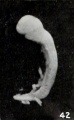
Figs. 42-50. Various forms of cyemata classed as fetus compressus: Nos. 1463 (X2).
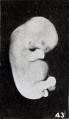
Figs. 42-50. Various forms of cyemata classed as fetus compressus: Nos. 1806 (X 1.5).
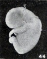
Figs. 42-50. Various forms of cyemata classed as fetus compressus: Nos. 1301 (X8.5).
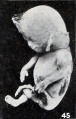
Figs. 42-50. Various forms of cyemata classed as fetus compressus: Nos. 1513 (X0.58).
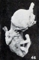
Figs. 42-50. Various forms of cyemata classed as fetus compressus: Nos. 1495c (X0.58).
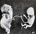
Figs. 42-50. Various forms of cyemata classed as fetus compressus: Nos. 896 (X.55).
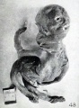
Figs. 42-50. Various forms of cyemata classed as fetus compressus: Nos. 1925 (X.65).
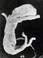
Figs. 42-50. Various forms of cyemata classed as fetus compressus: Nos. 1239 (X0.55).
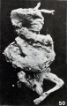
Figs. 42-50. Various forms of cyemata classed as fetus compressus: Nos. 1515 (X0.58).
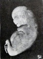
Fig. 51. Distortion due to maceration and retention in utero. No. 1931. XI.
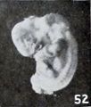
Figs. 52-63. Illustrating the absence of fundamental differences between the group of fetus compressus and grade 3 of the normal specimens. No. 12956 (fig. 52) pathological.
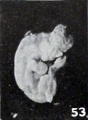
Figs. 52-63. Illustrating the absence of fundamental differences between the group of fetus compressus and grade 3 of the normal specimens. No. 532 (fig. 53) normal. Xl.8.
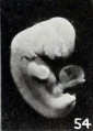
Figs. 52-63. Illustrating the absence of fundamental differences between the group of fetus compressus and grade 3 of the normal specimens. No. 1475 (fig. 54) pathological. X1.5.
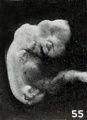
Figs. 52-63. Illustrating the absence of fundamental differences between the group of fetus compressus and grade 3 of the normal specimens. No. 852 (fig. 55) normal. X2.
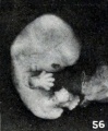
Figs. 52-63. Illustrating the absence of fundamental differences between the group of fetus compressus and grade 3 of the normal specimens. No. 1750 (fig. 56) normal. X1.5. (To be compared with No. 1806, fig. 43.)
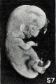
Figs. 52-63. Illustrating the absence of fundamental differences between the group of fetus compressus and grade 3 of the normal specimens. No. 1779 (fig. 57) normal. XI.
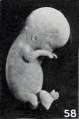
Figs. 52-63. Illustrating the absence of fundamental differences between the group of fetus compressus and grade 3 of the normal specimens. No. 651a (fig. 58) pathological. XI.
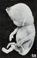
Figs. 52-63. Illustrating the absence of fundamental differences between the group of fetus compressus and grade 3 of the normal specimens. No. 1513 (fig. 59) pathological. X0.58.
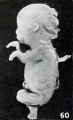
Figs. 52-63. Illustrating the absence of fundamental differences between the group of fetus compressus and grade 3 of the normal specimens. No. 1350 (fig. 60) normal. X0.58.
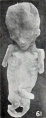
Figs. 52-63. Illustrating the absence of fundamental differences between the group of fetus compressus and grade 3 of the normal specimens. No. 1474<z (fig. 61) normal. X0.58. (To be compared with No. 896, fig. 47 )
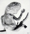
Figs. 52-63. Illustrating the absence of fundamental differences between the group of fetus compressus and grade 3 of the normal specimens. No. 1758 (fig. 62) pathological. X0.5. No. 1532 (fig. 63) normal. X0.5.
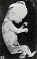
Figs. 52-63. Illustrating the absence of fundamental differences between the group of fetus compressus and grade 3 of the normal specimens. No. 1532 (fig. 63) normal. X0.5.
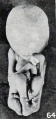
Fig. 64. Fusion of epidermis on lower extremities during maceration. No. 1859. X0.5. Fig.
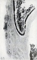
Fig. 65. Epidermal thickenings. No. 316. X7.5.
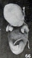
Fig. 66. Early drooping and distortion of extremities. No. 1710. X 1.
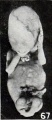
Fig. 67. Showing diversity in position and distortion of extremities. No. 1751. XO 5
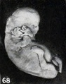
Fig. 68. A similar but younger specimen. No. 1931. XI.
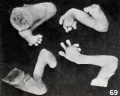
Fig. 69. Distortion of digits. No. 1751. XI.
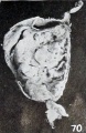
Fig. 70. Unilateral maceration bleb on cord. No. 1523. X0.5.
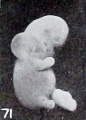
Fig. 71. Bleb formation on neck, same specimen. XI.
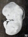
Fig. 72. Bleb formation in whole nuchal region. No. 2261. XI. 5.


Plate 6

Fig. 73. Nodular fibrosis of the amnion. No. 868. Xl-35.
Fig. 74. Nodular fibrosis of the amnion. No. 1048. X0.66.
Fig. 75. Large, clear, normal decidual cells. No. 1599. X200.
Fig. 76. Maceration, hemorrhage, and lysis, with early transformation of the decidua. No. 872. X200.
Fig. 77. Further transformation of the polygonal decidual cells into the spindle and fibroblast type of cells. Same specimen as figure 76. X200.
Fig. 78. Longitudinal section of right ovary, showing carcinomatous nodule to the right. No. 865. X2.
Fig. 79. Same specimen. Portion of carcinomatous nodule. X27.
Fig. 80. Left ovary from same case in longitudinal section, showing disk-shaped teratoma in section. X2. Fig.
Fig. 81. Teratoma in section, same case. X11.5.
Fig. 82. Remarkable vascular proliferation in the chorionic membrane. No. 309. X 120.
- Plate 6: Fig. 73 | Fig. 74 | Fig. 75 | Fig. 76 | Fig. 77 | Fig. 78 | Fig. 79 | Fig. 80 | Fig. 81 | Fig. 82 | Chapter 4 Pathologic analysis
| Embryology - 27 Apr 2024 |
|---|
| Google Translate - select your language from the list shown below (this will open a new external page) |
|
العربية | català | 中文 | 中國傳統的 | français | Deutsche | עִברִית | हिंदी | bahasa Indonesia | italiano | 日本語 | 한국어 | မြန်မာ | Pilipino | Polskie | português | ਪੰਜਾਬੀ ਦੇ | Română | русский | Español | Swahili | Svensk | ไทย | Türkçe | اردو | ייִדיש | Tiếng Việt These external translations are automated and may not be accurate. (More? About Translations) |
Mall FP. and Meyer AW. Studies on abortuses: a survey of pathologic ova in the Carnegie Embryological Collection. (1921) Contrib. Embryol., Carnegie Inst. Wash. Publ. 275, 12: 1-364.
- In this historic 1921 pathology paper, figures and plates of abnormal embryos are not suitable for young students.
1921 Carnegie Collection - Abnormal: Preface | 1 Collection origin | 2 Care and utilization | 3 Classification | 4 Pathologic analysis | 5 Size | 6 Sex incidence | 7 Localized anomalies | 8 Hydatiform uterine | 9 Hydatiform tubal | Chapter 10 Alleged superfetation | 11 Ovarian Pregnancy | 12 Lysis and resorption | 13 Postmortem intrauterine | 14 Hofbauer cells | 15 Villi | 16 Villous nodules | 17 Syphilitic changes | 18 Aspects | Bibliography | Figures | Contribution No.56 | Contributions Series | Embryology History
| Historic Disclaimer - information about historic embryology pages |
|---|
|