Carnegie stage 10
| Embryology - 19 Apr 2024 |
|---|
| Google Translate - select your language from the list shown below (this will open a new external page) |
|
العربية | català | 中文 | 中國傳統的 | français | Deutsche | עִברִית | हिंदी | bahasa Indonesia | italiano | 日本語 | 한국어 | မြန်မာ | Pilipino | Polskie | português | ਪੰਜਾਬੀ ਦੇ | Română | русский | Español | Swahili | Svensk | ไทย | Türkçe | اردو | ייִדיש | Tiếng Việt These external translations are automated and may not be accurate. (More? About Translations) |
Introduction

Facts
Week 4, 22 - 23 days, 2 - 3.5 mm, Somite number 4 - 12
Gestational Age GA - week 6
Features
- Somite Number 4 - 12, rostral neuropore, neural folds in region of developing brain, neural tube, somites, caudal neuropore, neural fold fuses, remnant of amniotic sac
Summary
- Ectoderm: Neural fold deeepens, edges approach midline, neural fold fuses, neural plate folds ventrally in brain region
- Mesoderm: Somitogenesis, continued segmentation of paraxial mesoderm (4 - 12 somite pairs)
See also Events
Identify
The rostral neuropore, neural folds in region of developing brain, neural tube, somites (note the different number formed), caudal neuropore, neural fold fuses, cut edge of amniotic sac.
| Stage 10 Links: Week 4 | Gastrulation | Lecture | Practical | Image Gallery | Carnegie Embryos | Embryos | Category:Carnegie Stage 10 | Next Stage 11 |
| Historic Papers: 1910 | 1917 | 1926 | 1939 | 1943 | 1957 | 1985 |
| Week: | 1 | 2 | 3 | 4 | 5 | 6 | 7 | 8 |
| Carnegie stage: | 1 2 3 4 | 5 6 | 7 8 9 | 10 11 12 13 | 14 15 | 16 17 | 18 19 | 20 21 22 23 |
- Carnegie Stages: 1 | 2 | 3 | 4 | 5 | 6 | 7 | 8 | 9 | 10 | 11 | 12 | 13 | 14 | 15 | 16 | 17 | 18 | 19 | 20 | 21 | 22 | 23 | About Stages | Timeline
Bright Field


Scanning EM


Image Source: Scanning electron micrographs of the Carnegie stages of the early human embryos are reproduced with the permission of Prof Kathy Sulik, from embryos collected by Dr. Vekemans and Tania Attié-Bitach. Images are for educational purposes only and cannot be reproduced electronically or in writing without permission.
Kyoto Collection
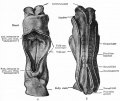
Facts: Week 4, 22 - 23 days, 2 - 3.5 mm, Somite Number 4 - 12
View: This is a dorsal view of the human embryo, the amniotic membrane has been removed. Top embryo is an early stage 10, bottom is late stage 10.
| Early | Late | |
|---|---|---|

|

|

|

| Stage 10 Embryo (12202) - transverse section |
|---|
|


Virtual Slide
| Stage 10 - Dorsal View
|
| Stage 10 | Embryo Slides |
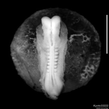
Image source: The Kyoto Collection images are reproduced with the permission of Prof. Kohei Shiota and Prof. Shigehito Yamada, Anatomy and Developmental Biology, Kyoto University Graduate School of Medicine, Kyoto, Japan for educational purposes only and cannot be reproduced electronically or in writing without permission.
Carnegie Collection


| Carnegie Collection - Stage 10 | ||||||||||
|---|---|---|---|---|---|---|---|---|---|---|
| Serial No. | Pairs of somites | Size (mm) | Grade | Fixative | Embedding Medium | Thinness (µm) | Stain | Year | Notes | |
| 391 | 8 | E, 2 Ch., 14 | Good | Formalin | P | 10 | Al. coch. | 1907 | Monograph by Dandy (1910)[1] | |
| 1201 | 7 | E,2 Ch.. 144 | Good | Formalin | P | 8 | H. & or. G. | 1915 | Univ. Chicago No. H 87 | |
| 2795 | 4-5 | E,2 | Poor | Alc. | P | 6 | Al coch,or.G. | 1919 | ||
| 3707 | 12 | E, 1 5 | Good | Formalin | P | 12.5 | I. H. | 1921 | Univ. Calif. No. H 197 | |
| 3709 | 4 | E. 1.4 Ch.. 14.8 | Poor | Formalin | P | 10 | Erythrosin | 1921 | Univ. Chicago No H 279 | |
| 3710 | 12 | E., 3.6 Ch., 19.0 | Good | Formalin | C-P | 10 | H. & or. G. | 1921 | Univ. Chicago No. H 392 | |
| 4216 | 8 | E, 2 Ch, 9.8 | Good | Formalin | P | 15 | ? | 1923 | Monograph by Payne (1925)[2] | |
| 5074 | 10 | E., 3.3 Ch., 10.8 | Exc. | Bouin | P | 10 | Al. coch. | 1925 | Univ. Rochester No. H 10. Monograph by Corner (1929)[3] | |
| 6330 | 7 | E, 2.83 | Good | P | 5 | Ehr. H. | 1931 | Univ. Chicago No. H 1404 | ||
| 6740 | 12 | E., 2.2 | Good | p | C-P | 8 | ? | 1933 | Litzenberg embryo. Studied by Boyden (1940) | |
| 7251 | 8 | E., 1.27 | Good | Formalin | C-P | 10 | (Stain - Haematoxylin Eosin) | 1941 | "Singapore embryo." Univ. Cambridge No. H 98. Studied by Wilson (1914)[4] | |
| 8244 | 6 | E., 1.55 Ch, 8,5 | Good | Alc. | C-P | 8 | (Stain - Haematoxylin Eosin) phlox. | 1944 | ||
| 9870 | 12 | Ch, ca. 8 | Good | Zenker | P | 5 | Various, chiefly carmine | 1952 | Univ. Chicago. No. H 637. Dicephaly | |
Abbreviations
| ||||||||||
| iBook - Carnegie Embryos | |
|---|---|

|
|
Events
For a detailed description of the stage 10 human embryos see the papers published Dandy 1910[1], Corner 1929,[5], Baxter 1939 [6], review by Heuser in 1957[7] and in chapter by O'Rahilly and Müller in 1987.[8]
- Hearing - 10 somites first indication of otic placode invagination[5] 12 somites cells migrate from the otic disc.[9]
- Vision - optic primordia appear.[10]
- Meninges (Spinal Cord) - paraxial mesoderm forms discrete somites, and wherever migration from them has occurred, the cells pass ventromedially toward the notochord. The notochord is, however, still continuous with the gut and in contact, dorsally, with the neural tube (groove). The spinal ganglia and the meninx primitiva will develop in the cell-free spaces between somites and neural tube.[11]
Embryo Examples
Based on O'Rahilly and Müller (1987).[8]
- 4 somites
- Carnegie No. 3709 (University of Chicago H 279). Characterized with outline sketches, by Bartelmez and Evans (1926).
- Histologisch-Embryologisches Institut, Embryo A, Vienna. Fully described by Sternberg (1927).
- Histologisch-Embryologisches Institut, Embryo Ca, Vienna. Fully described by Orts Llorca (1934).
- 4–5 somites
- Florian's Florian Embryo Bi II. The whereabouts of this, the following embryo, and the 10-somite Bi XI (see below) are not known; Florian's collection has not been found since his untimely death during World War II. The embryo Bi II was briefly characterized by Studnicka (1929), cited and partly illustrated by Florian (1928, 1930a).
- Florian's Florian Embryo Bi III. (See note on previous embryo.) Briefly characterized by Studnicka (1929) and cited by Florian (1928).
- Carnegie No. 2795. Cited and briefly characterized by Bartelmez and Evans (1926). The specimen is distorted and somewhat macerated.
- 5 somites
- Anatomisches Institut, Zürich, GM 1954. Described and illustrated by Schenck (1954).
- No. 103, Department of Anatomy, Tohoku University, Sendai. Distribution of alkaline phosphatase studied by Mori (1959a) in this and in another (No. 101), possibly 8-somite, embryo.
- 5–6 somites - Pfannenstiel “Klb” (originally at Giessen; was in Keibel's Institute at Freiburg i. Br. about 1911, may now be in Berlin). This well known embryo is No. 3 in the Keibel and Elze Normentafel (1908). Models by Kroemer (1903). A partial set of tracings made by H. M. Evans is in the Carnegie Collection, No. Template:CE5463.
- 6 somites
- Carnegie No. 8244. Somewhat distorted; histologically fair.
- University of Michigan No. 71, Ann Arbor. Briefly described by Arey and Henderson (1943). A full description in an unpublished doctoral dissertation is in the files of L. B. Arey at Northwestern University, Chicago.
- Carnegie No 8818 (University of Chicago H 338) Pathological, not used in present study. Listed here because cited by Bartelmez and Evans (1926).
- 6–7 somites
- His’s Embryo “SR.” Cited by His (1880) and by Bartelmez and Evans (1926). Has been studied only in the gross.
- Embryo LM (present location unknown). Cited here from manuscript notes at Carnegie laboratory, made from Russian text of Burow (1928). Condition said to be poor.
- 7 somites
- Embryo “Ludwig,” Berlin. Described by Streiter (1951). This specimen, which is somewhat macerated, is in certain characteristics considerably in advance of others of similar somitic number.
- Carnegie No. 6330 (University of Chicago H 1404). Extensive manuscript notes on this specimen, made under the supervision of G. W. Bartelmez, are in the files of the Carnegie laboratory.
- 8 somites
- Carnegie No. 4216. Described by Payne (1925), and very frequently cited.
- Dublin. Described by West (1930); see also Arey (1938). Photographs and models are in the Carnegie Collection, No. 4923. Bartelmez (personal communication) thinks that this distorted embryo had only 5–6 somites.
- Carnegie Embryo 391. Described by Dandy (1910)[1] and frequently cited (cf. Bartelmez and Evans, 1926, with additional illustrations). There were neither camera drawings nor photographs of the intact specimen, and therefore the reconstructions are not entirely satisfactory. The plaster models now at the Carnegie laboratory were made by O. Heard under the supervision of Bartelmez for the paper by Bartelmez and Evans (1926). The apparent lack of fusion of the neural folds described by Dandy is an artifact produced by a crack.
- Carnegie No. 1201 (University of Chicago H 87). Described briefly by Evans and Bartelmez (1917); cited, with illustrations, by Bartelmez and Evans (1926).
- Embryologisches Institut, Embryo Ct, Vienna. Fully described by Politzer (1930). Arey (1938) counts 8 paired somites in this embryo instead of 7 as stated by Politzer.
- University of Cambridge, Department of Anatomy H 98. Photographs and models in Carnegie Collection, No. 7251. Described by J. T. Wilson (1914). Cited by Bartelmez and Evans (1926), who consider it slightly abnormal in form although good histologically.
- 9 somites
- Embryo “Esch I,” Marburg. Elaborately described by Veit and Esch (1922), and cited, with illustrations, by Bartelmez and Evans (1926), who count 9 somites instead of 8 as stated by the original authors. Chorionic villi studied in detail by Ortmann (1938). Photographs and models are in the Carnegie Collection, No. 4251.
- Embryo “Du Ga,” Geneva. Described by Eternod (1896); models by Ziegler were distributed commercially. Cited by Bartelmez (1922) and Bartelmez and Evans (1926), with illustrations. Tracings made by H. M. Evans at Geneva and models are in the Carnegie Collection, No. 4439.
- Embryo Unger, Keibel Collection, Freiburg i. Br., No. 4 of Keibel and Elze (1908). Listed by Bartelmez and Evans (1926).
- Embryo “Jacobsen,” formerly at Kiel (Graf Spee's collection was destroyed in World War II). Described by von Spee (1887). Listed by Bartelmez and Evans (1926) as having “at least” 9 somites.
- Embryo Ca of Orts Llorca, Madrid. Various details described by Mari Martinez (1950) and Martinez Rovira (1953).
- 9-10 somites - Embryo R. Meyer 335. (Robert Meyer's collection was purchased by the late Hedwig Frey and bequeathed by him to the Anatomisches Institut, University of Zurich.) Listed by Bartelmez and Evans (1926), and cited by Felix (1912).
- 10 somites
- Da2, Anatomical Institute, Basel. Described by Ludwig (1929). Plastic reconstructions. Neural groove closure extends rostral to otic discs.
- Carnegie No. 5074 (University of Rochester H 10). Fully described by Corner (1929), and subjected to volumetric analysis by Boyden (1940). Excellent specimen.
- Grosser's Embryo Schwz (present location unknown). Briefly described, without illustrations, by Treutler (1931). Preservation said to be not altogether satisfactory.
- Florian’s Embryo Bi XI (See note on Bi II above.) Briefly described, with illustrations, by Politzer and Sternberg (1930); cited and partly illustrated by Florian (1930a).
- Anatomy Department, University of South Wales, Cardiff. Partly described and illustrated by Baxter and Boyd (1939).[6]
- 11 somites
- Embryo T 152, University of Toronto, Department of Anatomy. Cited by Arey (1938).
- Embryo G-dt, Uppsala. Described by Holmdahl (1943) as having 11 well-differentiated pairs of somites with beginning delimitation of 4 more.
- 11–12 somites - Carnegie No. 8970 (University of Chicago H 637). Somewhat damaged. Cited, with illustrations, by Bartelmez (1922) and Bartelmez and Evans (1926).
- 12 somites
- Carnegie No. 3710 (University of Chicago H 392). Cited by Bartelmez (1922) and Bartelmez and Evans (1926).
- Carnegie No. 3707 (University of California H 197). “Legge embryo.” Cited, with illustrations, by Bartelmez and Evans (1926). The coital history accompanying this specimen, which was declared to be reliable, would give it a postovulatory age of either 18 or 39 days; the former seems rather brief but the latter is much too long.
- Litzenberg embryo, University of Minnesota, Minneapolis. Briefly described by J. C. Litzenberg (1933); characterized and subjected to volumetric analysis by Boyden (1940), who counts 12 somites instead of 13–14 as in the original description. Photographs and model in Carnegie Collection, No. 6740.
- M. 24, University of Michigan, Ann Arbor. Cited by Arey (1938).
| Other Stage 10 Embryo Examples | |||||||||||||||||||||||||||||||||||||||||||||||||||||||||||||||||||||||||||||||||||||||||||||||||||||||||||||||||||||||||||||||||||||||||||||||||||||||||||||||||
|---|---|---|---|---|---|---|---|---|---|---|---|---|---|---|---|---|---|---|---|---|---|---|---|---|---|---|---|---|---|---|---|---|---|---|---|---|---|---|---|---|---|---|---|---|---|---|---|---|---|---|---|---|---|---|---|---|---|---|---|---|---|---|---|---|---|---|---|---|---|---|---|---|---|---|---|---|---|---|---|---|---|---|---|---|---|---|---|---|---|---|---|---|---|---|---|---|---|---|---|---|---|---|---|---|---|---|---|---|---|---|---|---|---|---|---|---|---|---|---|---|---|---|---|---|---|---|---|---|---|---|---|---|---|---|---|---|---|---|---|---|---|---|---|---|---|---|---|---|---|---|---|---|---|---|---|---|---|---|---|---|---|
Embryo list below based on O'Rahilly and Müller (1987).
| |||||||||||||||||||||||||||||||||||||||||||||||||||||||||||||||||||||||||||||||||||||||||||||||||||||||||||||||||||||||||||||||||||||||||||||||||||||||||||||||||
| O'Rahilly R. and Müller F. Developmental Stages in Human Embryos. Contrib. Embryol., Carnegie Inst. Wash. 637 (1987). | |||||||||||||||||||||||||||||||||||||||||||||||||||||||||||||||||||||||||||||||||||||||||||||||||||||||||||||||||||||||||||||||||||||||||||||||||||||||||||||||||
See also Carnegie Collection embryo table.
| |||||||||||||||||||||||||||||||||||||||||||||||||||||||||||||||||||||||||||||||||||||||||||||||||||||||||||||||||||||||||||||||||||||||||||||||||||||||||||||||||
References
- ↑ 1.0 1.1 1.2 1.3 1.4 Dandy WE. A human embryo with seven pairs of somites measuring about 2 mm in length. (1910) Amer. J Anat. 10: 85-109.
- ↑ Payne, F. 1925. General description of a 7-somite human embryo. Carnegie Instn. Wash. Publ. 361, Contrib. Embryol., 16,115-124.
- ↑ 3.0 3.1 Corner GW. A well-preserved human embryo of 10 somites. (1929) Contrib. Embryol., Carnegie Inst. Wash. Publ. 394, 20:81-102.
- ↑ 4.0 4.1 Wilson JT. Observations upon young human embryos. (1914) J Anat Physiol., 48(3): 315-51 PMID 17233002 PMC1288949
- ↑ 5.0 5.1 Corner GW. A well-preserved human embryo of 10 somites. (1929) Carnegie Instn. Wash. Publ. 394, Contrib. Embryol., Carnegie Inst. Wash. 20: 81-102.
- ↑ 6.0 6.1 6.2 Baxter JS. and Boyd JD. Observations on the neural crest of a ten-somite human embryo. (1939) J Anat. 73: 318–326. PMID 17104759
- ↑ Heuser CH. and Corner GW. Developmental horizons in human embryos. Description of age group X, 4 to 12 somites. (1957) Carnegie Instn Wash Publ 611, Contrib. Embryol., 36: 29-39.
- ↑ 8.0 8.1 O'Rahilly R. and Müller F. Developmental Stages in Human Embryos. Contrib. Embryol., Carnegie Inst. Wash. 637 (1987).
- ↑ Bartelmez GW. and Evans HM. Development of the human embryo during the period of somite formation, including embryos with 2 to 16 pairs of somites. (1926) Contrib. Embryol., Carnegie Inst. Wash. Publ. 362, 17: 1-67.
- ↑ Pearson AA. (1980). The development of the eyelids. Part I. External features. J. Anat. , 130, 33-42. PMID: 7364662
- ↑ Sensenig EC. The early development of the meninges of the spinal cord in human embryos. (1951) Contrib. Embryol., Carnegie Inst. Wash. Publ. 611.
- ↑ Payne, F. 1925. General description of a 7-somite human embryo. Carnegie Instn. Wash. Publ. 361, Contrib. Embryol., 16,115-124.
Additional Images
Historic
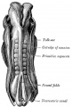
Historic drawing of human embryo, 2.11 mm. in length. (After Eternod.)
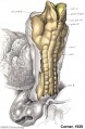
A well-preserved human embryo of 10 somites by George W. Corner
Eternod. From models by Ziegler.
Eternod. From models by Ziegler.


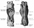
Dandy WE. A human embryo with seven pairs of somites measuring about 2 mm in length. (1910) Amer. J Anat. 10: 85-109.
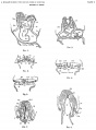
Plate 1 Sections
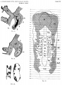
Plate 2 Sections
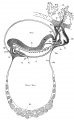
Plate 3 Sagittal view of embryo vascular system
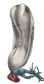
Plate 4 Dorsol-lateral view of embryo model
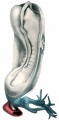
Plate 5 Dorsol-lateral view somites and mesoderm
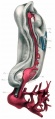
Plate 6 Dorsol-lateral view brain vesicles and aorta


Veit O. Head ganglia of an embryo of eight somite pairs {Kopfganglienleisten Bei Einem Menschlichen Embryo Von 8 Somitenpaaren). (1918) Anat. Hefte, Bd. 56, S. 305-320.
Baxter JS. and Boyd JD. Observations on the neural crest of a ten-somite human embryo. (1939) J Anat. 73: 318–326. PMID 17104759
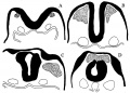
Text-fig 1 reconstruction of cephalic portion of nervous system
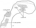
Text-fig 2 acoustico-facial neural crest primordia in certain human embryos
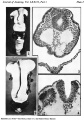
Plate 1
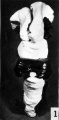
Fig 1 Reconstruction of the future brain region viewed from above and behind.
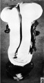
Fig 2 Reconstruction of the future brain region viewed from in front
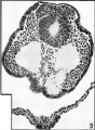
Fig 3 cranial part of the acoustico-facial primordium
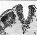
Fig 4 level of the eighth somite
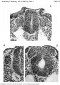
Plate 2
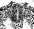
Fig 5 caudal part of the acoustico-facial primordium
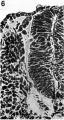
Fig 6 level of the first somite
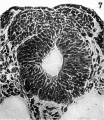
Fig 7 cranial part of the acoustico-facial neural crest primordium


Heuser CH. and Corner GW. Developmental horizons in human embryos. Description of age group X, 4 to 12 somites. (1957) Carnegie Instn Wash Publ 611, Contrib. Embryol., 36: 29-39.
- Carnegie Stages: 1 | 2 | 3 | 4 | 5 | 6 | 7 | 8 | 9 | 10 | 11 | 12 | 13 | 14 | 15 | 16 | 17 | 18 | 19 | 20 | 21 | 22 | 23 | About Stages | Timeline
Cite this page: Hill, M.A. (2024, April 19) Embryology Carnegie stage 10. Retrieved from https://embryology.med.unsw.edu.au/embryology/index.php/Carnegie_stage_10
- © Dr Mark Hill 2024, UNSW Embryology ISBN: 978 0 7334 2609 4 - UNSW CRICOS Provider Code No. 00098G