Introduction
This page links to sample embryology images that have been digitised to a new format that allows the viewer to both zoom and move the image on the screen as you would with an online map programs and online virtual slides. These images have also been optimised and formatted for viewing on all mobile devices.
UNSW Students please note these are not the Histology and Pathology Virtual Slide Set (located here).
- Embryo Virtual Slide Links: Embryo | Electron Microscopy | Historic | Permalinks Help
Instructions
The collapsed tables below show controls and navigation for viewing on different platforms.
| Mobile
|
- Simple zoom in (+) and zoom out (-) control (shown at the top left of each image).
- Mobile devices can also just double tap the screen to zoom.
|
|
| Desktop
|
|
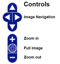
Image Controls (shown at the top left of each image)
- Desktop version, click on either the thumbnail image below or the image label to open the new image.
- The image controls are located at the top left, or zoom can be controlled by double clicking or with mouse wheel.
- Use the browser back button to return to this page.
Permalink (shown at the bottom right of each image) Allows a permanent link to be created to the current image view. (More? how to use Permalink)
|
|
| Original
|
- Opens the Preview version of the original online image file (scaled for preview).
- Click the Preview Image to open the full size image (Note, this may be a large file, image can also be zoomed on mobile and desktop devices).
- The Virtual Slide should also be available in the summary information shown below the image.
|
|
Want to make your own link to a specific part of a virtual slide? Read how to use Permalink.
| Technical Note
|
| Both the Mobile and Desktop links appear as external links as the image files are located outside the Wiki database.
When you have opened a Virtual Slide, you can use your browser back button to return to this current page.
|
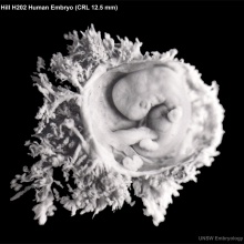
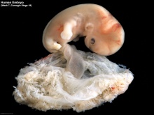
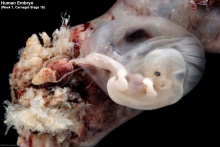
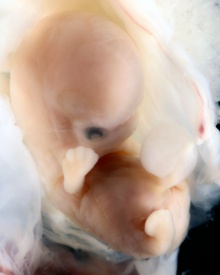
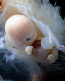
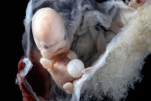
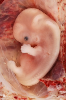
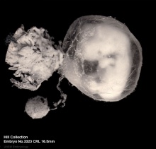
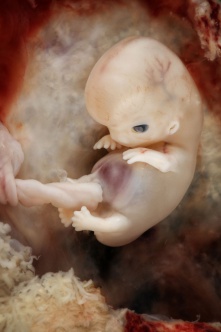
Embryo Stages
Embryonic Development
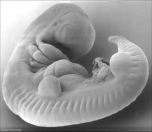
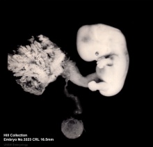
Stage 7
Stage 10
Stage 13
Stage 14
Stage 15
Stage 17
Stage 18
Stage 19
Stage 20
Stage 21
Stage 22

|
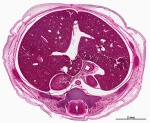
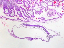
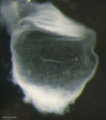
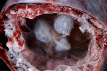
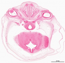
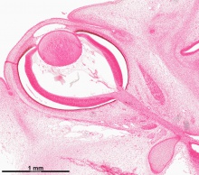
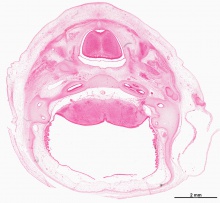
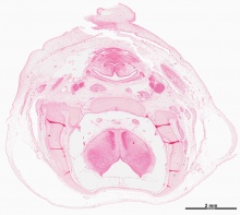
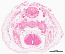
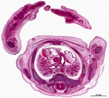
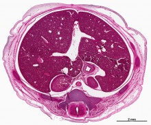
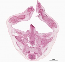
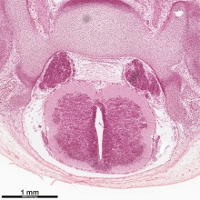
These are high resolution histological sections (Stain - Haematoxylin Eosin) through regions of the Stage 22 human embryo from the serial section set. Image bottom is dorsal and top is ventral, sections are viewing the embryo from below.
|
Feature Links
| Virtual Slide Features - Stage 22 Liver
|
|
Virtual Slide - Stage 22 Liver and Ductus Venosus All Virtual Slides
The links shown in the table below are to specific features shown on the Human embryo (stage 22) Liver and Ductus Venosus virtual slide. See also notes on Liver Development
Clicking the text will open the slide at a detailed view with the structure generally located in the centre of the view. The slide then can also be zoomed out from the set magnification using the controls in the upper left or the mouse.
Use your browser back button to return to this table.
|
You can also make your own selected feature view.
- Set the virtual slide to the region and zoom of interest.
- Click the Permalink (lower righthand corner).
- Then bookmark in your browser, or copy the web address.
See also Permalink help
|
| Cardiovascular
|
Liver
|
Endocrine
|
Musculoskeletal
|
Neural
|
Gastrointestinal
stomach (pylorus)
|
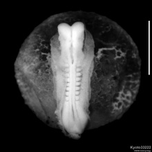
| Virtual Slide Features - Stage 22 Spinal Cord
|
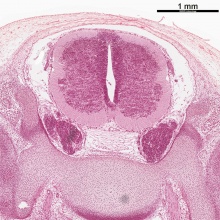
Spinal Cord Features
|
The links shown are to specific features shown on the Human embryo (stage 22) Spinal Cord virtual slide. See also notes on Spinal Cord Development
Clicking the text will open the slide at a detailed view with the structure generally located in the centre of the view. The slide then can also be zoomed out from the set magnification using the controls in the upper left or the mouse. Use your browser back button to return to this table.
Other Features
|
| All Virtual Slides
|
Making your own Link - You can also make your own selected feature view. (See also Permalink help)
- Set the virtual slide to the region and zoom of interest.
- Click the Permalink (lower righthand corner).
- Then bookmark in your browser, or copy the web address.
|
|
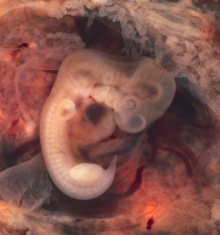
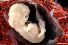
Stage 23
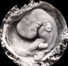
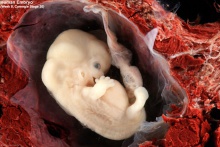
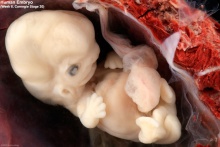
Fetal
Fetal Development
Week 9-10
Ovary
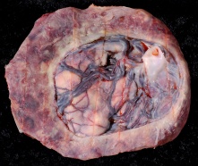
Placenta
Glossary Links
- Glossary: A | B | C | D | E | F | G | H | I | J | K | L | M | N | O | P | Q | R | S | T | U | V | W | X | Y | Z | Numbers | Symbols | Term Link
Cite this page: Hill, M.A. (2024, April 16) Embryology Embryo Virtual Slides. Retrieved from https://embryology.med.unsw.edu.au/embryology/index.php/Embryo_Virtual_Slides
- What Links Here?
- © Dr Mark Hill 2024, UNSW Embryology ISBN: 978 0 7334 2609 4 - UNSW CRICOS Provider Code No. 00098G


