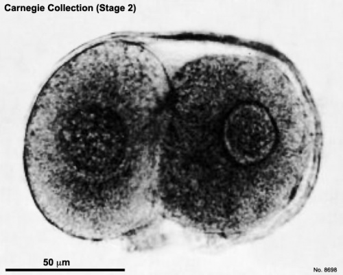
Carnegie stage 2
| Embryology - 16 Apr 2024 |
|---|
| Google Translate - select your language from the list shown below (this will open a new external page) |
|
العربية | català | 中文 | 中國傳統的 | français | Deutsche | עִברִית | हिंदी | bahasa Indonesia | italiano | 日本語 | 한국어 | မြန်မာ | Pilipino | Polskie | português | ਪੰਜਾਬੀ ਦੇ | Română | русский | Español | Swahili | Svensk | ไทย | Türkçe | اردو | ייִדיש | Tiếng Việt These external translations are automated and may not be accurate. (More? About Translations) |
Introduction
|
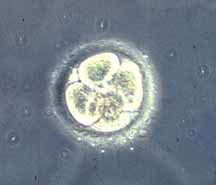
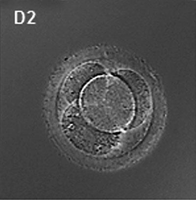
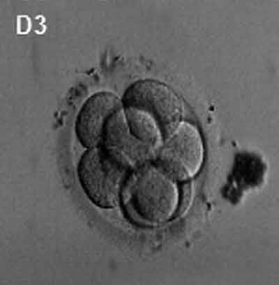
First cell mitotic divisions of the zygote forming initially 2 blastomeres that continue to divide to form the morula, a "berry", a solid mass of cells.
This early mitosis is a unique embryonic cell cycle (M, S, M phases) compared to adult (M, G1, S, G2, M phase). With virtually no G1 or G2 phases this results in a reduction in cytoplasmic volume of each daughter cell with each cell division. Summary
|
| Week: | 1 | 2 | 3 | 4 | 5 | 6 | 7 | 8 |
| Carnegie stage: | 1 2 3 4 | 5 6 | 7 8 9 | 10 11 12 13 | 14 15 | 16 17 | 18 19 | 20 21 22 23 |
- Carnegie Stages: 1 | 2 | 3 | 4 | 5 | 6 | 7 | 8 | 9 | 10 | 11 | 12 | 13 | 14 | 15 | 16 | 17 | 18 | 19 | 20 | 21 | 22 | 23 | About Stages | Timeline
|
|
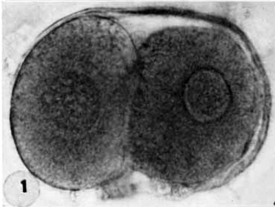
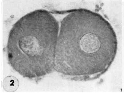
| Two-cell segmenting egg | Mid-serial section two-cell |
|
|
| Human Embryo (day 2) | Human Embryo (day 3) |
See also Human oocyte to blastocyst
Carnegie Collection
Two cell blastomere (1-2 days)
| Carnegie Collection - Stage 2 | ||||||||||
|---|---|---|---|---|---|---|---|---|---|---|
| Serial No. | Normality | No. of Blastomeres | Size of Fixed Specimen (µm) | Fixative | Embedding Medium | Thinness (µm) | Stain | Year | Notes | |
| 8190 | abnormal | 9 | 160x104 | Alc. & Bouin | C-P | 6 | Iron h., or G | 1943 | Hertig et al. (1954) | |
| 8260 | in vitro | 2 | 50x75 | Bouin | C-P | 8 | (Stain - Haematoxylin Eosin) | 1944 | Menkin and Rock (1948) | |
| 8450 | abnormal | 8 | 100x96 | Alc. & Bouin | C-P | 6 | (Stain - Haematoxylin Eosin), phlox. | 1947 | Hertig et al. (1954) | |
| 8452 | abnormal | 12 | 110x93 | Alc. & Bouin | C-P | 6 | (Stain - Haematoxylin Eosin), phlox. | 1946 | Hertig et al. (1954) | |
| 8500.1 | in vitro | 3 | 50x86 | Bouin | C-P | 8 | (Stain - Haematoxylin Eosin) | 1947 | Menkin and Rock (1948) | |
| 8630 | abnormal | 5 | 104x94 | Alc. & Bouin | C-P | 6 | (Stain - Haematoxylin Eosin) | 1948 | Hertig et al. (1954) | |
| 8698 | normal | 2 | 122x88 | Alc. & Bouin | C-P | 6 | (Stain - Haematoxylin Eosin) | 1949 | Hertig et al. (1954) | |
| 8904 | normal | 12 | 115 | Specimen lost | 1951 | Hertig et al. (1954) | ||||
Abbreviations
| ||||||||||
| ||||||||||
Events
Some Recent Findings
|
References
Hertig AT. Rock J. and Adams EC. A description of 34 human ova within the first 17 days of development. (1956) Amer. J Anat., 98:435-493.
References
Hertig AT. Rock J. and Adams EC. A description of 34 human ova within the first 17 days of development. (1956) Amer. J Anat., 98:435-493.
Additional Images
- Carnegie Stages: 1 | 2 | 3 | 4 | 5 | 6 | 7 | 8 | 9 | 10 | 11 | 12 | 13 | 14 | 15 | 16 | 17 | 18 | 19 | 20 | 21 | 22 | 23 | About Stages | Timeline
Cite this page: Hill, M.A. (2024, April 16) Embryology Carnegie stage 2. Retrieved from https://embryology.med.unsw.edu.au/embryology/index.php/Carnegie_stage_2
- © Dr Mark Hill 2024, UNSW Embryology ISBN: 978 0 7334 2609 4 - UNSW CRICOS Provider Code No. 00098G
