File:Thomson1919 fig04.jpg
Original file (1,251 × 1,089 pixels, file size: 316 KB, MIME type: image/jpeg)
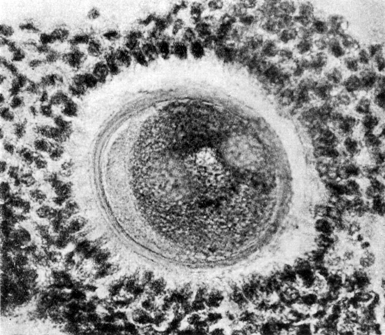
Fig. 4. Human ovum in discus proligerus in anaphase of first division and commencement of metaphase of second division
x600.
Fig. 4, thickness of section 0.01 mm, stained with Weigert’s Iron Haematoxylin and Van Giesen. This represents the appearance displayed by the contiguous section to fig. 3; it calls for less detailed description since it represents many of the same details of structure already referred to in fig. 3. The essential points of difference are: first, that the section has passed above or below the position of the polar body, thus giving us a clue to its size. As the sections are 0.01 mm thick it follows that the largest diameter of the polar body cannot exceed that measure; second, the material (coagulum?) filling the space between the cytoplasm and the Zona pellucida is uniform in structure, and the large vacuoles have disappeared; third, the size of the three spherical vesicles within the cytoplasm is more nearly equal, suggesting that they have been cut in the axis of the amphiaster. Associated with each lateral sphere, which, as we have already noted, is to be regarded as a centrosphere, may be seen a darkly stained granule, occupying a position, if not quite in the centre, at least near it. ’ These centrospheres are not so precisely defined from the surrounding cytoplasm as in fig. 3, nor are their contents so clear; but these are appearances which may be due to the density of the staining, or to the slight variation in the thickness of the sections.
The nuclear material of the daughter cell, which lies intermediate in position between the two centrospheres, exhibits a granular appearance due to the presence of chromatin grains, some of which are arranged in thread-like fashion.
| Historic Disclaimer - information about historic embryology pages |
|---|
 |
- Human Ovum Links: Fig 1. Prophase I | Fig 2. | | Fig 3. | Plate 10 | Plate 11 | Plate 12
| Online Editor Notes |
|---|
|
Nature Obituary 1935 - Prof. Arthur Thomson (1858 - 1935)
Nature 135, 295-295 (23 February 1935) | doi:10.1038/135295a0 |
|
Reference
Cite this page: Hill, M.A. (2024, May 9) Embryology Thomson1919 fig04.jpg. Retrieved from https://embryology.med.unsw.edu.au/embryology/index.php/File:Thomson1919_fig04.jpg
- © Dr Mark Hill 2024, UNSW Embryology ISBN: 978 0 7334 2609 4 - UNSW CRICOS Provider Code No. 00098G
File history
Click on a date/time to view the file as it appeared at that time.
| Date/Time | Thumbnail | Dimensions | User | Comment | |
|---|---|---|---|---|---|
| current | 13:58, 6 August 2015 | | 1,251 × 1,089 (316 KB) | Z8600021 (talk | contribs) | |
| 13:57, 6 August 2015 | 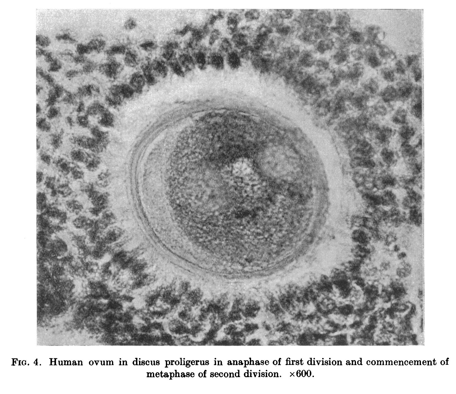 | 1,482 × 1,281 (486 KB) | Z8600021 (talk | contribs) |
You cannot overwrite this file.
File usage
The following page uses this file: