Category:Human Embryo
From Embryology
This Embryology category shows pages and media related to human embryonic development occurring in the first 8 weeks of human development.
Subcategories
This category has the following 72 subcategories, out of 72 total.
C
- Carnegie Collection
- Carnegie Embryo 1267
- Carnegie Embryo 1324
- Carnegie Embryo 1332
- Carnegie Embryo 1534
- Carnegie Embryo 175
- Carnegie Embryo 190
- Carnegie Embryo 199
- Carnegie Embryo 2114
- Carnegie Embryo 219
- Carnegie Embryo 2256
- Carnegie Embryo 293
- Carnegie Embryo 368
- Carnegie Embryo 390
- Carnegie Embryo 409
- Carnegie Embryo 423
- Carnegie Embryo 424
- Carnegie Embryo 426
- Carnegie Embryo 431
- Carnegie Embryo 4361
- Carnegie Embryo 437
- Carnegie Embryo 452
- Carnegie Embryo 484
- Carnegie Embryo 490
- Carnegie Embryo 491
- Carnegie Embryo 508
- Carnegie Embryo 509
- Carnegie Embryo 511
- Carnegie Embryo 576
- Carnegie Embryo 6150
- Carnegie Embryo 626
- Carnegie Embryo 657
- Carnegie Embryo 709
- Carnegie Embryo 7900
- Carnegie Embryo 84
- Carnegie Embryo 8913
- Carnegie Stage 1
- Carnegie Stage 10
- Carnegie Stage 11
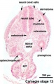
- Carnegie Stage 12
- Carnegie Stage 13
- Carnegie Stage 15
- Carnegie Stage 16
- Carnegie Stage 17
- Carnegie Stage 18
- Carnegie Stage 19
- Carnegie Stage 20
- Carnegie Stage 21
- Carnegie Stage 22
- Carnegie Stage 23
- Carnegie Stage 3
- Carnegie Stage 6
- Carnegie Stage 7
- Carnegie Stage 8
- Carnegie Stage 9
E
Pages in category 'Human Embryo'
The following 200 pages are in this category, out of 261 total.
(previous page) (next page)A
- Template:Abdominal Wall Muscle Timeline table
- Abnormal Development - Biological Toxins
- Abnormal Development - Chemicals
- Abnormal Development - Drugs
- Abnormal Development - Herbal Drugs
- Abnormal Development - Illegal Drugs
- Abnormal Development - Thalidomide
- Alpha-Fetoprotein
- Amniocentesis
- Assisted Reproductive Technology
- Assisted Reproductive Technology - Glossary
- Australian Drug Categories
- Australian In Vitro Fertilization and Donor Questions
B
C
- Carnegie stage 1
- Carnegie stage 10
- Carnegie stage 10 gallery
- Carnegie stage 11
- Carnegie stage 12
- Carnegie stage 13
- Carnegie stage 14
- Carnegie stage 15
- Carnegie stage 16
- Carnegie stage 17
- Carnegie Stage 17 Movie
- Carnegie stage 18
- Carnegie stage 19
- Carnegie stage 2
- Carnegie stage 20
- Carnegie stage 21
- Carnegie stage 22
- Carnegie stage 22 - selected serial sections
- Carnegie stage 22 - serial sections
- Carnegie stage 23
- Carnegie stage 3
- Carnegie stage 4
- Carnegie stage 5
- Carnegie stage 6
- Carnegie stage 7
- Carnegie stage 8
- Carnegie stage 9
- Carnegie stage 9 gallery
- Template:Carnegie stage table 1
- Template:Carnegie stages
- Carnegie Stages
- Template talk:Carnegie stages
- Chorionic villus sampling
- Comparative Genomic Hybridization
- Template:CRL
- Template:CS1
- Template:CS10
- Template:CS11
- Template:CS12
- Template:CS13
- Template:CS14
- Template:CS15
- Template:CS16
- Template:CS17
- Template:CS18
- Template:CS19
- Template:CS2
- Template:CS20
- Template:CS21
- Template:CS22
- Template:CS23
- Template:CS3
- Template:CS4
- Template:CS5
- Template:CS6
- Template:CS7
- Template:CS8
- Template:CS9
D
E
F
G
H
- Template:Harvard Collection table01
- Human Abnormal Development
- Human Chorionic Gonadotropin
- Template:Human embryo Hertwig G31 table
- Template:Human embryo Klb table
- Template:Human embryo Meyer 300 table
- Template:Human embryo Pfannenstiel III table
- Template:Human embryo Strahl 4mm table
- Template:Human Eyelid timeline table
M
O
P
- Paper - A fifteen-somite human embryo
- Paper - A human embryo of the pre-somite period from the uterine tube
- Paper - A Human Embryo of Thirteen Somites
- Paper - A Human Embryo with Seven Pairs of Somites Measuring about 2 mm in Length
- Paper - A Human Ovum at the Previllous Stage
- Paper - A previllous human embryo (1946)
- Paper - Congenital absence of ventrolateral abdominal musculature (1946)
- Paper - Description of a 4 mm human embryo (1906)
- Special:Badtitle/NS501:Paper - Description of a Human Embryo of 13-14 Mesodermic Somites
- Paper - Development of the human placenta in the first three months of gestation (1960)
- Paper - Development of the trigone of the bladder and the termination of the mesonephric ducts (1946)
- Paper - Growth allometry of the myocardium in human embryos from stages 15 to 23
- Paper - Hydatiform degeneration in an early human embryo (1946)
- Paper - Observations on the neural crest of a ten-somite human embryo (1939)
- Paper - On measuring human embryos
- Paper - On Ossification Centers in Human Embryos
- Paper - On The Age Of Human Embryos
- Paper - Principles of growth and repair in membrane bones (1946)
- Paper - Sequential innervation of the intestinal loop in the human embryo
- Paper - Simple formulae for estimating the age and size of human embryos
- Paper - Structural organization of the human cerebral cortex prior to the appearance of the cortical plate (1983)
- Paper - The Anatomy of a 17.8 mm Human Embryo
- Paper - The Anatomy of the Head End of a 20 mm Human Embryo
- Paper - The chondrocranium of a 20 mm human embryo
- Paper - The development of the human brain stage 12
- Paper - The development of the islands of Langerhans in the human embryo (1903)
- Paper - The Development of the Nose and of the Pharynx and its Derivatives in Man
- Paper - The development of the subcutaneous vascular plexus in the head of the human embryo (1923)
- Paper - The early development of the eye in staged human embryos
- Paper - The early development of the otic vesicle in staged human embryos
- Paper - The first appearance of the neural tube and optic primordium in the human embryo at stage 10
- Paper - The human brain at stages 18-20 including the choroid plexuses and the amygdaloid and septal nuclei (1990)
- Paper - The Nuclei of Origin of the Cranial Nerves in the 10 mm Human Embryo
- Paper - The Origin of the Otic and Optic Primordia in Man
- Paper - The timing and sequence of events in the development of the human endocrine system during the embryonic period proper
- Paper The development of the subcutaneous vascular plexus in the head of the human embryo (1923)
- Template:Pearce1903 table1
- Preimplantation Genetic Diagnosis
- Preimplantation Genetic Screening
- Prenatal Diagnosis
- Prenatal Genetic Diagnosis
R
- Template:Ref-Arey1925
- Template:Ref-Atwell1926b
- Template:Ref-Bartelmez1924
- Template:Ref-Bossy1981
- Template:Ref-Bremer1906
- Template:Ref-Chidester1908
- Template:Ref-FlorianHill1935
- Template:Ref-Hamilton1946b
- Template:Ref-Hamilton1946c
- Template:Ref-HamiltonBoyd1960
- Template:Ref-Ingalls1920
- Template:Ref-Ingalls1929
- Template:Ref-Johnson1914
- Template:Ref-Jordan1909a
Media in category 'Human Embryo'
The following 200 files are in this category, out of 738 total.
(previous page) (next page) 050653-01.jpg 1,122 × 1,490; 124 KB
 600px stage14.jpg 421 × 600; 27 KB
 600px stage23.jpg 421 × 600; 26 KB
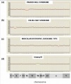
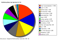 Abnormal AusData81-92.png 523 × 358; 10 KB
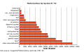 Abnormal AusData81-92Graph.png 509 × 320; 7 KB
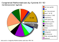 Abnormal81-92-heart.png 481 × 344; 6 KB
 Abnormal81-92-neuron.png 481 × 344; 9 KB
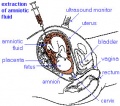 Amniocentesis.jpg 284 × 250; 31 KB
 B2020649-130-1.jpg 917 × 1,311; 123 KB
 B2020649-130-2.jpg 900 × 1,311; 126 KB
 B2020649-160-1.jpg 919 × 1,394; 113 KB
 B2020649-160-2.jpg 919 × 1,394; 137 KB
 B2020649-170-1.jpg 903 × 1,506; 143 KB
 B2020649-170-2.jpg 929 × 1,506; 142 KB
 Barniville1914 fig01.jpg 765 × 758; 59 KB
 Barniville1914 fig21.jpg 1,023 × 1,569; 301 KB
 Barniville1914 figA.jpg 1,000 × 1,237; 269 KB
 Barniville1914 plate01.jpg 2,042 × 2,271; 817 KB
 Barniville1914 plate02.jpg 1,458 × 2,509; 791 KB
 Bartelmez1922-fig01.jpg 900 × 770; 131 KB
 Bartelmez1922-fig02.jpg 1,203 × 1,700; 317 KB
 Bartelmez1922-fig03.jpg 885 × 1,000; 169 KB
 Bartelmez1922-fig04.jpg 1,300 × 750; 108 KB
 Bartelmez1922-fig05.jpg 800 × 750; 164 KB
 Bartelmez1922-fig06.jpg 749 × 1,000; 179 KB
 Bartelmez1922-fig07.jpg 1,200 × 620; 116 KB
 Bartelmez1922-fig08.jpg 1,000 × 1,255; 251 KB
 Bartelmez1922-fig09.jpg 1,200 × 1,558; 287 KB
 Bartelmez1922-fig10.jpg 1,000 × 1,136; 209 KB
 BaxterBoyd1939-fig01.jpg 361 × 736; 43 KB
 BaxterBoyd1939-fig02.jpg 459 × 851; 65 KB
 BaxterBoyd1939-fig03.jpg 732 × 1,000; 170 KB
 BaxterBoyd1939-fig04.jpg 642 × 615; 109 KB
 BaxterBoyd1939-fig05.jpg 1,000 × 864; 263 KB
 BaxterBoyd1939-fig06.jpg 489 × 917; 141 KB
 BaxterBoyd1939-fig07.jpg 795 × 917; 242 KB
 BaxterBoyd1939-plate01.jpg 1,680 × 2,400; 786 KB
 BaxterBoyd1939-plate02.jpg 1,681 × 2,400; 860 KB
 BaxterBoyd1939-text-fig01.jpg 1,283 × 1,000; 138 KB
 BaxterBoyd1939-text-fig02.jpg 1,200 × 861; 133 KB
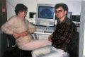 Beverley and Mark 1997.jpg 800 × 542; 88 KB
 Bl130758-01.jpg 753 × 1,091; 59 KB
 Bl170452-01.jpg 1,430 × 1,915; 211 KB
 Boyd1950 fig01.jpg 1,280 × 769; 247 KB
 Boyd1950 fig07.jpg 884 × 1,000; 159 KB
 Boyden1931 fig04.jpg 1,280 × 652; 130 KB
 Boyden1931 fig05.jpg 1,000 × 646; 97 KB
 Carnegie stage 13 caudal trunk.jpg 400 × 625; 53 KB
 CNS primary vesicles.jpg 987 × 562; 49 KB
 CNS secondary vesicles.jpg 987 × 562; 81 KB
 Comparative genomic hybridization 01.jpg 453 × 536; 42 KB
 CSt3.jpg 500 × 377; 20 KB
 Cvs.jpg 336 × 222; 28 KB
 Dandy1910-plate01.jpg 1,738 × 2,359; 541 KB
 Dandy1910-plate02.jpg 1,754 × 2,400; 951 KB
 Dandy1910-plate03.jpg 1,000 × 1,617; 224 KB
 Dandy1910-plate04.jpg 1,000 × 1,875; 174 KB
 Dandy1910-plate05.jpg 1,000 × 2,034; 238 KB
 Dandy1910-plate06.jpg 1,000 × 2,166; 265 KB
 Dickie1914 fig01.jpg 463 × 611; 57 KB
 Dickie1914 fig02.jpg 443 × 526; 58 KB
 Dickie1914 fig03.jpg 672 × 516; 64 KB
 Dickie1914 fig04.jpg 755 × 578; 71 KB
 Dickie1914 fig05.jpg 613 × 567; 51 KB
 Dickie1914 fig06.jpg 504 × 607; 57 KB
 Dickie1914 fig07.jpg 407 × 577; 74 KB
 Dickie1914 fig08.jpg 538 × 523; 93 KB
 Dickie1914 fig09.jpg 351 × 526; 31 KB
 Dickie1914 fig10.jpg 337 × 525; 33 KB
 Embryo stages 01.gif 319 × 400; 355 KB
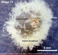 Embryo-membranes stage 11.jpg 600 × 568; 61 KB
 External ear stages-14-23-adult a.jpg 800 × 524; 30 KB
 Finley1923 fig01.jpg 494 × 968; 53 KB
 Finley1923 fig02.jpg 700 × 800; 77 KB
 Finley1923 fig03.jpg 600 × 512; 56 KB
 Finley1923 fig04.jpg 700 × 627; 61 KB
 Finley1923 fig05.jpg 674 × 800; 146 KB
 Finley1923 fig06.jpg 729 × 800; 95 KB
 Finley1923 fig07.jpg 314 × 800; 40 KB
 Finley1923 fig08.jpg 296 × 800; 32 KB
 Finley1923 fig09.jpg 456 × 800; 49 KB
 Finley1923 fig10.jpg 221 × 802; 17 KB
 Finley1923 fig11.jpg 432 × 800; 38 KB
 Finley1923 fig12.jpg 593 × 800; 48 KB
 Finley1923 fig13.jpg 594 × 800; 51 KB
 Finley1923 Plate 1.jpg 776 × 1,000; 151 KB
 Finley1923 Plate 2.jpg 864 × 1,200; 153 KB
 Frazer002 bw600.jpg 600 × 501; 45 KB
 Frazer043a 600.jpg 534 × 650; 58 KB
 Gestational age distribution NIPT.jpg 671 × 493; 41 KB
 Ginkgo biloba.jpg 240 × 180; 10 KB
 Gray0020.jpg 1,038 × 1,536; 446 KB
 Gray0027.gif 300 × 298; 10 KB
 Gray0031.jpg 1,371 × 1,309; 270 KB
 Gray0458.gif 442 × 600; 34 KB
 Gray0458.jpg 693 × 911; 83 KB
 Gray0459.jpg 1,195 × 961; 258 KB
 Gray0472.jpg 550 × 653; 57 KB
 Gray0502.jpg 1,000 × 1,329; 215 KB
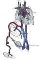
 Heart human embryo CRL10mm 01.jpg 1,000 × 1,389; 537 KB
 His1897 fig15.jpg 867 × 1,072; 182 KB
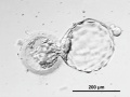
 Human blastocyst day 1-5.jpg 500 × 450; 46 KB
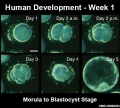
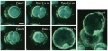 Human blastocyst day 1-6.jpg 708 × 338; 45 KB
 Human blastocyst day 3-6.mov ; 4.26 MB
- Human blastocyst day 5-6 small.mov ; 509 KB
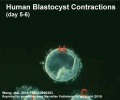 Human blastocyst day 5-6.jpg 498 × 414; 24 KB
- Human blastocyst day 5-6.mov ; 979 KB
- Human blastocyst hatching day 5-6.mov ; 1.11 MB
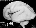 Human brain growth 01.jpg 1,022 × 800; 119 KB
 Human Carnegie stage 1-23.jpg 1,000 × 563; 98 KB
 Human Carnegie stage 10-23.jpg 1,024 × 768; 95 KB
 Human Carnegie stage 13 GJA1 expression 2.jpg 705 × 384; 76 KB
 Human Carnegie stage 13 GJA1 expression.jpg 706 × 470; 96 KB
 Human Carnegie stage 13 SOX11 MAZ GJA1 expression.jpg 777 × 1,000; 192 KB
 Human Carnegie stage 13 stem cell markers.jpg 657 × 585; 78 KB
 Human Carnegie stage 15 HOXC5 expression.jpg 670 × 504; 90 KB
 Human CS13 otic vesicle 01.jpg 1,028 × 774; 112 KB
 Human CS13-15 otic vesicle 01.jpg 1,574 × 1,779; 364 KB
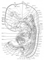
 Human Embryo 17.8mm GIT.jpg 600 × 401; 38 KB
 Human Embryo 17.8mmCNS GIT.jpg 500 × 678; 105 KB
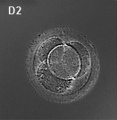 Human embryo day 2.jpg 400 × 409; 19 KB
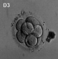 Human embryo day 3.jpg 400 × 409; 7 KB
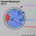
 Human embryo day 5 label.gif 500 × 506; 243 KB
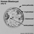 Human embryo day 5 label.jpg 500 × 506; 37 KB
 Human embryo day 5 label2.jpg 500 × 506; 43 KB
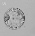 Human embryo day 5.jpg 400 × 409; 6 KB
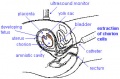
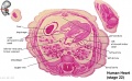
 Human embryo head week 6 to 8.jpg 540 × 780; 66 KB
 Human embryo olfactory 01.jpg 2,044 × 1,552; 986 KB
 Human neural crest cell migration-in vitro.jpg 1,280 × 959; 163 KB
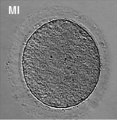 Human oocyte-metaphase I.jpg 400 × 409; 32 KB
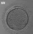 Human oocyte-metaphase II.jpg 400 × 409; 12 KB
 Human oocyte.jpg 216 × 193; 6 KB
 Human Stage13 sagittal upper half01.jpg 1,518 × 2,048; 269 KB
 Human Stage13 sagittal upper half02.jpg 1,518 × 2,048; 289 KB
 Human Stage14 neural01.jpg 1,375 × 2,048; 283 KB
 Human Stage14 neural02.jpg 1,375 × 2,048; 506 KB
 Human Stage14-16 CN5-01.jpg 1,028 × 681; 44 KB
 Human Stage16 neural01.jpg 1,352 × 2,048; 247 KB
 Human Stage16 neural02.jpg 1,352 × 2,048; 286 KB
 Human Stage16 neural03.jpg 1,352 × 2,048; 245 KB
 Human Stage21 neural01.jpg 2,048 × 1,533; 211 KB
 Human Stage21 neural02.jpg 2,048 × 1,533; 230 KB
 Human zygote two pronuclei 01.jpg 528 × 472; 34 KB
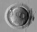 Human zygote two pronuclei 02.jpg 519 × 457; 28 KB
 Human zygote two pronuclei 02.png 433 × 422; 121 KB
 Human zygote two pronuclei 03.jpg 503 × 477; 33 KB
 Human zygote two pronuclei 22.jpg 519 × 457; 38 KB
 Human- Stage 22 integument 01.jpg 1,000 × 750; 205 KB
 Human- Stage 22 integument 02.jpg 800 × 600; 147 KB
 Human- Stage 22 integument 03.jpg 600 × 450; 95 KB
 Human- Stage 22 integument 04.jpg 400 × 300; 48 KB
 Human- Stage 22 thymus 01.jpg 1,200 × 900; 456 KB
 Human-heart-E3L.jpg 639 × 393; 79 KB
 Human-oocyte to blastocyst.jpg 600 × 402; 49 KB
 Human-oocyte.jpg 400 × 409; 29 KB
 Implanting human conceptus 01.jpg 1,056 × 834; 246 KB
 Inner cell mass cartoon.jpg 831 × 800; 78 KB
 Keibel Mall 034.jpg 684 × 800; 80 KB
 Keibel Mall 034b.jpg 813 × 1,000; 120 KB
 Keibel Mall 049-051.jpg 680 × 1,000; 66 KB
 Keibel Mall 049.jpg 405 × 417; 17 KB
 Keibel Mall 050.jpg 242 × 443; 14 KB
 Keibel Mall 051.jpg 250 × 444; 12 KB
 Keibel Mall 052.jpg 604 × 600; 37 KB
 Keibel Mall 053.jpg 436 × 600; 33 KB
 Keibel Mall 054-056.jpg 1,020 × 600; 69 KB
 Keibel Mall 057.jpg 478 × 792; 42 KB
 Keibel Mall 058.jpg 319 × 700; 30 KB
 Keibel Mall 059-060.jpg 1,000 × 586; 54 KB
 Keibel Mall 061-062.jpg 662 × 700; 55 KB
 Keibel Mall 063.jpg 405 × 800; 45 KB
 Keibel Mall 066-071.jpg 610 × 800; 58 KB
 Keibel Mall 077.jpg 424 × 700; 33 KB
 Keibel Mall 078.jpg 278 × 236; 18 KB
 Keibel Mall 079-082.jpg 683 × 906; 147 KB
 Keibel Mall 088-091.jpg 673 × 564; 75 KB
 Keibel Mall 092.jpg 685 × 813; 129 KB
 Keibel Mall 093.jpg 787 × 512; 97 KB
 Keibel Mall 094.jpg 680 × 791; 98 KB
 Keibel Mall 095.jpg 677 × 670; 74 KB
 Keibel Mall 096.jpg 878 × 723; 143 KB
 Keibel Mall 097.jpg 690 × 491; 66 KB
 Keibel Mall 098.jpg 688 × 495; 83 KB
 Keibel Mall 099.jpg 680 × 546; 70 KB
 Keibel Mall 100.jpg 689 × 468; 71 KB
 Keibel Mall 101.jpg 681 × 616; 63 KB
 Keibel Mall 102.jpg 682 × 503; 67 KB
 Keibel Mall 103.jpg 686 × 454; 65 KB
 Keibel Mall 104.jpg 678 × 599; 58 KB
 Keibel Mall 105.jpg 672 × 643; 81 KB
 Keibel Mall 106.jpg 634 × 476; 53 KB
 Keibel Mall 107.jpg 732 × 558; 88 KB
 Keibel Mall 108.jpg 722 × 564; 92 KB
 Keibel Mall 109.jpg 677 × 709; 97 KB
 Keibel Mall 110.jpg 673 × 418; 39 KB
 Keibel Mall 111.jpg 687 × 674; 62 KB
 Keibel Mall 112.jpg 670 × 677; 97 KB
 Keibel Mall 113.jpg 679 × 502; 87 KB
 Keibel Mall 114.jpg 704 × 496; 96 KB
 Keibel Mall 115.jpg 686 × 506; 91 KB


