BGD Lecture - Face and Ear Development
| Embryology - 26 Apr 2024 |
|---|
| Google Translate - select your language from the list shown below (this will open a new external page) |
|
العربية | català | 中文 | 中國傳統的 | français | Deutsche | עִברִית | हिंदी | bahasa Indonesia | italiano | 日本語 | 한국어 | မြန်မာ | Pilipino | Polskie | português | ਪੰਜਾਬੀ ਦੇ | Română | русский | Español | Swahili | Svensk | ไทย | Türkçe | اردو | ייִדיש | Tiếng Việt These external translations are automated and may not be accurate. (More? About Translations) |
Introduction

|
The face is the anatomical feature which is truly unique to each human, though the basis of its general development is identical for all humans and similar to that seem for other species. The face has a complex origin arising from a number of head structures and sensitive to a number of teratogens during critical periods of its development. The related structures of upper lip and palate significantly contribute to the majority of face abnormalities.
Head The head and neck structures are more than just the face, and are derived from pharyngeal arches 1 - 6 with the face forming from arch 1 and 2 and the frontonasal prominence. Each arch contains similar Arch components derived from endoderm, mesoderm, neural crest and ectoderm. Because the head contains many different structures also review notes on sensory, respiratory, Integumentary (tooth), endocrine (thyroid, parathyroid, pituitary, thymus) and cleft lip/cleft palate. Hearing We use the sense of balance and hearing to position ourselves in space, sense our surrounding environment, and to communicate. Importantly hearing is linked into postnatal neurological development (milestones) involved with language and learning.
|
Lecture Objectives
| Lecture Archive |
|---|
| 2019 PDF | 2018 | 2018 PDF | 2017 | 2017 PDF | 2016 |
2015 | 2014 | 2014 PDF | 2013 | 2012 | 2009 | Face and Ear Practical | DB |
To introduce the developmental embryology of both the face and ear, and their associated abnormalities.
|
 |


| Head Movies | ||||||||||||||||||||||||||||
|---|---|---|---|---|---|---|---|---|---|---|---|---|---|---|---|---|---|---|---|---|---|---|---|---|---|---|---|---|
|
| |||||||||||||||||||||||||||
|
|
|
|
|
| |||||||||||||||||||||||
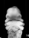
Week 3
| Buccopharyngeal Membrane and Pharynx |
|---|
| <html5media height="360" width="360">File:Endoderm 003.mp4</html5media> |
Buccopharyngeal Membrane
These images of the Week 4 embryo (23 - 26 days, Stage 11) show the breakdown of the buccopharyngeal (oral) membrane.
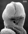
Low power ventral view of the Buccopharyngeal Membrane
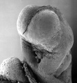
Higher power ventrolateral view of the Buccopharyngeal Membrane
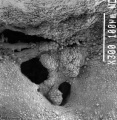
Close up view of the degenerating Buccopharyngeal Membrane
Buccopharyngeal Membrane
Buccal and Nasal Cavities


The Pharynx


The cavity within the pharyngeal arches forms the pharynx.
- begins at the buccopharyngeal membrane (oral membrane), apposition of ectoderm with endoderm (no mesoderm between)
- expands behind pharyngeal arches
- narrows at glottis and bifurcation of gastrointestinal (oesophagus) and respiratory (trachea) systems
- regions on roof, walls and floor have important contributions to endocrine in oral and neck regions
- also contributes to tongue development
Week 4

| Week 4 - Arches (Carnegie stage 11) |
|---|

|
Pharyngeal Arch Components

Major features to identify for each: arch, pouch, groove and membrane. Contribute to the formation of head and neck and in the human appear at the 4th week. The first arch contributes the majority of upper and lower jaw structures.
Pharyngeal Arch Development
Pharyngeal (branchial) arch (Greek. branchia = gill) consists of all 3 trilaminar embryo layers
- ectoderm - outside surface and core neural crest
- mesoderm - core of mesenchyme
- endoderm - inside pharynx
|
|

Neural Crest
- Mesenchyme invaded by neural crest generating connective tissue components
- cartilage, bone, ligaments
- arises from midbrain and hindbrain region
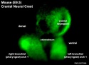
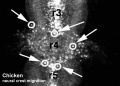
| Neural Crest Migration | |||||||
|---|---|---|---|---|---|---|---|
| <html5media height="340" width="410">File:Chicken-neural crest migration 01.mp4</html5media> | Chicken Embryo - DiI-labelled neural crest cells towards the branchial arches.
White rings indicate migration of individual cells. Each image represents 10 confocal sections separated by 10 microns. | ||||||
| <html5media height="350" width="400">File:Mouse cranial neural crest migration 01.mp4</html5media> | 
Mouse Embryo - GFP-labelled cranial neural crest cells in embryonic mouse E9.5. | ||||||
|
| ||||||
Arch Features
Each arch contains: artery, cartilage, nerve, muscular component
Arches and Phanynx Form the face, tongue, lips, jaws, palate, pharynx and neck cranial nerves, sense organ components, glands
- Humans have 5 arches - 1, 2, 3, 4, 6 (Arch 5 does not form or regresses rapidly)
- form in rostro-caudal sequence, Arch 1 to 6 (from week 4 onwards)
- arch 1 and 2 appear at time of closure of cranial neuropore
- Face - mainly arch 1 and 2
- Neck components - arch 3 and 4 (arch 4 and 6 fuse)
- arch
- groove - (cleft) externally separates each arch (only first pair persist as external auditory meatus)
- pouch - internally separates each arch (pockets out from the pharynx)
- membrane - ectoderm and endoderm contact regions (only first pair persist as tympanic membrane )
Pharyngeal Arch 1 (Mandibular Arch) has 2 prominences
- smaller upper - maxillary forms maxilla, zygomatic bone and squamous part of temporal
- larger lower - mandibular, forms mandible
Pharyngeal Arch 2 (Hyoid Arch)
- forms most of hyoid bone
Arch 3 and 4
- neck structures
| Arch Arteries | |
|---|---|
Embryo Blood Flow - placental vein -> liver -> heart -> truncus arteriosus -> aortic sac -> arch arteries -> dorsal aorta -> placental artery |

|
| Arch Cartilage | |
|---|---|
|

Pharyngeal arch cartilages
Merkel's cartilage (first pharyngeal arch) |

| Arch Muscle |
|---|
|
| Arch Nerve |
|---|
| Arch Pouches |
|---|
|
| Pharyngeal Arch - Summary Table | ||||||||||||||||||||||||||||||||||||
|---|---|---|---|---|---|---|---|---|---|---|---|---|---|---|---|---|---|---|---|---|---|---|---|---|---|---|---|---|---|---|---|---|---|---|---|---|
|
Endocrine
The arch pouches contribute to endocrine organ development, except for the thyroid and pituitary. Note endocrine development will be covered in detail in another later BGD lecture.
| thyroid | Anterior pituitary |
|---|---|
|
|


Face Development
Begins week 4 centered around stomodeum, external depression at oral membrane
5 initial primordia from neural crest mesenchyme (week 4)
- single frontonasal prominence (FNP) - forms forehead, nose dorsum and apex
- nasal placodes develop later bilateral, pushed medially
- paired maxillary prominences - form upper cheek and upper lip
- paired mandibular prominences - lower cheek, chin and lower lip


Week 8
|
|


Head/Skull


Cranium (Neurocranium) surrounds brain.
- dermatocranium intramembranous ossification - skull calvarial vault
- chondrocranium (endochondral ossification) - skull base
- 8 bones - occipital, 2 parietals, frontal, 2 temporals, sphenoidal, ethmoidal.
Face (Viscerocranium) development of the facial bones
- 14 bones - 2 nasals, 2 maxillæ, 2 lacrimals, 2 zygomatics, 2 palatines, 2 inferior nasal conchæ, vomer, mandible.

Calveria - bone has no cartilage (direct ossification of mesenchyme)
Head Growth
Bones do not fuse, fibrous sutures
- allow distortion to pass through birth canal
- allow growth of the brain
- 6 fontanelles - posterior closes at 3 months, anterior closes at 18 months
- puberty growth of face


- Links: skull
Sensory Placodes

- During week 4 a series of thickened surface ectodermal patches "placodes" form in pairs rostro-caudally in the head region.
- These sensory placates later contribute key components of each of our special senses (vision, hearing and smell).
- Initial placode postion on the developing head is significantly different to their final position in the future sensory system
| Placode Research |
|---|
| Recent research suggests that all sensory placodes may arise from common panplacodal primordium origin around the neural plate, and then differentiate to eventually have different developmental fates.
Other species have a number of additional placodes which form other sensory structures (fish, lateral line receptor). |
Otic Placode
- Carnegie stage 12 still visible on embryo surface.inner ear
- Carnegie stage 13/14 embryo (shown below) the otic placode has sunk from the surface ectoderm to form a hollow epithelial ball, the otocyst, which now lies beneath the surface surrounded by mesenchyme (mesoderm). The epithelia of this ball varies in thickness and has begun to distort, it will eventually form the inner ear membranous labyrinth.
Lens Placode
- (optic placode) lies on the surface, adjacent to the outpocketing of the nervous system (which will for the retina) and will form the lens.
Nasal Placode
- Has 2 components (medial and lateral) and will form the nose olefactory epithelium.
Hearing Development

Inner Ear
| Week 5 | Week 8 |
|---|---|

Otocyst (Week 5, Stage 13) |

Inner Ear (Week 8, Stage 22) |
- Inner Ear Labyrinth
- Cochlea - Otic vesicle - Otic placode (ectoderm)
- Semicircular canals - Otic vesicle - Otic placode (ectoderm)
- Saccule and utricle - Otic vesicle - Otic placode (ectoderm)
- Cranial Nerve VIII
- Auditory component - Otic vesicle and neural crest (ectoderm)
- Vestibular component - Otic vesicle and neural crest (ectoderm)
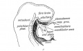
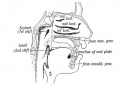
Middle Ear
|
Pharyngeal arch cartilages |
External Ear

- Auricle - Pharyngeal Arches 1 and 2 (ectoderm, mesoderm)
- form from 6 hillocks (week 5) 3 on each of arch 1 and 2
- External Auditory Meatus - Pharyngeal Arch 1 groove or cleft (ectoderm)
- Tympanic Membrane - Pharyngeal Arch 1 membrane (ectoderm, mesoderm, endoderm)
Postnatal Changes

|
Adult - longer (twice as long), wider and runs at approximately 45 degrees to the horizontal, tube is opened by two separate muscles (tensor palati and levator palati)
|
- Auditory tube = Eustachian, otopharyngeal or pharyngotympanic tube.
- Connects middle ear cavity to nasopharynx portion of pharynx
- Ventilation - pressure equalization in the middle ear
- Clearance - allow fluid drainage from the middle ear Tube is normally closed and opened by muscles
- Links: Hearing Development
Palate
Embryonic
| Primary palate, fusion in the human embryo between week 6-7 (stage 17 and 18, GA Week 8-9), from an epithelial seam to the mesenchymal bridge.
|

|

Fetal

Secondary palate, fusion in the human embryo in week 9 (GA week 11). This requires the early palatal shelves growth, elevation and fusion during the early embryonic period. The fusion event is to both each other and the primary palate. palatal shelf elevation | secondary palate
Tongue Development


- Ectoderm of the first arch surrounding the stomodeum forms the epithelium lining the buccal cavity.
- Also the salivary glands, enamel of the teeth, epithelium of the body of the tongue.
- As the tongue develops "inside" the floor of the oral cavity, it is not readily visible in the external views of the embryonic (Carnegie) stages of development.
- Contributions from all arches, which changes with time
- begins as swelling rostral to foramen cecum, median tongue bud
- Arch 1 - oral part of tongue (ant 3/2)
- Arch 2 - initial contribution to surface is lost
- Arch 3 - pharyngeal part of tongue (post 1/3)
- Arch 4 - epiglottis and adjacent regions
Tongue Muscle

- Skeletal muscle originate from the somites.
- Tongue muscles develop before masticatory muscles and is completed by birth.
Masticatory muscles
- Originate from the somitomeres. These muscles develop late and are not complete even at birth.
- paraxial mesoderm in cranial region forms somitomeres that do not become somites.
Salivary Glands
- epithelial buds in oral cavity (wk 6-7) extend into mesenchyme
- parotid, submandibular, sublingual
Abnormalities
Will be covered in detail in the associated practical class.
Cleft Lip and Palate

|

|

|
| cleft palate | unilateral cleft lip and palate | bilateral cleft lip and palate |
- 300+ different abnormalities, different cleft forms and extent, upper lip and ant. maxilla, hard and soft palate
Statistics - The ten most frequently reported birth defects in Victoria between 2003-2004.
| Victoria - 10 most reported birth anomalies | |
|---|---|
| Based upon statistics from the Victorian Perinatal Data Collection Unit in Victoria between 2003-2004. | |

|
hypospadias (More? External Genital Male Development Movie) |

|
Obstructive Defects of the Renal Pelvis (obstructive defects of the renal pelvis, uteropelvic junction obstruction, pelvo-uterero junction obstruction) Term describing a developmental renal abnormality due to partial or complete blockage of the drainage of the kidney pelvis requiring surgical correction. The blockage can also have several causes including: unusual ureter twisting or bending, ureter compression by a blood vessel, malformations of the muscular wall. The blockage leads to an accumulation of urine in the affected region, with several potential effects: nephron damage from compression (hydronephrosis); decreased urine output leading to lack of amniotic fluid (oligohydramnios); respiratory development effects due to the lack of amniotic fluid.
(More? renal abnormalities | renal) |

|
ventricular septal defect (More? ventricular septal defect)
Heart Development Timeline (see Basic Cardiac Embryology) |

|
Developmental dysplasia of the hip or Congenital Dislocated Hip
(Developmental dysplasia of the hip (DDH), congenital hip dislocation, congenital hip dysplasia) Term describes a spectrum of musculoskeletal disorders of hip instability due either to the femoral head being able to move outside the acetabulum (luxation or dislocation), or abnormally within the acetabulum (subluxation or partial dislocation). This includes presentation following a normal examination of the hips in the newborn period (Ortolani and Barlow tests). When detected can be managed with splinting (Denis-Browne splint) allows the hip joint to develop normally and does not require surgery. If undetected and left untreated, the hip joint develops abnormally and surgical reduction is required. (More? Pelvis Development) |

|
Trisomy 21 or Down syndrome - The most common genetic abnormality. (More? Trisomy 21) |

|
hydrocephalus rapid increase in head circumference or an unusually large head size due to excessive accumulation of cerebrospinal fluid in the brain.(More? hydrocephalus | Neural Abnormalities | NINDS - Hydrocephalus Fact Sheet | Hydrocephalus Support Association | USA National Hydrocephalus Foundation) |
|
|
cleft palate - The palate separates the nasal cavity from the oral cavity, the abnormality has many different causes, and occurs more frequently in females (57%) than in males (43%). (More? cleft palate) |

|
Trisomy 18 or Edward Syndrome - multiple abnormalities of the heart, diaphragm, lungs, kidneys, ureters and palate 86% discontinued (More? Trisomy 18) |
| Renal Agenesis/Dysgenesis - reduction in neonatal death and stillbirth since 1993 may be due to the more severe cases being identified in utero and being represented amongst the increased proportion of terminations (approximately 31%). (More? Renal Abnormalities - Renal Agenesis) | |
|
|
cleft lip and palate - occur with another defect in 33.7% of cases.(More? cleft lip and palate) |
| Links: Human Abnormal Development | Australian Statistics | Victoria 2004 | USA 2006 | Europe 2010 | |

| USA Statistics | ||||||||||||||||||||||||||||||||||||||||||||||||||||||||||||||||||||||||
|---|---|---|---|---|---|---|---|---|---|---|---|---|---|---|---|---|---|---|---|---|---|---|---|---|---|---|---|---|---|---|---|---|---|---|---|---|---|---|---|---|---|---|---|---|---|---|---|---|---|---|---|---|---|---|---|---|---|---|---|---|---|---|---|---|---|---|---|---|---|---|---|---|
| ||||||||||||||||||||||||||||||||||||||||||||||||||||||||||||||||||||||||
Cleft Palate
- Cleft palate has the International Classification of Diseases code 749.0.
- In Australia the national rate (1982-1992) for this abnormalitity in births was 4.8 - 6/10,000 births, which represented 1,530 infants 5.5% were stillborn and 11.5% liveborn died during neonatal period and slightly more common in twin births than singleton.
Cleft Lip
- The International Classification of Diseases code 749.1 for isolated cleft lip and 749.2 for cleft lip with cleft palate.
- In Australia the national rate (1982-1992) for this abnormalitity was 8.1 - 9.9 /10,000 births. Of 2,465 infants 6.2% were stillborn and 7.8% liveborn died during neonatal period and the rate was similar in singleton and twin births.
| Palate Links: palate | cleft lip and palate | cleft palate | head | Category:Palate |
First Arch Syndrome
- There are 2 major types of associated first arch syndromes, Treacher Collins (Mandibulofacial dysostosis) and Pierre Robin (Pierre Robin complex or sequence), both result in extensive facial abnormalites.
Treacher Collins Syndrome
Pierre Robin Syndrome
- Hypoplasia of the mandible, cleft palate, eye and ear defects.
- Initial defect is small mandible (micrognathia) resulting in posterior displacement of tongue and a bilateral cleft palate.
DiGeorge Syndrome
- absence of thymus and parathyroid glands, 3rd and 4th pouch do not form
- disturbance of cervical neural crest migration
Cysts
- Many different types
Facial Clefts
- extremely rare
- Holoprosencephaly
- shh abnormality
Maternal Effects
- Retinoic Acid - present in skin ointments
- 1988 associated with facial developmental abnormalities
Fetal Alcohol Syndrome

Due to alcohol in early development (week 3+) leading to both facial and neurological abnormalities
- lowered ears, small face, mild+ retardation
- Microcephaly - leads to small head circumference
- Short Palpebral fissure - opening of eye
- Epicanthal folds - fold of skin at inside of corner of eye
- Flat midface
- Low nasal bridge
- Indistinct Philtrum - vertical grooves between nose and mouth
- Thin upper lip
- Micrognathia - small jaw
Exposure of embryos in vitro to ethanol simulates premature differentiation of prechondrogenic mesenchyme of the facial primordia (1999)
- Links: Fetal Alcohol Syndrome
Table - Structures derived from Arches
| Arch | Nerve | Skeletal Structures | Muscles | Ligaments |
| 1 (maxillary/mandibular) | trigeminal (V) | mandible, maxilla, malleus, incus | ant lig of malleus, sphenomandibular ligament | |
| 2 (hyoid) | facial (VII) | stapes, styloid process, lesser cornu of hyoid, upper part of body of hyoid bone | stylohyoid ligament | |
| 3 | glossopharyngeal (IX) | greater cornu of hyoid, lower part of body of hyoid bone | ||
| 4 & 6 | superior laryngeal and recurrent laryngeal branch of vagus (X) | thyroid, cricoid, arytenoid, corniculate and cuneform cartilages |
Structures derived from Pouches
Each pouch is lined with endoderm and generates specific structures.
| Overall Structure | Specific Structures | |
| tubotympanic recess | tympanic membrane, tympanic cavity, mastoid antrum, auditory tube | |
| intratonsillar cleft | crypts of palatine tonsil, lymphatic nodules of palatine tonsil | |
| inferior parathyroid gland, thymus gland | ||
| superior parathyroid gland, ultimobranchial body | ||
| becomes part of 4th pouch |
Structures derived from Grooves
Only the first groove differentiates into an adult structure and forms part of the external acoustic meatus.
Structures derived from Membranes
At the bottom of each groove lies the membrane which is formed from the contact region of ectodermal groove and endodermal pouch.
Only the first membrane differentiates into an adult structure and forms the tympanic membrane.
References
| Additional Resources | |
|---|---|
| Online Textbooks | Search |
|
Developmental Biology by Gilbert, Scott F. Sunderland (MA): Sinauer Associates, Inc.; c2000 Figure 1.3. Pharyngeal arches | Table 13.2. Some derivatives of the pharyngeal arches | The Cranial Neural Crest Madame Curie Bioscience Database Chapters taken from the Madame Curie Bioscience Database (formerly, Eurekah Bioscience Database) Cranial Neural Crest and Development of the Head Skeleton | Neural Crest Cells and the Community of Plan for Craniofacial Development: Historical Debates and Current Perspectives |
Bookshelf pharyngeal arch | head development | face development Pubmed pharyngeal arch | head development | face development | |
Terms
| Terms |
|---|
|
| Hearing Terms | ||
|---|---|---|
Hearing and Balance Development
|
External Links
External Links Notice - The dynamic nature of the internet may mean that some of these listed links may no longer function. If the link no longer works search the web with the link text or name. Links to any external commercial sites are provided for information purposes only and should never be considered an endorsement. UNSW Embryology is provided as an educational resource with no clinical information or commercial affiliation.
| Embryo Images Unit |
|---|
| Craniofacial Development | Cell Populations | Pharyngeal Arches | Tongue | Nose and Upper Lip | Palate Development |
BGDB: Lecture - Gastrointestinal System | Practical - Gastrointestinal System | Lecture - Face and Ear | Practical - Face and Ear | Lecture - Endocrine | Lecture - Sexual Differentiation | Practical - Sexual Differentiation | Tutorial
Glossary Links
- Glossary: A | B | C | D | E | F | G | H | I | J | K | L | M | N | O | P | Q | R | S | T | U | V | W | X | Y | Z | Numbers | Symbols | Term Link
Cite this page: Hill, M.A. (2024, April 26) Embryology BGD Lecture - Face and Ear Development. Retrieved from https://embryology.med.unsw.edu.au/embryology/index.php/BGD_Lecture_-_Face_and_Ear_Development
- © Dr Mark Hill 2024, UNSW Embryology ISBN: 978 0 7334 2609 4 - UNSW CRICOS Provider Code No. 00098G