Menstrual Cycle - Histology
| Embryology - 8 May 2024 |
|---|
| Google Translate - select your language from the list shown below (this will open a new external page) |
|
العربية | català | 中文 | 中國傳統的 | français | Deutsche | עִברִית | हिंदी | bahasa Indonesia | italiano | 日本語 | 한국어 | မြန်မာ | Pilipino | Polskie | português | ਪੰਜਾਬੀ ਦੇ | Română | русский | Español | Swahili | Svensk | ไทย | Türkçe | اردو | ייִדיש | Tiếng Việt These external translations are automated and may not be accurate. (More? About Translations) |
Introduction

During the menstrual cycle, the uterus undergoes a series of histologically recognisable cyclic changes under the influence of changing circulating hormonal levels. Menstrual cycle stages can be clinically characterised by menstrual histology of smears and tissue analysis, first devised by Papanicolaou in 1933.[1]
This page presents clinical histology images from vaginal smears and uterine endometrium dilatation and curettage samples during different phases of the human menstrual cycle.
Beneath these clinical images are a number of more detailed uterus histology images.
| Histology Links: stains | fixatives | artifacts | menstrual histology | placenta histology | heart histology | liver histology | Pancreas | Gall Bladder | Colon | Renal | Respiratory Histology | Bone | Category:Histology | UNSW Histology |
| Historic Histology Textbooks: 1941 Histology] | 1944 Oral Histology |
History of the Pap Smear
(Papanicolaou smear, pap test, cervical smear) The text below is from the ABC - Great Moments In Science.
- "Luckily, we have the famous Pap Smear - an excellent way to find cancer of the cervix before it digs in locally and/or spreads throughout the body. The Pap Smear is named after a certain Dr. Papanicolaou - who did a Pap Smear on his wife virtually every day for 20 years.
- George Nikolas Papanicolaou was born in 1883 in Kymi, a small town overlooking the Aegean Sea on the Island of Euboea in Greece. His father, Nikolas Papanicolaou was both the Major of Kymi and a medical doctor. His older brother, Naso, had studied law, so his father convinced George to continue in the family medical tradition. So George studied medicine, and did well, graduating with a degree in honours in 1904............"
- Drawings of cells in human vaginal smears
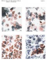
Plate 8
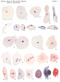
Plate 9
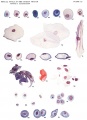
Plate 10


Papanicolaou (1933)[1] Hand coloured photomicrographs from the publication showing the human reproductive cycle as revealed by vaginal smears.
Papanicolaou Stain
This histology technique was originally described in a publication by George Nikolas Papanicolaou in 1942.[2] There are 5 different stains used in the technique and it has been used for many different human bodily fluids (CSF, semen, aspirations). The original protocol has also been modified several ways (Bismarck brown Y deleted).
- Links: Papanicolaou Stain | Histology Stains | Histology
Cervical Screening Program
In Australia, the "Pap Smear" test will be replaced in 2017 by a new "National Cervical Screening Program". This new program will use new technologies to detect HPV DNA rather than pathological screening for abnormal cells from a "Pap Smear". For more information see the external link below.
- "The two yearly Pap test for women aged 18 to 69 will change to a five yearly human papillomavirus (HPV) test for women aged 25 to 74. Women will be due for the first Cervical Screening Test two years after their last Pap test."
- Links: National Cervical Screening Program | Compass Trial |
ABC radio program Monday 27 March 2017 - Death of the pap smear? | ABC Audio - Death of the pap smear?
Dilation and Curettage
Medical procedure where uterine endometrium is collected by Dilation and Curettage (D&C or DnC). This medical procedure is used to diagnose or treat uterine abnormalities or in association with a miscarriage.
- Links: Medline Plus D&C | PMID 15725872
Histology
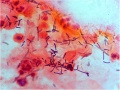
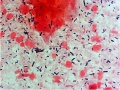
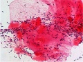
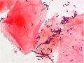
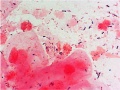
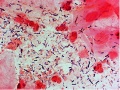
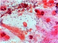
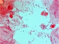
| Phase | Days (range) | Smear | Smear Description | Uterine Endometrium (D&C) |
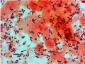
| Menstrual | 1 - 4 | Click on image to see full size. | Both stratum corneum (red) and stratum spinosum (blue) epithelial cells will mostly blood.
Leukocytes and bacteria may also be present. |

|
| Early Proliferative | 5 - 9 | 
|
Mainly large and small basophilic (blue) stratum spinosum cells. | |
| Mid Proliferative | 9 - 13 | 
|
Stratum spinosum (blue) increase in size.
Dark precipate outside cells are bacteria. |

|
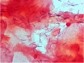
| Late Proliferative, Ovulatory | 13-14 | 
|
mainly stratum corneum (red) which are large and flat.
Appear due to high estrogen levels. |

|
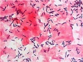
| Secretory | 15 - 22 | 
|
stratum spinosum cells (blue) which are folded or with curled edges.
Appear immediately after ovulation due to increase in progesterone. Leukocytes (small black cells) becoming more numerous. |

|
| Late Secretory, (Ischemic) Premenstrual | 23 - 28 | 
|
stratum spinosum cells (blue) mainly with a few stratum corneum cells (red).
Clustering of cells occurs at this stage. Both leukocytes and bacteria are prevelant. |

|
Human vaginal smear histology images in sequence: early proliferative | mid-proliferative | late proliferative | secretory | late secretory
Human Uterus (D and C histology images) in sequence: menstrual | mid-proliferative | late proliferative | secretory | late secretory
See also Uterus Development
Histology Images
- Uterus Histology Links: Labeled - proliferative phase | Labeled - gland proliferative phase | Labeled - secretory phase | Unlabeled - secretory phase | Unlabeled - late secretory phase | Labeled - gland secretory phase | Menstrual Cycle | Uterine Gland | Uterus Development
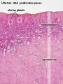
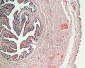
Uterine body endometrium and myometrium during the proliferative phase of the menstrual cycle overview
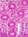
Uterine body endometrium during the proliferative phase of the menstrual cycle
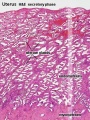
Uterine body endometrium during the secretory phase of the menstrual cycle overview
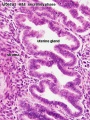
Uterine body endometrium during the secretory phase of the menstrual cycle
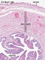
Uterine tube
Uterine tube


Abnormalities
Bacterial Vaginosis
- The normal vaginal flora (lactobacillus morphotypes) is replaced by a mixed microbial flora consisting of Gardnerella vaginalis, Mycoplasma hominis and anaerobes.
- Originally described by Gardner and Dukes (1955).[3]
- clinical features - malodorous, thin homogeneous vaginal discharge with elevated vaginal pH above 4.5.
- Nugent's criteria[4] - microbiological diagnosis of bacterial vaginosis, by counting bacterial cell types on Gram stained slides of vaginal smears.
Proposed Gram stain based categorisation[5]
- grade I - when only Lactobacillus cell types (large Gram positive rods) were present.
- grade II (intermediate) when both Lactobacillus and Gardnerella or Bacteroides-Prevotella cell types were present.
- grade III (bacterial vaginosis) when Lactobacillus cell types were absent and only Gardnerella, Bacteroides-Prevotella or Mobiluncus cell types were present.
- grade IV when Gram positive cocci were predominantly present.
L. crispatus
L. crispatus
non-L. crispatus with thin lactobacilli
non-L. crispatus with thin lactobacilli
mixture non-L. crispatus with L. crispatus

mixture non-L. crispatus with L. crispatus
irregular-shaped Gram positive rod
irregular-shaped Gram positive rod
mixture Lactobacillus and bacterial vaginosis-associated
mixture Lactobacillus and bacterial vaginosis-associated
bacterial vaginosis
bacterial vaginosis


- Smear Image Links: L. crispatus | L. crispatus | non-L. crispatus with thin lactobacilli | non-L. crispatus with thin lactobacilli | mixture non-L. crispatus with L. crispatus | mixture non-L. crispatus with L. crispatus | irregular-shaped Gram positive rod | irregular-shaped Gram positive rod | mixture Lactobacillus and bacterial vaginosis-associated | mixture Lactobacillus and bacterial vaginosis-associated | bacterial vaginosis | bacterial vaginosis
- Links: Menstrual Cycle - Histology | Histology - Gram Stain | Bacterial Vaginosis | CDC (USA) Fact Sheet - Bacterial Vaginosis
References
- ↑ 1.0 1.1 Papanicolaou GN. The sexual cycle in the human female as revealed by vaginal smears. (1933) Amer. J Anat. 52: 519–637.
- ↑ <pubmed>17842594</pubmed>| Science
- ↑ <pubmed>14361525</pubmed>
- ↑ <pubmed>1706728</pubmed>| PMC269757
- ↑ <pubmed>16225680</pubmed>| PMC1266370 | BMC Microbiol.
Search Pubmed
Search Pubmed Now: Menstrual Cycle Histology | uterine histology | vaginal smear | pap smear |
External Links
External Links Notice - The dynamic nature of the internet may mean that some of these listed links may no longer function. If the link no longer works search the web with the link text or name. Links to any external commercial sites are provided for information purposes only and should never be considered an endorsement. UNSW Embryology is provided as an educational resource with no clinical information or commercial affiliation.
- Medline Plus DnC | D and C - series | D and C video
- Better Health Victoria Dilatation and curettage
- Sociedad Argentina de Citología Papanicolaou staining protocol
Glossary Links
- Glossary: A | B | C | D | E | F | G | H | I | J | K | L | M | N | O | P | Q | R | S | T | U | V | W | X | Y | Z | Numbers | Symbols | Term Link
Cite this page: Hill, M.A. (2024, May 8) Embryology Menstrual Cycle - Histology. Retrieved from https://embryology.med.unsw.edu.au/embryology/index.php/Menstrual_Cycle_-_Histology
- © Dr Mark Hill 2024, UNSW Embryology ISBN: 978 0 7334 2609 4 - UNSW CRICOS Provider Code No. 00098G