Gastrointestinal Tract - Liver Histology
| Embryology - 18 Apr 2024 |
|---|
| Google Translate - select your language from the list shown below (this will open a new external page) |
|
العربية | català | 中文 | 中國傳統的 | français | Deutsche | עִברִית | हिंदी | bahasa Indonesia | italiano | 日本語 | 한국어 | မြန်မာ | Pilipino | Polskie | português | ਪੰਜਾਬੀ ਦੇ | Română | русский | Español | Swahili | Svensk | ไทย | Türkçe | اردو | ייִדיש | Tiếng Việt These external translations are automated and may not be accurate. (More? About Translations) |
Introduction
This section of notes gives an overview mainly of adult liver histology, see also Liver Development notes.
 Page also provides further background information for Medicine phase 1 Health Maintenance B Hepatobiliary System 1 Practical.
This page content is not part of the HMB practical class.
Virtual Slides
Hepatobiliary System 1 (requires Moodle log-in)
Lab Audio
![]() Wednesday 17 August 2011 3-4 pm G2G4.
Wednesday 17 August 2011 3-4 pm G2G4.
- Links: listen 1 | download 1 | listen 2 | download 2 | listen 3 | download 3 | listen 4 | download 4 | listen 5 | download 5 | Liver Histology Background
Images
- Liver Histology: Central vein (label) | Central vein (unlabel) | Portal triad 1 (label) | Portal triad 2 (label) | Portal triad (unlabel) | Hepatocytes (unlabel) | Hepatocytes polyploid (label) | Liver - reticular connective tissue (LP) | Liver - reticular connective tissue (HP) | Liver - fetal (HP) | Liver - fetal (HP) | Liver Development | GIT Histology
Liver Structure
Liver Lobule
| This looped animation shows the different ways of interpreting the cellular structure of the liver lobule. | 
|
Liver Blood Flow
Dual blood supply of the liver merges upon entry into the liver lobule at the portal field.
|

|
Hepatocytes
| These are the adult functional cells forming the majority of the liver (80% of the cells).
Many different functions including:
|

|
Histology Images
Development Histology
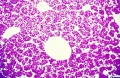
Histology - embryonic liver (week 8)
Histology - embryonic liver (week 8)
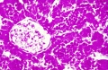
Histology - fetal liver HEx40
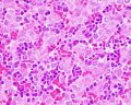
Histology - fetal liver x100


Adult Histology
- Liver Histology: Central vein (label) | Central vein (unlabel) | Portal triad 1 (label) | Portal triad 2 (label) | Portal triad (unlabel) | Hepatocytes (unlabel) | Hepatocytes polyploid (label) | Liver - reticular connective tissue (LP) | Liver - reticular connective tissue (HP) | Liver - fetal (HP) | Liver - fetal (HP) | Liver Development | GIT Histology

|

|

|

|

Unlabeled Large Images
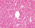
Human liver showing central vein
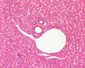
Portal triad
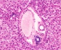
Portal triad
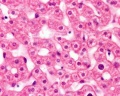
Hepatocytes


Kupffer Cells and Reticular Fibres

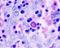
Hepatocyte Polyploidy
Human hepatocytes

Mouse hepatocytes in vitro and in vivo[1]
Liver Histology - Super Resolution

Liver sinusoidal endothelial cell fenestrations[2]
Liver Histology - Electron Micrograph
The electron micrographs below show the cellular, vascular and bilary organisation of the liver.


References
- ↑ Duncan AW, Taylor MH, Hickey RD, Hanlon Newell AE, Lenzi ML, Olson SB, Finegold MJ & Grompe M. (2010). The ploidy conveyor of mature hepatocytes as a source of genetic variation. Nature , 467, 707-10. PMID: 20861837 DOI.
- ↑ Mönkemöller V, Øie C, Hübner W, Huser T & McCourt P. (2015). Multimodal super-resolution optical microscopy visualizes the close connection between membrane and the cytoskeleton in liver sinusoidal endothelial cell fenestrations. Sci Rep , 5, 16279. PMID: 26549018 DOI.
Terms
- Glisson's capsule (Glisson's sheath) - a collagenous capsule covering the external surface of the liver the outer layer comprising a single layer of mesothelial cells. The capsule also extends into the liver as "sheaths" around the hepatic ducts, hepatic arteries and portal tributaries. Named after Francis Glisson (1599 – 1677) a British anatomist.
- Kupffer cells - liver macrophage located in sinusoidal space. Named after Karl Wilhelm von Kupffer (1829 - 1902 ) a German anatomist.
- sinusoids (vascular sinusoids, liver sinusoids) - the spaces between the hepatocytes that are distensible vascular channels lined with fenestrated endothelial cells forming a discontinuous simple squamous epithelium.
- stellate cells (Ito cells) - Named after Toshio Ito, a twentieth century Japanese physician PMID 11450594
External Links
External Links Notice - The dynamic nature of the internet may mean that some of these listed links may no longer function. If the link no longer works search the web with the link text or name. Links to any external commercial sites are provided for information purposes only and should never be considered an endorsement. UNSW Embryology is provided as an educational resource with no clinical information or commercial affiliation.
- Blue Histology Liver
- UNSW Virtual Slides Medicine phase 1 Health Maintenance B Hepatobiliary System 1 Practical (requires login for access).
- UIOWA Virtual Slidebox of Histology Liver and biliary system
Glossary Links
- Glossary: A | B | C | D | E | F | G | H | I | J | K | L | M | N | O | P | Q | R | S | T | U | V | W | X | Y | Z | Numbers | Symbols | Term Link
Cite this page: Hill, M.A. (2024, April 18) Embryology Gastrointestinal Tract - Liver Histology. Retrieved from https://embryology.med.unsw.edu.au/embryology/index.php/Gastrointestinal_Tract_-_Liver_Histology
- © Dr Mark Hill 2024, UNSW Embryology ISBN: 978 0 7334 2609 4 - UNSW CRICOS Provider Code No. 00098G