File:Ossification endochondral 01.jpg
From Embryology

Size of this preview: 799 × 599 pixels. Other resolution: 817 × 613 pixels.
Original file (817 × 613 pixels, file size: 198 KB, MIME type: image/jpeg)
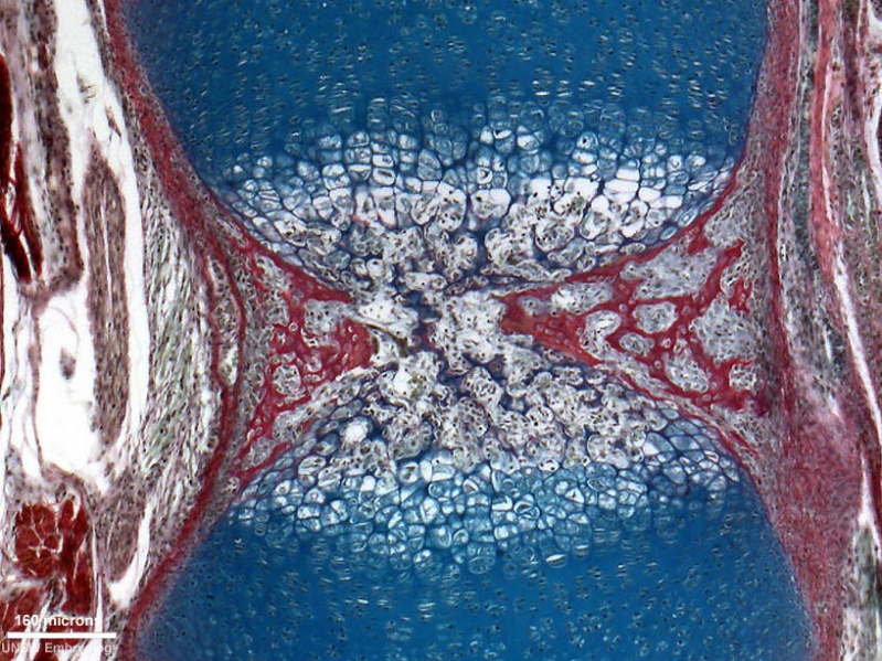
Developing Vertebra - Endochondral Ossification
Histological image of a the periosteal collar of the developing vertebra (neonatal rat), scale bar 160 microns.
Legend
|
See also adjacent region image of Developing Intervertebral Disc |
- Links: axial skeleton | Image - Intervertebral Disc | Image - Vertebra | bone | Cartilage Histology | Bone Histology
Cite this page: Hill, M.A. (2024, May 8) Embryology Ossification endochondral 01.jpg. Retrieved from https://embryology.med.unsw.edu.au/embryology/index.php/File:Ossification_endochondral_01.jpg
- © Dr Mark Hill 2024, UNSW Embryology ISBN: 978 0 7334 2609 4 - UNSW CRICOS Provider Code No. 00098G
File history
Click on a date/time to view the file as it appeared at that time.
| Date/Time | Thumbnail | Dimensions | User | Comment | |
|---|---|---|---|---|---|
| current | 12:33, 23 March 2012 | | 817 × 613 (198 KB) | Z8600021 (talk | contribs) | ==Developing Vertebra - Endochondral Ossification== * Histological image of a developing vertebra and intervertebral disc (neonatal rat). * intervertebral disc - nucleus pulposus and annular fibrocartilage (bottom of image) * vertebra - cartilage templa |
You cannot overwrite this file.
File usage
The following 2 pages use this file: