File:Mouse organ of corti 04.jpg
Original file (1,280 × 1,024 pixels, file size: 202 KB, MIME type: image/jpeg)
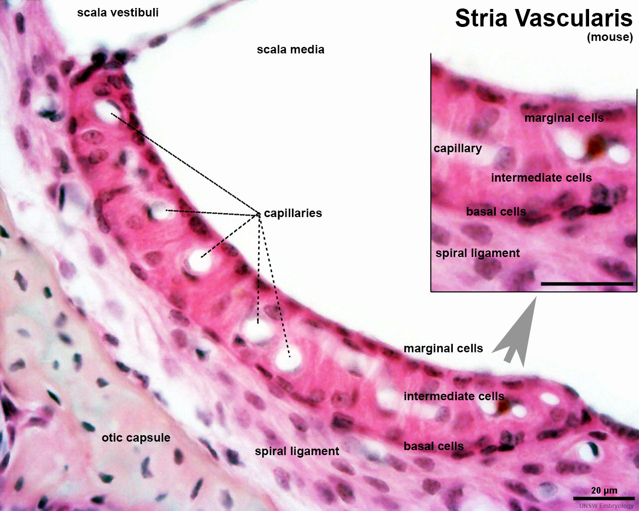
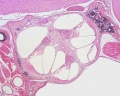
Organ of Corti - Stria Vascularis (mouse)
This histology image is a section through one turn of the cochlea showing the stria vascularis.
The stria vascularis:
- produces endolymph for the scala media.
- is highly vascularised
- consists of three layers of distinct cell types
| Marginal cells | Intermediate cells | Basal cells |
|---|---|---|
| line the lumen of the cochlear duct | melanocyte-like cells lie between the marginal and basal cell layers | mesenchymal spiral ligament fibrocytes |
| derived from epithelia | derived from the neural crest | derived from otic mesenchyme PMID 21925491 |
| Neural Crest Links: neural crest | Lecture - Early Neural | Lecture - Neural Crest Development | Lecture Movie | Schwann cell | adrenal | melanocyte | peripheral nervous system | enteric nervous system | cornea | cranial nerve neural crest | head | skull | cardiac neural crest | Nicole Le Douarin | Neural Crest Movies | neural crest abnormalities | Category:Neural Crest | |||
|
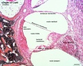
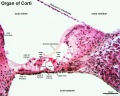
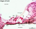
Within the cochlea, the specialised structure required for converting mechanical vibration into an electrical signal occurs at the organ of Corti. Named after Alfonso Giacomo Gaspare Corti (1822–1876), an Italian anatomist who discovered this structure in 1851.
- Cochlea Links: stria vascular histology | stria vascularis 1 | stria vascularis 2 | stria vascularis 3 | human vascularis development | Neural Crest Development | Inner Ear Development

stria vascular histology
stria vascular 1
stria vascular 2
stria vascular 3
vascularis development


Inner Ear Histology: image - cochlea | image - cochlear duct | image - organ of corti | image - organ of corn detaili | image - stria vascularis | Inner Ear | Histology
cochlea
cochlear duct
organ of corti
organ of corti (detail)
stria vascularis


Links: Histology | Histology Stains | Blue Histology images copyright Lutz Slomianka 1998-2009. The literary and artistic works on the original Blue Histology website may be reproduced, adapted, published and distributed for non-commercial purposes. See also the page Histology Stains.
Cite this page: Hill, M.A. (2024, May 15) Embryology Mouse organ of corti 04.jpg. Retrieved from https://embryology.med.unsw.edu.au/embryology/index.php/File:Mouse_organ_of_corti_04.jpg
- © Dr Mark Hill 2024, UNSW Embryology ISBN: 978 0 7334 2609 4 - UNSW CRICOS Provider Code No. 00098G
File history
Click on a date/time to view the file as it appeared at that time.
| Date/Time | Thumbnail | Dimensions | User | Comment | |
|---|---|---|---|---|---|
| current | 14:15, 18 May 2016 | | 1,280 × 1,024 (202 KB) | Z8600021 (talk | contribs) | |
| 13:33, 18 May 2016 | 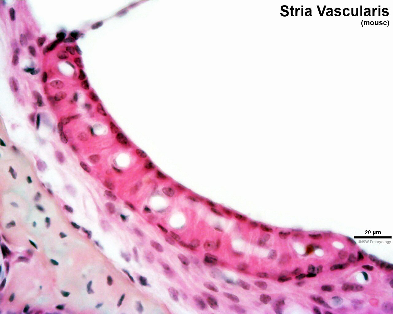 | 1,280 × 1,024 (144 KB) | Z8600021 (talk | contribs) |
You cannot overwrite this file.
File usage
The following 14 pages use this file:
- Hearing - Inner Ear Development
- Neural Crest - Melanocyte Development
- Neural Crest Development
- File:Cochlea stria vascularis cartoon 01.jpg
- File:Cochlea stria vascularis cartoon 02.jpg
- File:Cochlea stria vascularis cartoon 03.jpg
- File:Human cochlea stria vascularis 01.jpg
- File:Mouse organ of corti 01.jpg
- File:Mouse organ of corti 02.jpg
- File:Mouse organ of corti 03.jpg
- File:Mouse organ of corti 04.jpg
- File:Mouse organ of corti 05.jpg
- Template:Inner Ear Histology
- Template:Stria vascularis links