File:Mouse interdigit apoptosis 02.jpg
Original file (764 × 764 pixels, file size: 61 KB, MIME type: image/jpeg)
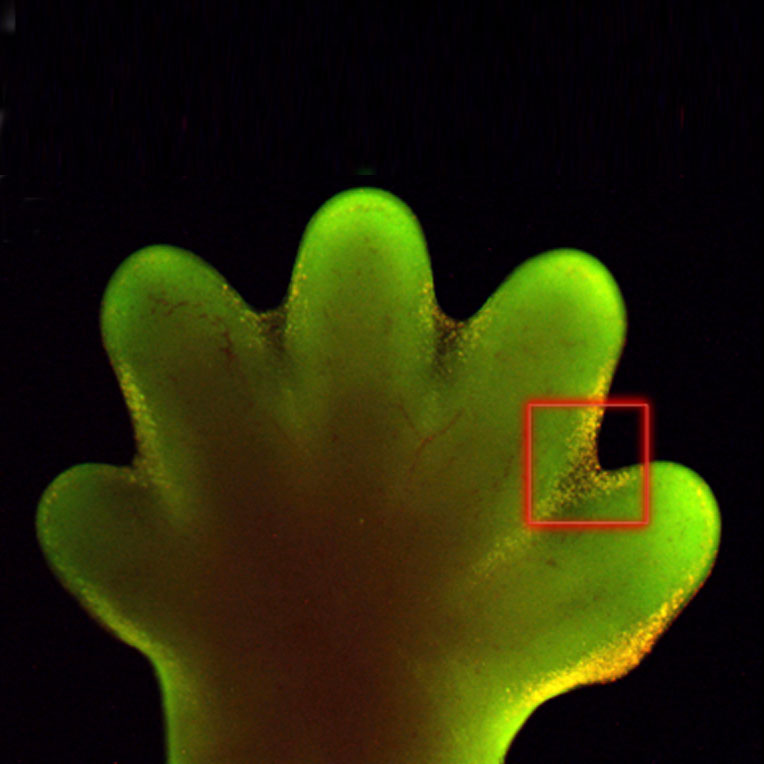
Mouse Interdigit Apoptosis
Mouse embryo E15.5 hindlimb (wild-type) showing apoptotic cells in the interdigital mesenchyme. Insert is enlarged view of selected region (red box). Apoptosis was identified by acridine orange stain that appears yellow in figure.
Reference
<pubmed>17194222</pubmed>| PMC1713256 | PLoS Genet.
Copyright : © 2006 Bandyopadhyay et al. This is an open-access article distributed under the terms of the Creative Commons Attribution License, which permits unrestricted use, distribution, and reproduction in any medium, provided the original author and source are credited.
Original image name: Figure 2 (panel E and G extracted and resized from full image)
File history
Click on a date/time to view the file as it appeared at that time.
| Date/Time | Thumbnail | Dimensions | User | Comment | |
|---|---|---|---|---|---|
| current | 15:49, 14 November 2011 | | 764 × 764 (61 KB) | S8600021 (talk | contribs) | ==Mouse Interdigit Apoptosis== Mouse embryo E15.5 hindlimb (wild-type) showing apoptotic cells in the interdigital mesenchyme. Insert is enlarged view of selected region (red box). Apoptosis was identified by acridine orange stain that appears yellow in |
You cannot overwrite this file.
File usage
There are no pages that use this file.