File:Mesonephric duct position week 6-11.jpg

Original file (697 × 800 pixels, file size: 72 KB, MIME type: image/jpeg)
Human Mesonephric duct position (week 6 to 11)
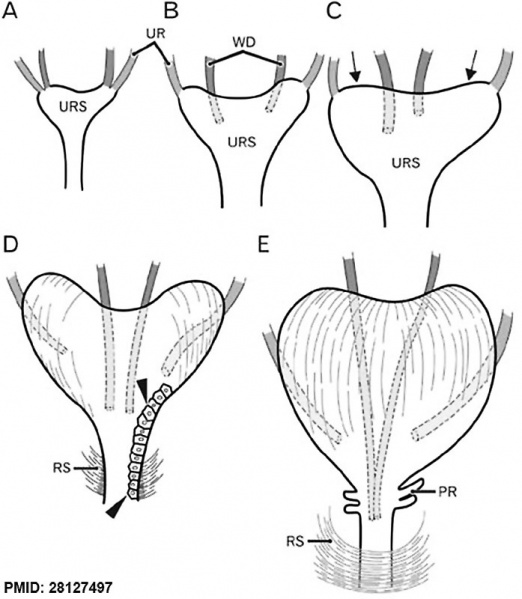
Schematic representations of the descent of the mesonephric duct (Wolffian duct, WD) or vas deferens. Anterior view.
At 6 weeks (A), the WD opens to the urogenital sinus at a site adjacent to the ureteral orifice (UR).
At 7–8 weeks (B, C), rather than descent, there are individual variations in the WD position along the mediolateral axis as well as in left/right difference in morphology of the urogenital sinus (URS). The future bladder and urethra are not discriminated in the sinus.
At 8–9 weeks, the bilateral upper angles (arrows) of the URS start upward growth toward the umbilicus.
At 9 weeks (D), depending on development of smooth muscles in the bladder as well as rhapdosphincter muscles of the urethra (RS), the descent of the vas deferens becomes evident. However, the epithelium is still same (arrowheads) between the future bladder and urethra.
At 10–11 weeks (E), a drastic upward growth of bladder smooth muscles as well as a developing prostate (PR) accelerates the descent of the vas.
- Links: Urinary Bladder Development
Reference
<pubmed>28127497</pubmed>
https://acbjournal.org/DOIx.php?id=10.5115/acb.2016.49.4.231
Copyright
This is an Open Access article distributed under the terms of the Creative Commons Attribution Non-Commercial License (http://creativecommons.org/licenses/by-nc/4.0/) which permits unrestricted non-commercial use, distribution, and reproduction in any medium, provided the original work is properly cited.
Fig. 9. Acb-49-231-g009-l.jpg
Cite this page: Hill, M.A. (2024, May 15) Embryology Mesonephric duct position week 6-11.jpg. Retrieved from https://embryology.med.unsw.edu.au/embryology/index.php/File:Mesonephric_duct_position_week_6-11.jpg
- © Dr Mark Hill 2024, UNSW Embryology ISBN: 978 0 7334 2609 4 - UNSW CRICOS Provider Code No. 00098G
File history
Click on a date/time to view the file as it appeared at that time.
| Date/Time | Thumbnail | Dimensions | User | Comment | |
|---|---|---|---|---|---|
| current | 16:10, 9 March 2017 | | 697 × 800 (72 KB) | Z8600021 (talk | contribs) | Fig. 9 Schematic representations of the descent of the Wolffian duct (WD) or vas deferens. Anterior view. At 6 weeks (A), the WD (red) opens to the urogenital sinus at a site adjacent to the ureteral orifice (UR; green). At 7–8 weeks (B, C), rather... |
You cannot overwrite this file.
File usage
The following page uses this file: