File:Human sperm pathologies EM01.jpg
Original file (761 × 759 pixels, file size: 148 KB, MIME type: image/jpeg)
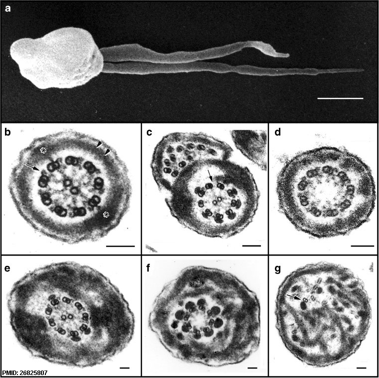
Electron microscopic analysis of human sperm pathologies
a Scanning electron micrograph of a dysplasia of the fibrous sheath (DFS) in human spermatozoa. Note the two thick, irregular and very short tails (length less than 10 μm, normal 50–60 μm).
b Transverse section of a normal flagellum at the distal principal piece (compare with Fig. 4). At this point, the axoneme is composed of nine doublet microtubules around a central pair microtubule apparatus (CPMA), nine radial spokes projecting toward the CPMA, and two dynein arms (outer and inner) anchored to each A-tubule and oriented in a clockwise direction toward the B-tubule of the neighboring doublet microtubule (arrow), as viewed from the base to the tip of the flagellum. The FS is composed of two lateral columns inserting into doublet microtubules #3 and #8 (asterisks), and two semi-circumferential ribs (double arrow head). By this point along the flagellum, the ODFs have terminated.
c, d Spermatozoa from two patients with primary ciliary dyskinesia. In c, the fused complete and incomplete axonemes are due to the failure of neighboring spermatids to separate from their cytoplasmic bridges, and here, there is partial lack of dynein arms (arrow) and FS distortions. In d, the CPMA is missing (i.e., a 9 + 0 axoneme) but radial spokes are still present.
e–g Three transverse sections of DFS spermatozoa with marked FS abnormalities: the FS forms thick disordered periaxonemal rings and the lateral columns are misplaced in e and f; the axoneme in e is preserved, but in f, there is lack of one doublet microtubule and the CPMA is missing (8 + 0 axoneme). In g, note the complete disorientation of the axoneme, where one doublet microtubule appears to lack dynein arms (arrow). Diameters of pathological flagella may range from 1 to 1.2 μm (normal flagellar diameter ≅ 0.4 μm).
Scale bars 1 μm in a and 0.1 μm in b–g
Reference
<pubmed>26825807</pubmed>
Copyright
© The Author(s) 2016
Open Access - This article is distributed under the terms of the Creative Commons Attribution 4.0 International License (http://creativecommons.org/licenses/by/4.0/), which permits unrestricted use, distribution, and reproduction in any medium, provided you give appropriate credit to the original author(s) and the source, provide a link to the Creative Commons license, and indicate if changes were made.
Fig. 7 10815_2016_652_Fig7_HTML.jpg Original image resized and labelled with PMID.
Cite this page: Hill, M.A. (2024, April 27) Embryology Human sperm pathologies EM01.jpg. Retrieved from https://embryology.med.unsw.edu.au/embryology/index.php/File:Human_sperm_pathologies_EM01.jpg
- © Dr Mark Hill 2024, UNSW Embryology ISBN: 978 0 7334 2609 4 - UNSW CRICOS Provider Code No. 00098G
File history
Click on a date/time to view the file as it appeared at that time.
| Date/Time | Thumbnail | Dimensions | User | Comment | |
|---|---|---|---|---|---|
| current | 11:36, 25 January 2017 | | 761 × 759 (148 KB) | Z8600021 (talk | contribs) | PMID 26825807 10815_2016_652_Fig7_HTML.jpg |
You cannot overwrite this file.
File usage
The following page uses this file: