File:Heart innervation 01.jpg
Original file (1,280 × 599 pixels, file size: 92 KB, MIME type: image/jpeg)
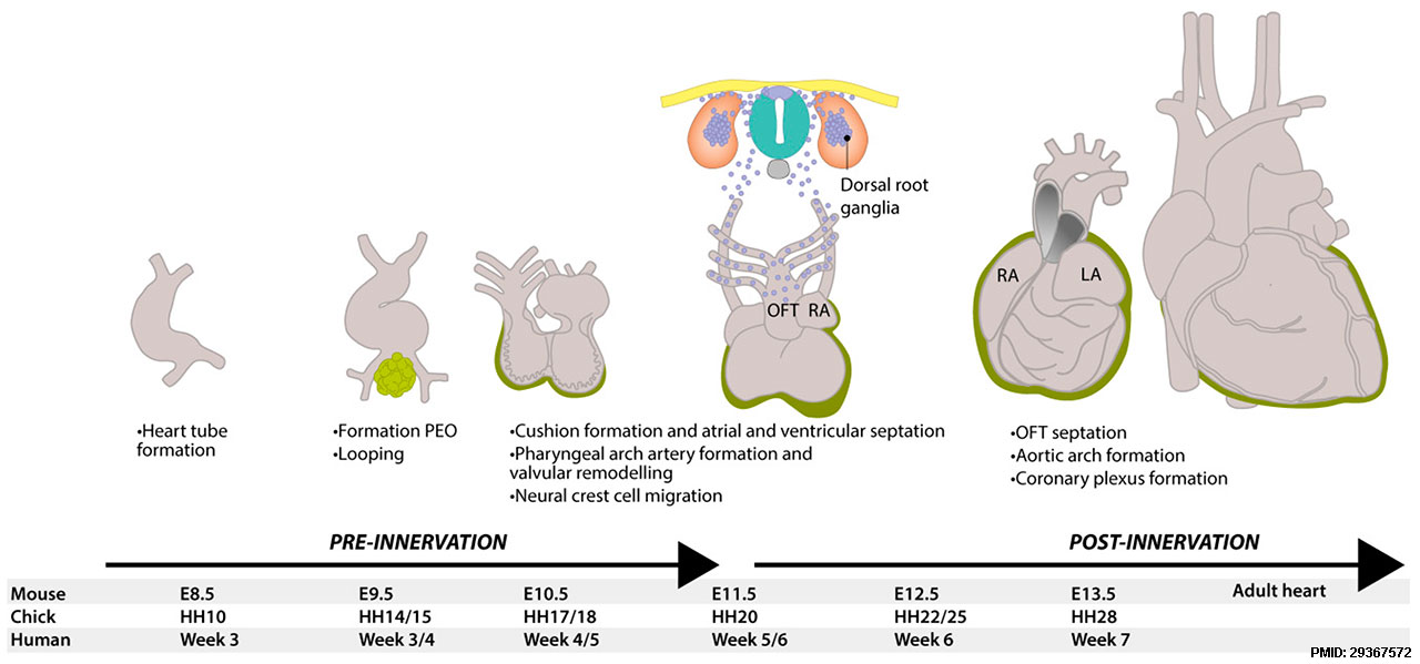
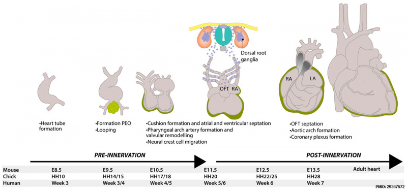
Development of the heart and neural tube in mice
E8.5 - mouse heart is a tube with blood flowing in a peristaltic manner from the caudal venous pole to the cranial arterial (outflow) tract.
E9.5 - heart starts looping and, at the same time, epicardial cells from the proepicardial organ (green) start to migrate and cover the outside of the heart.
E11.5 - neural crest derived cells (blue) delaminate from the neural tube and start migrating ventrally and caudally, contributing to many structures, including dorsal root ganglia. They contribute to valvular remodeling and the septation of the pulmonary and aortic vessels, as well as to delivering neurons to innervate the heart. When the heart has finished looping, the inflow and outflow tract are both found at the base of the heart and the electrical conduction now has an apex-to-base direction.
DRG = dorsal root ganglion, HH = Hamburger and Hamilton stage, LA = left atrium, LV = left ventricle, OFT = outflow tract, RA = right atrium, RV = right ventricle, PEO = proepicardial organ.
Reference
Végh AMD, Duim SN, Smits AM, Poelmann RE, Ten Harkel ADJ, DeRuiter MC, Goumans MJ & Jongbloed MRM. (2016). Part and Parcel of the Cardiac Autonomic Nerve System: Unravelling Its Cellular Building Blocks during Development. J Cardiovasc Dev Dis , 3, . PMID: 29367572 DOI.
Copyright
© 2016 by the authors; licensee MDPI, Basel, Switzerland. This article is an open access article distributed under the terms and conditions of the Creative Commons Attribution (CC-BY) license (http://creativecommons.org/licenses/by/4.0/).
Figure 1. https://www.mdpi.com/2308-3425/3/3/28
Cite this page: Hill, M.A. (2024, May 13) Embryology Heart innervation 01.jpg. Retrieved from https://embryology.med.unsw.edu.au/embryology/index.php/File:Heart_innervation_01.jpg
- © Dr Mark Hill 2024, UNSW Embryology ISBN: 978 0 7334 2609 4 - UNSW CRICOS Provider Code No. 00098G
File history
Click on a date/time to view the file as it appeared at that time.
| Date/Time | Thumbnail | Dimensions | User | Comment | |
|---|---|---|---|---|---|
| current | 06:16, 27 November 2018 | | 1,280 × 599 (92 KB) | Z8600021 (talk | contribs) | Development of the heart and neural tube in mice. At E8.5, the murine heart is a tube with blood flowing in a peristaltic manner from the caudal venous pole to the cranial arterial (outflow) tract. At E9.5, the heart starts looping and, at the same tim... |
You cannot overwrite this file.
File usage
The following 2 pages use this file: