File:Wheeler-plate04.jpg

Original file (829 × 1,113 pixels, file size: 108 KB, MIME type: image/jpeg)
Plate 4
Original plate images magnification shown in brackets.
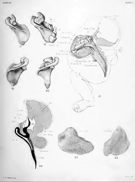
Fig. 17. Dorsal superior view of a normal left scapula of a new-born
Fig. 18. .Same view of left scapula of specimen 862a
shows the irregular vertebral and superior margins with the abnormal spicule of bone attached. It also shows the sheets of fascia attached to the vertebral and median margins of the scapula and the insertions of the rhomboideus and levator scapular muscles on this fascia.
(Natural size).
Fig. 19. Dorsal superior view of a normal right scapula of a new-born
Fig. 20. Same view of right scapula of this specimen
showing irregular vertebral margin. (Natural size).
Fig. 21. Diagram of left thoracic and deep dorsal musculature
Diagram of left thoracic and deep dorsal musculature on (he left side) of the mounted axial skeleton. The occiput and model of cerebral-spinal cavity are in place. The median outline of the .specimen is also given in relation to these structures. Those nuiscles approximately normal arc either sectioned or oidy drawn at their origin or insertion. They are the m. pectoralis major and minor, the rectus, the external oblique, the latissimus dorsi, the quadratus lumborum, and the levator scapulae. The abnormal muscles are shown entire, except for the serratus anterior, whose origin is indicated by broken lines. The largest mass of abnormal muscles consists of a longitudinal bundle extending from the sacrum to the occiput and labeled m. sacrospin. From about the center of this bundle the serratus posterior inferior projects onto the lower three ribs. The muscles at the upper end of the bundle are quite irregular. Along the fourth and fifth ribs a mass of muscle extends nearly to their costal cartilages. At the distal termination of these fibers lie several small irregularly placed bundles. In the upper thoracic region is a narrow band of muscle overlving the others. (half size).
Fig. 22. Lateral view of abnormal right lung formed of but one lobe
(half size)
Fig. 23. Lateral view of normal two-lobed left lung
(half size)
Fig. 24. Diagram of those structures of the central nervous system
which lie near the midline and which can be identified. The outline of the subdural space used was obtained from the .sagittal section. Posteriorly this passes near to the median margin of the left encephalocele. The cerebrum designated by a dotted line is shown protruding below the foramen magnum into the encephalocele. A small portion of the cerebellum, represented by line-hatching, is seen to lie very much flattened on top of the cord. The brain-stem and cord, much bent, arc shown in solid black. Those cranial nerves which were identified are shown by lines. Only the first spinal nerve is shown. The floor of the fourth ventricle lies inverted on top of a flat cord.
File history
Click on a date/time to view the file as it appeared at that time.
| Date/Time | Thumbnail | Dimensions | User | Comment | |
|---|---|---|---|---|---|
| current | 08:47, 16 February 2011 | | 829 × 1,113 (108 KB) | S8600021 (talk | contribs) | ==Plate 4== Original plate images magnification shown in brackets. ===Fig. 17. Dorsal superior view of a normal left scapula of a new-born=== ===Fig. 18. .Same view of left scapula of specimen 862a shows the irregular vertebral and superior margins wit |
You cannot overwrite this file.
File usage
The following page uses this file: