File:Papanicolaou1933-plate09.jpg
From Embryology
Size of this preview: 442 × 599 pixels. Other resolution: 1,582 × 2,144 pixels.
Original file (1,582 × 2,144 pixels, file size: 257 KB, MIME type: image/jpeg)
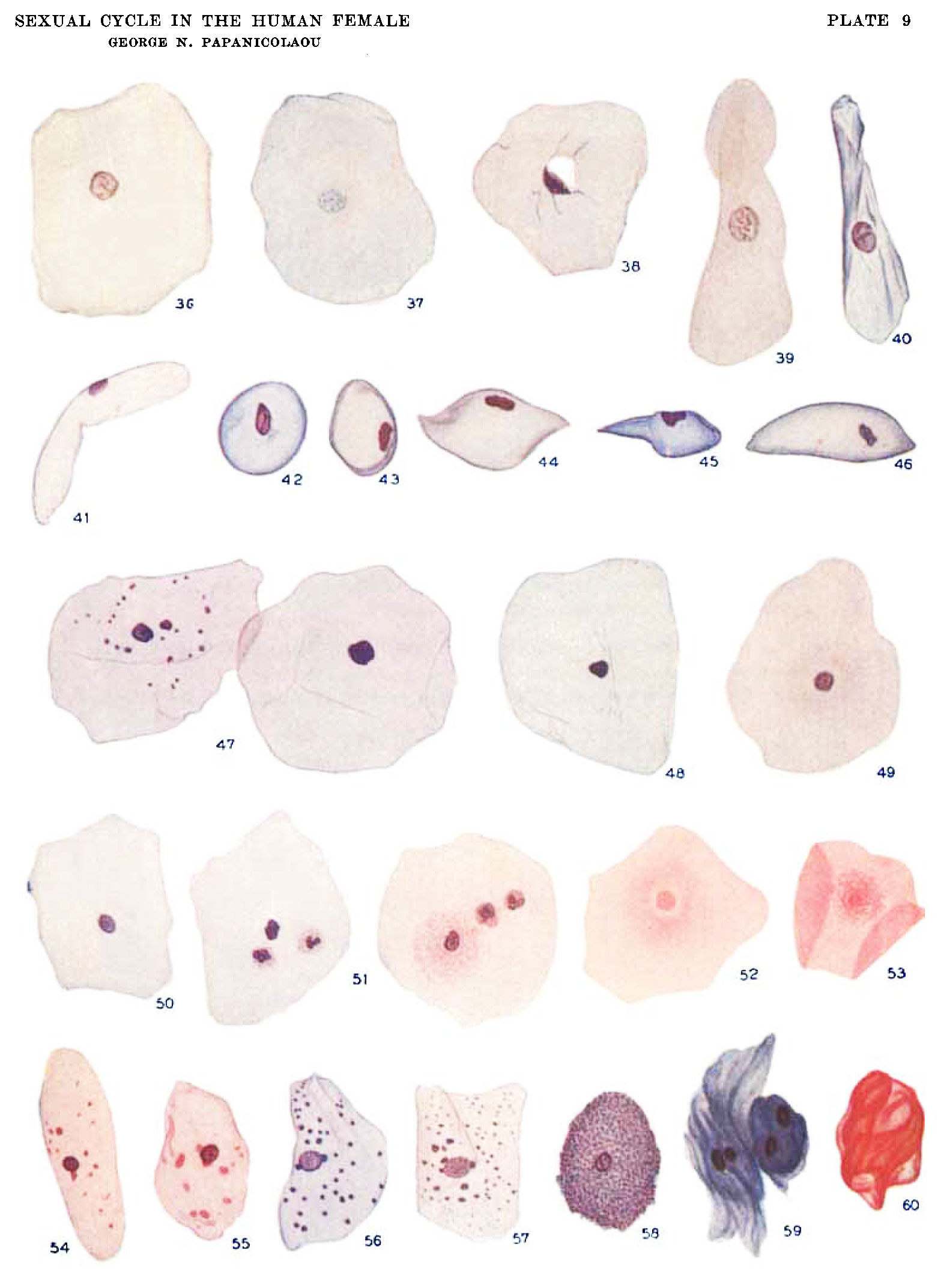
Plate 9. Drawings of various types of cells found in normal human vaginal smears
All cells are equally magnified and stained by the same method, as described in this paper. Compens. 0c. 6, mm. oil obj. 2 mm.
- 36 to 60 Cells from human vaginal smears at different stages of the normal menstrual cycle.
Reference
Papanicolaou GN. The Sexual Cycle in the Human Female as revealed by Vaginal Smears. Am J Anat. 1933;52: 519–637.
Cite this page: Hill, M.A. (2024, May 9) Embryology Papanicolaou1933-plate09.jpg. Retrieved from https://embryology.med.unsw.edu.au/embryology/index.php/File:Papanicolaou1933-plate09.jpg
- © Dr Mark Hill 2024, UNSW Embryology ISBN: 978 0 7334 2609 4 - UNSW CRICOS Provider Code No. 00098G
File history
Click on a date/time to view the file as it appeared at that time.
| Date/Time | Thumbnail | Dimensions | User | Comment | |
|---|---|---|---|---|---|
| current | 10:22, 8 September 2015 | | 1,582 × 2,144 (257 KB) | Z8600021 (talk | contribs) |
You cannot overwrite this file.
File usage
The following 2 pages use this file: