File:Oesophageal atresia x-ray 01.jpg
Original file (894 × 588 pixels, file size: 102 KB, MIME type: image/jpeg)
Esophageal Atresia x-ray
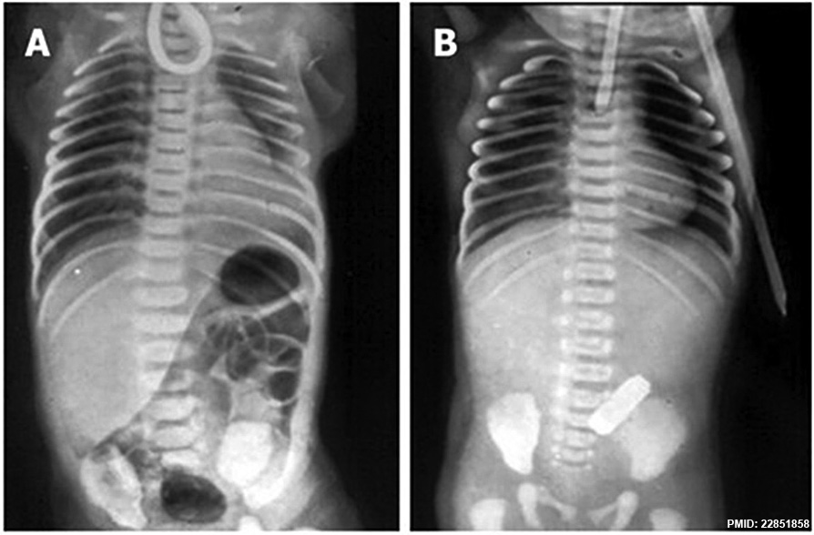
Plain X-rays of the chest and abdomen of two neonates with esophageal (oesophageal) atresia.
- A The non-progression of an orogastric catheter in the blind esophageal pouch and the presence of air in the stomach diagnose esophageal atresia with distal tracheoesophageal fistula.
- B The radiopaque tube in the blind esophageal pouch and the absence of air in the stomach identify esophageal atresia without tracheoesophageal fistula.
- Links: Oesophageal atresia x-ray | Classification of Oesophageal atresia | Gastrointestinal Tract - Abnormalities
Reference
Pinheiro PF, Simões e Silva AC & Pereira RM. (2012). Current knowledge on esophageal atresia. World J. Gastroenterol. , 18, 3662-72. PMID: 22851858 DOI.
Copyright
©1995-2014 Baishideng Publishing Group Inc. All rights reserved. Articles published by this open-access journal are distributed under the terms of the Creative Commons Attribution-Noncommercial License, which permits use, distribution, and reproduction in any medium, provided the original work is properly cited, the use is non commercial and is otherwise in compliance with the license.
Cite this page: Hill, M.A. (2024, May 11) Embryology Oesophageal atresia x-ray 01.jpg. Retrieved from https://embryology.med.unsw.edu.au/embryology/index.php/File:Oesophageal_atresia_x-ray_01.jpg
- © Dr Mark Hill 2024, UNSW Embryology ISBN: 978 0 7334 2609 4 - UNSW CRICOS Provider Code No. 00098G
File history
Click on a date/time to view the file as it appeared at that time.
| Date/Time | Thumbnail | Dimensions | User | Comment | |
|---|---|---|---|---|---|
| current | 17:09, 9 May 2015 | | 894 × 588 (102 KB) | Z8600021 (talk | contribs) |
You cannot overwrite this file.
File usage
The following page uses this file: