File:Mouse pax7 neural fold 01.jpg

Original file (513 × 915 pixels, file size: 100 KB, MIME type: image/jpeg)
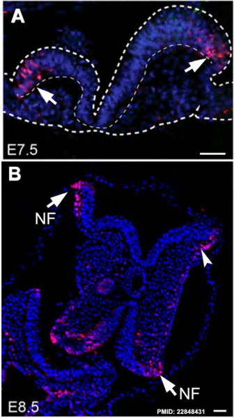
Mouse Pax7 Neural Fold
Spatiotemporal expression of neural crest markers including Pax7 in E7.5 to E8.5 mouse embryos.
In C57BL/6 embryos, immunohistochemistry detects Pax7 (red) in
- A,B - E7.5 to E8.5 lateral neural folds (NF; arrows);
- B - E8.5 cephalic mesenchyme (arrowhead).
Dapi (blue) is a nuclear stain.
Scale bars −100 um.
- Links: E7.5 E8.5 neural fold | E9.5 Pax7 heart | E11.5 Trunk neural crest 1 | E11.5 Trunk neural crest 2 | E15.5 Pax7 limb | Mouse Development | Neural Crest Development | Pax
Reference
<pubmed>22848431</pubmed>| PMC2634972 | PLoS One.
Copyright
© 2012 Murdoch et al. This is an open-access article distributed under the terms of the Creative Commons Attribution License, which permits unrestricted use, distribution, and reproduction in any medium, provided the original author and source are credited.
Figure 1. doi:10.1371/journal.pone.0041089.g001
File history
Click on a date/time to view the file as it appeared at that time.
| Date/Time | Thumbnail | Dimensions | User | Comment | |
|---|---|---|---|---|---|
| current | 11:21, 17 February 2013 | | 513 × 915 (100 KB) | Z8600021 (talk | contribs) | {{Mouse neural crest pax7 links}} |
You cannot overwrite this file.
File usage
The following page uses this file: