File:Mouse embryo E11 and tomography 01.jpg
From Embryology

Size of this preview: 800 × 507 pixels. Other resolution: 1,184 × 751 pixels.
Original file (1,184 × 751 pixels, file size: 120 KB, MIME type: image/jpeg)
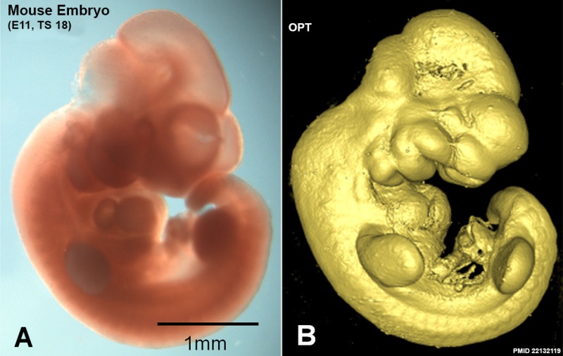
Mouse Embryo (E11)
- A - External view of an E11 Theiler stage (TS) control embryo.
- B- Surface rendered view of the Optical Projection Tomography (OPT) 3D reconstruction of the same embryo showing morphological features used for staging purposes, staged as TS18.
- Links: Mouse Development | Category:Mouse E11.0 | Computed Tomography | Image - Mouse embryo E11 tomography | Image - Mouse embryo E11 HNF3beta notochord marker
Reference
<pubmed>22132119</pubmed>| PLoS One.
Copyright
© 2011 Hajduk et al. This is an open-access article distributed under the terms of the Creative Commons Attribution License, which permits unrestricted use, distribution, and reproduction in any medium, provided the original author and source are credited.
Figure 1. doi:10.1371/journal.pone.0027635.g001 Panel A and B cropped from full figure, reorganised, resized and relabelled.
File history
Click on a date/time to view the file as it appeared at that time.
| Date/Time | Thumbnail | Dimensions | User | Comment | |
|---|---|---|---|---|---|
| current | 12:41, 18 August 2014 | | 1,184 × 751 (120 KB) | Z8600021 (talk | contribs) | ==Mouse Embryo (E11)== * A - External view of an E11 Theiler stage (TS) control embryo. * B - Surface rendered view of the Optical Projection Tomography (OPT) 3D reconstruction of the same embryo showing morphological features used for staging purpos... |
You cannot overwrite this file.
File usage
The following 2 pages use this file: