File:Mouse blastocyst development 01.jpg
Original file (1,280 × 682 pixels, file size: 149 KB, MIME type: image/jpeg)
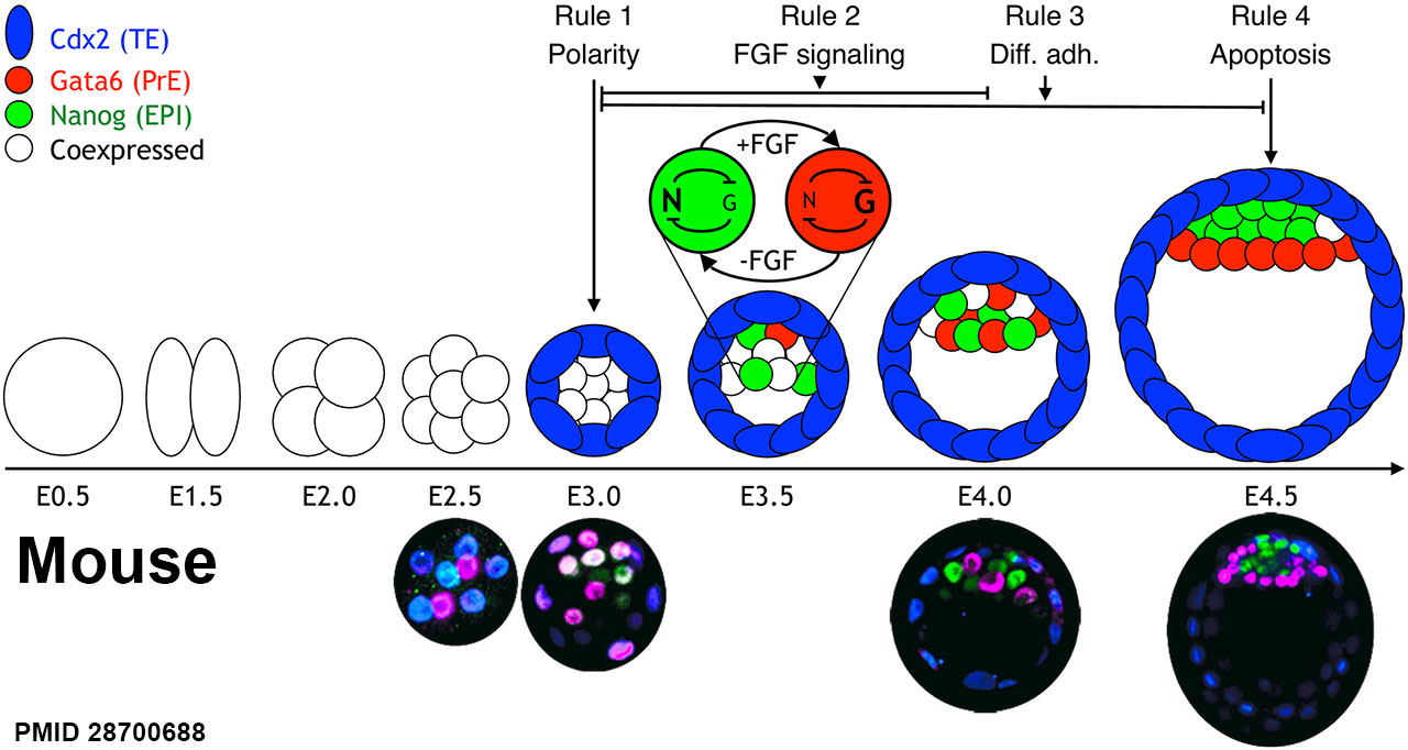
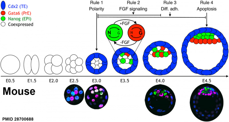
Schematic of Mouse Early Embryonic development
E0.5 - The zygote begins and undergoes 3 rounds of cleavage divisions
E2.5. - 8 cells. During the next round of division, the blastomeres undergo compaction and become polarized, resulting in the outer trophectoderm (TE) (blue) and the inner cell mass (ICM) (still coexpressing Gata6 and Nanog). The TE expresses the transcription factor caudal-related homeobox 2 (Cdx2).
E3.5 - a cavity has formed, and the ICM is positioned at 1 side of the embryo. At this stage, the ICM transcription factors, Gata6 (red) and Nanog (green), are expressed in a mutually exclusive salt-and-pepper pattern in some cells.
E4.5 - Nanog- and Gata6-expressing cells have physically segregated into 2 distinct layers and are developmentally restricted to either the epiblast (EPI) or primitive endoderm (PrE). The lower panel shows immunostaining of embryos at different stages during preimplantation development. Color coding is the same as in the panel above. The timing of the 4 different rules that we apply is indicated on top of the diagram.
- Links: Blastocyst Development | Mouse Development
Reference
<pubmed>28700688</pubmed>
Copyright
© 2017 Nissen et al. This is an open access article distributed under the terms of the Creative Commons Attribution License, which permits unrestricted use, distribution, and reproduction in any medium, provided the original author and source are credited.
Fig 1. Journal.pbio.2000737.g001.jpg Original PNG resized and relabelled.
Cite this page: Hill, M.A. (2024, May 15) Embryology Mouse blastocyst development 01.jpg. Retrieved from https://embryology.med.unsw.edu.au/embryology/index.php/File:Mouse_blastocyst_development_01.jpg
- © Dr Mark Hill 2024, UNSW Embryology ISBN: 978 0 7334 2609 4 - UNSW CRICOS Provider Code No. 00098G
File history
Click on a date/time to view the file as it appeared at that time.
| Date/Time | Thumbnail | Dimensions | User | Comment | |
|---|---|---|---|---|---|
| current | 15:59, 9 August 2017 | | 1,280 × 682 (149 KB) | Z8600021 (talk | contribs) | ===Reference=== Journal.pbio.2000737.g001.jpg {{Footer}} |
You cannot overwrite this file.
File usage
The following page uses this file: