File:Liver- Kupffer cell and reticular fibre.jpg

Original file (600 × 800 pixels, file size: 49 KB, MIME type: image/jpeg)
Liver Histology
Liver, rabbit - trichrome & carbon and Liver - reticulin
The first slide will allow you to identify the macrophages which adhere to the wall of the liver sinusoids.
They are represented by the accumulations of small brown/black dots, the carbon particles ingested by the macrophages.
- Links: Liver Histology | Liver Development
Links: Histology | Histology Stains | Blue Histology images copyright Lutz Slomianka 1998-2009. The literary and artistic works on the original Blue Histology website may be reproduced, adapted, published and distributed for non-commercial purposes. See also the page Histology Stains.
Cite this page: Hill, M.A. (2024, April 29) Embryology Liver- Kupffer cell and reticular fibre.jpg. Retrieved from https://embryology.med.unsw.edu.au/embryology/index.php/File:Liver-_Kupffer_cell_and_reticular_fibre.jpg
- © Dr Mark Hill 2024, UNSW Embryology ISBN: 978 0 7334 2609 4 - UNSW CRICOS Provider Code No. 00098G
Liv41re.jpg
File history
Click on a date/time to view the file as it appeared at that time.
| Date/Time | Thumbnail | Dimensions | User | Comment | |
|---|---|---|---|---|---|
| current | 13:04, 9 March 2018 | | 600 × 800 (49 KB) | Z8600021 (talk | contribs) | updated size and contrast |
| 09:39, 22 December 2010 | 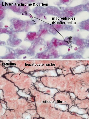 | 300 × 400 (45 KB) | S8600021 (talk | contribs) | ==Liver Histology== Liver, rabbit - trichrome & carbon and Liver - reticulin The first slide will allow you to identify the macrophages which adhere to the wall of the liver sinusoids. They are represented by the accumulations of small brown/black dots, |
You cannot overwrite this file.