File:Leydig cell PMID13693345 EM03.jpg

Original file (1,359 × 957 pixels, file size: 325 KB, MIME type: image/jpeg)
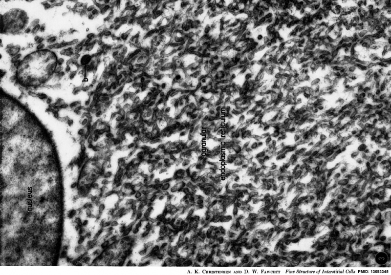
Leydig Cell Electron Micrograph
Opossum testicular interstitial cells showing extensive smooth endoplasmic reticulum involved in testosterone synthesis.
In this historic 1961 paper, Smooth Endoplasmic Reticulum (SER) is called "agranular endoplasmic reticulum", because of the absence of attached ribosomes (RER). SER is abundant in cells involved in steroidal hormone synthesis.
Figure legend
A view at higher power of interstitial cell cytoplasm, showing the elaborate meshwork of interconnecting tubules which constitutes the agranular endoplasmic reticulum. There is biochemical evidence indicating that this reticulum is involved in the biosynthesis of the steroid hormones secreted by these cells. At the upper right is one of the small bodies (b) commonly seen in the cytoplasm. original image X 33,000.
Reference
CHRISTENSEN AK & FAWCETT DW. (1961). The normal fine structure of opossum testicular interstitial cells. J Biophys Biochem Cytol , 9, 653-70. PMID: 13693345
Copyright
Rockefeller University Press - Copyright Policy This article is distributed under the terms of an Attribution–Noncommercial–Share Alike–No Mirror Sites license for the first six months after the publication date (see http://www.jcb.org/misc/terms.shtml). After six months it is available under a Creative Commons License (Attribution–Noncommercial–Share Alike 4.0 Unported license, as described at https://creativecommons.org/licenses/by-nc-sa/4.0/ ). (More? Help:Copyright Tutorial)
Original figure 3 rotated, relabelled and adjusted in size and contrast.
Cite this page: Hill, M.A. (2024, April 26) Embryology Leydig cell PMID13693345 EM03.jpg. Retrieved from https://embryology.med.unsw.edu.au/embryology/index.php/File:Leydig_cell_PMID13693345_EM03.jpg
- © Dr Mark Hill 2024, UNSW Embryology ISBN: 978 0 7334 2609 4 - UNSW CRICOS Provider Code No. 00098G
File history
Click on a date/time to view the file as it appeared at that time.
| Date/Time | Thumbnail | Dimensions | User | Comment | |
|---|---|---|---|---|---|
| current | 17:21, 7 August 2014 | | 1,359 × 957 (325 KB) | Z8600021 (talk | contribs) | |
| 17:14, 7 August 2014 | 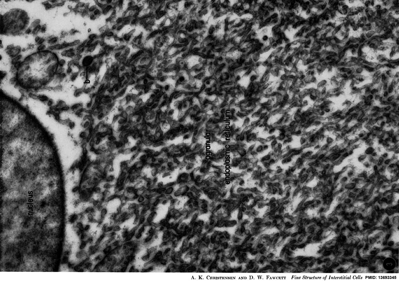 | 1,359 × 957 (288 KB) | Z8600021 (talk | contribs) | J Biophys Biochem Cytol. 1961 Mar;9:653-70. The normal fine structure of opossum testicular interstitial cells. CHRISTENSEN AK, FAWCETT DW. Abstract The interstitial tissue of the opossum testis includes interstitial or Leydig cells, macrophages, and s... |
You cannot overwrite this file.
File usage
The following page uses this file: