File:Keibel1897 plate01.jpg

Original file (1,504 × 2,000 pixels, file size: 365 KB, MIME type: image/jpeg)
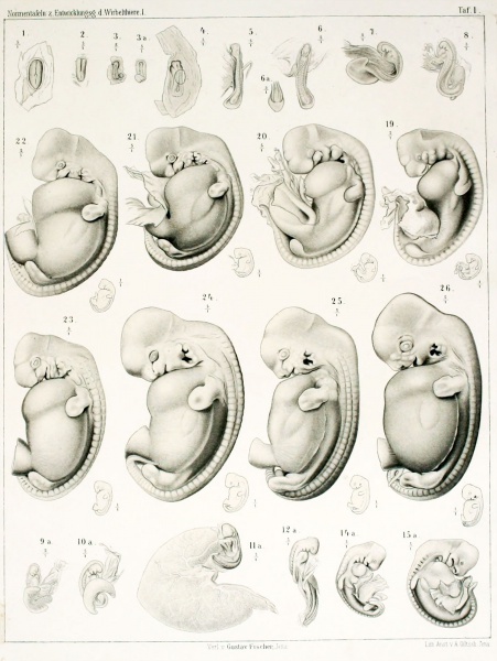
Plate 1.
Fig. 1
The embryonic disc p.7 b. 93 is from a 14-Days 10 hours before the slaughter sow occupied. After the embryonic disc was removed in the usual way with chrome acetic uterus, it was fixes to sublimate on. FIG. 1 illustrates the embryonic disc is how they presented itself to me after this treatment in 70% Alcohol. M., and M, are a draft after Ziegler model, which was produced with a basic layer reconstruction. We see, as the primitive streak has already been limited to the rear half of the embryonic disc. Before the primitive streak region we see the first installation of the medullary and the medullary. The medullary groove extends up close to the front end of the embryonic disc, rear end already on the point, the front end to embrace the primitive streak. The area in front of the front end of Primitiv.streifens is easy aufgewulstet, and this bead forms the base of the rear part, the medullary groove. At the front end of the embryonic disc a little bump already at surface observation stands right in front of the medullary groove clearly emerges. As can be seen from the average series, called these bumps, the front end of the head extension resp. the notochord, it corresponds to the front end of the embryo at all. The small protrusion that is noticeable at the rear end of the embryonic disc is caused by Mesodermwucherung and the mesodermal Allantoisanlage. It should be emphasized that in this embryonic disc, the posterior intestinal bay is already well trained. The occurrence of the amnion was already mentioned in the previous embryonic disc, the development of the amnion has made progress, we find, as in the previous stage, evenly developed in circumference around the embryonic disc.
Fig. 2
If the germinal disc shown in FIG. 2, the medullary groove in the fore significantly compared to the primitive streak. Even more clearly than in the previous stage it can be seen as the front gewulstete end of the primitive streak is encompassed by the Medullarfalten. Forward towards the medullary forks before the hump, which corresponds to the front end of the notochord and at all the embryo developed. The head of the embryo starts to stand out.
Full exact proportion can not be determined at this embryo because the rearmost end of the primitive streak is handled.
Fig. 3
The embryo, which is illustrated in FIG. 3 and 3a, was the mother animal taken 15 days one hour after mating and fixes with Pikrinschwefelchromsäure. I could see in the review of 3 on each side and each Urwirbelpaare accrued before and after yet another without limit cranial resp. caudal. Fig. 3 shows the ring intersected the chorion to the embryo, and you can see how the chorionic turns into the amnion. Only in the districts in which the amnion is still open, you can see directly on the dorsal side of the embryo, otherwise you see the P ^ mbryonalkörper only by the chorion shine through. The caudal end of the embryo is hook-shaped bent downwards. This embryo is the Kopfamnion less developed than the Schwanzamnion, cranial run the two lateral amniotic folds acute angle against each other; caudal they go in the bow into one another; by the still fairly wide opening of the Amnionnabels you see on the medullary and the somites.
Fig. 3a gives the ventral view, showing how the Kmliryo is tightly surrounded by the amnion. At the top end you can see in the liinein quite shallow foregut bay. At the caudal end of the embryo to recognize the Primitiv.streifen, the ends of the medullary and the mesodermal Allantoiswucherung. The front end of the primitive streak, rises before Sicli a flat bead (about the beginning of the head extension) is covered by the elapsing caudal Medullarwülsten. The mesodermal allantoic growth shows a slight lobing. It should be noted at this embryo that the hook-shaped bend of the Caudalendes about is not typical for this stage porcine embryos. It come at this stage before fully stretched embryos, but it was not possible for me to receive such a standard for the panel. It is perhaps not out of place to point out that such irregular bending of the embryos of this stage and even earlier stage in Keimscheiben- are nothing unusual. They are obtained even with the most careful preparation, and I can hardly assume that there are dealing with blemishes which embryos by removing the EM and seed discs are caused from the uterus. Rather, I believe that in these cases, the embryo and the embryonic disc is given directly if necessary to switch the many wrinkles and lines the uterus. That this necessity occurs precisely at these stages, is easily explained if one considers that as long as the amnion is still open and so long as the fixed conversion connected embryo the chorionic still fits tightly from amnion, to the same located on the surface of the egg , In later stages, when the l ^ is sunken mbryo deeper into the interior of the egg and is only through the Amnionnabelstrang with the chorion in connection, the embryo is removed from the direct influence of the uterus. Are things so, and I do not see another explanation well, so we have recorded the interesting fact that the pig embryo even when the coarser mechanical individual settings are by such folds in not entirely inconsiderable proportion modified, the development processes nevertheless take place usually in the normal manner.
Fig. 4
The embryo is the mother animal taken within 14 days after the mating; it was acid in chromium and sublimate fixes. FIG. 4 shows the embryo and the adjacent part of the egg. The embryo lies with its longitudinal axis according to the longitudinal axis of the P2ies. The amnion approaches the conclusions and therefore covered the greater part of the embryo; the yolk sac shimmers through slightly darker by Chorion. The recognizable by the brain membranes part of the embryo is distinguished by its width from the back soft medullary apart and fa.ssen the early part of the primitive strip between them. In the front third of the embryo is widened laterally; Here are the first systems of the heart, which now already occurs externally in the appearance in this way. At the caudal end of the embryo one sees the slightly lobed mesodermal Allantoiswucherung. The whole embryo is slightly curved on the ventral side. In a review of Hessen to detect 4-5 Urwirbelpaare; the series returned 4 on both sides delimited Urwirbelpaare, cranial them another, forward not demarcated Urwirbelpaar and caudal conceived in formation somites. The neural plate begins to distinguish itself from the rest in the Urwirbelregion ectoderm, while the first traces of dorsal root ganglia plants are noticeable. The very first installation of endodermal allantoic Hess be detected.
Fig. 5
5 u. 5 a. (SN 293. Bez. 16. 20. G6A. NT Tab. 44.) The Fig. 5 and 5a embryo represented the uterus 16 days 20 hours after the loading has been taken under this type of chromium acid and then become fixed in sublimate further. The embryo is almost stretched, the heart area is lifted in part. We see him on the Eigg. 5 and 5a give closely from amnion circumstances where a fairly long Amnionnabelstrang is clear. Both Eiguren also show that the Embrj ^ o still lying in the longitudinal axis of the egg with its longitudinal axis. The caudal end is slightly curved anteriorly and something turned in such a way that also part of the right side from the left can see, so you can see how the MeduUarrohr a short distance caudal is still open (about 0.5 mm) , Even the front neuropore is still open in the area of optic vesicles. The otic vesicle are very shallow dimples. Two gill pockets are on the ectoderm. Not only the Wolff's transition, but also the first plant of Excretionsapparates is clear. The heart is an S-shaped tube, which contains very little blood; on the yolk sac is the Blutl) ducation underway. The endodermal Allantoishörner are formed vigorously.
Fig. 6
The embryo, which in Figs. 6, () a, 61) and 6 c is shown, almost at the same stage as the embryo, which have been depicted in FIG. 5 etc.. He has as well as those 14 somites. The medullary tube is as far closed. The Allantoishörner are inflated a little more, and so stands, as seen from the ventral side, as the Fig. 6b and 6c show the area of the allantois, nevertheless the Allantoishörner still related to the mesoderm of Somatopleura, quite well off. Another difference is given by the fact that the embryo turned out in this case already perpendicular to the longitudinal axis of the egg. The caudal end of the embryo is not just something ventral, but also a little bow off to the right and at the same time is to him a very slight twist rotation from right to left identified; So just in the opposite direction as the embryo Fig. 5.
Fig. 7
Even though the internal development of the embryo 17. 96a. is little more than that of the 3 last discussed embryos, so he sees at first glance much different. The embryo has nearly a perfect spiral rotation from left to right, so that one in Figure 7, which represents the head of the profile exactly from the left, the tail end of the right and gets something from the ventral side to view. Also, a considerable part of the heart region has lifted from the yolk sac. The neural tube is still open at this embryo in the eye bubbles and caudal depending on a very small spot. From the primitive streak, the last remnants can be detected; the tail bud starts to form. The Ohrgrübchen have become much clearer, but still quite flat, between the primary optic vesicles and the ectoderm mesoderm is grown. The number of somites is 19.
Figure 8
(SX 294. Benz. S. II 9 oec. XT Tab. 56.) The illustrated in Fig. 8 embryo p.17, 96 c shows a very striking figure, which one is better regarded by the figure as through a long description is. One eighth in the figure on the spiral
I) As you may be aware of the direction the spiral rotation ever "oh the view expressed by n, to occupy different views known, so I ask to be noted that I, turned from right to IMKS turned the embryo of M hnks right. In both of these embryos all Verhaltnasse are so clear, is now IASS em mistake probably not fear further. drehuno- from left to right and aal the way the Caudalende is beaten to the right and above. Noteworthy is the fact that the connection between the intestine and Allantois no longer behind but ventral Lieo-t. The heart area is completely lifted, the primitive streak only rudimentary remains detectable and the tail bud has formed. The front neuropore is closed, the rear still open on 2 sections of 15. From the intestinal area should be emphasized that the first plant of the liver is indicated.
| Historic Disclaimer - information about historic embryology pages |
|---|
 |
Reference
Franz Keibel, Normentafeln zur Entwicklungsgeschichte der Wirbelthiere (Normal plates of the development of vertebrates) Volume Hft.1 (1897) Jena, G. Fischer, Germany.
Cite this page: Hill, M.A. (2024, April 26) Embryology Keibel1897 plate01.jpg. Retrieved from https://embryology.med.unsw.edu.au/embryology/index.php/File:Keibel1897_plate01.jpg
- © Dr Mark Hill 2024, UNSW Embryology ISBN: 978 0 7334 2609 4 - UNSW CRICOS Provider Code No. 00098G
File history
Click on a date/time to view the file as it appeared at that time.
| Date/Time | Thumbnail | Dimensions | User | Comment | |
|---|---|---|---|---|---|
| current | 09:49, 20 November 2013 | | 1,504 × 2,000 (365 KB) | Z8600021 (talk | contribs) |
You cannot overwrite this file.
File usage
The following 3 pages use this file: