File:Human uterine tube ciliated epithelium SEM.jpg

Original file (1,200 × 855 pixels, file size: 248 KB, MIME type: image/jpeg)
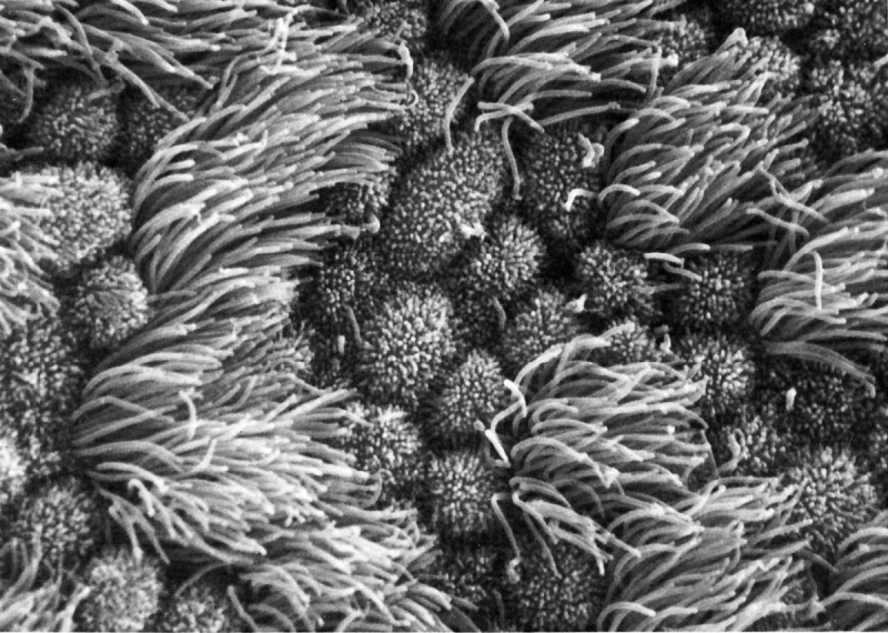
Human Uterine Tube Ciliated Epithelium SEM
Uterine epithelial cilia are responsible for the initial movement of the ooycte and conceptus (zygote, morula, blastocyst). In humans, this is during the first week of development. Uterine epithelial microvilli are involved with the implantation process. Hormones (estrogen and progesterone) regulate both cilia and microvilli.
The differences in size and shape of cilia and microvilli are well illustrated by scanning micrographs of the lumenal surface of the epithelium lining the mammalian oviduct.
- The tufts of cilia associated with individual ciliated cells project several microns above the convex apices of nonciliated cells covered with short microvilli.
- The number of ciliated cells in this epithelium is under hormonal control by estrogens.
| Cilia | Microvilli |
|---|---|
|
|
- Links: Implantation | Menstrual Cycle | Week 1 | Uterus Development
Reference
Don W. Fawcett M.D. The Cell Chapter 13 (Cilia and Flagella) Figure 316.
Copyright
Attribution Non-Commercial; No Derivatives:This image is licensed under a Creative Commons Attribution, Non-Commercial, No Derivatives License.
Original file name: 11618.jpg http://www.cellimagelibrary.org/images/11618
NCBI Organism Classification: Homo sapiens
Cell Type: ciliated epithelial cell, oviduct epithielial cell
Cellular Component: cilium, microvillus
File history
Click on a date/time to view the file as it appeared at that time.
| Date/Time | Thumbnail | Dimensions | User | Comment | |
|---|---|---|---|---|---|
| current | 07:44, 26 April 2011 | | 1,200 × 855 (248 KB) | S8600021 (talk | contribs) | ==Human uterine tube ciliated epithelium SEM== Figure 316 from Chapter 13 (Cilia and Flagella) of 'The Cell' by Don W. Fawcett M.D. The differences in size and shape of cilia and microvilli are well illustrated by scanning micrographs of the lumenal surf |
You cannot overwrite this file.
File usage
The following 4 pages use this file: