BGDA Lecture - Development of the Embryo/Fetus 1

From Embryology
Embryology - 19 Mar 2024    Facebook link Pinterest link Twitter link  Expand to Translate  
Google Translate - select your language from the list shown below (this will open a new external page)

العربية | català | 中文 | 中國傳統的 | français | Deutsche | עִברִית | हिंदी | bahasa Indonesia | italiano | 日本語 | 한국어 | မြန်မာ | Pilipino | Polskie | português | ਪੰਜਾਬੀ ਦੇ | Română | русский | Español | Swahili | Svensk | ไทย | Türkçe | اردو | ייִדיש | Tiếng Việt    These external translations are automated and may not be accurate. (More? About Translations)

BGDsmall.jpg

Introduction

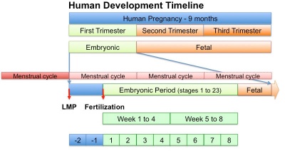
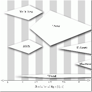
Human development timeline graph 02.jpg

In medicine foundations you were given a broad overview of human development. Now in BGDA we will be working through the human development process in more detail, focussing on key events.

2018 Lecture PDF

  • Begin by reviewing the recent Foundations Lecture and Practical.
  • This BGDA lecture covers conceptus development from fertilization to implantation to trilaminar embryo formation.
    • Note that fertilization and week 1 concepts have already been covered in an earlier BGDA lecture.
  • The lecture will also introduce early fetal membranes and placentation.


One-minute.gif

1 Minute Embryology | UNSW theBox

2019 Lecture PDF

Lecture Archive  
2018 PDF

2017 PDF | 2016 | 2015 | 2015 PDF | 2014 | 2014 PDF | 2013 |

Textbooks  

UNSW Embryology

Logo.png Hill, M.A. (2020). UNSW Embryology (20th ed.) Retrieved March 19, 2024, from https://embryology.med.unsw.edu.au

The Developing Human: Clinically Oriented Embryology

Moore, K.L., Persaud, T.V.N. & Torchia, M.G. (2015). The developing human: clinically oriented embryology (10th ed.). Philadelphia: Saunders. (links only function with UNSW connection)

The Developing Human, 10th edn.jpg
  1. Introduction to the Developing Human
  2. First Week of Human Development
  3. Second Week of Human Development
  4. Third Week of Human Development
The Developing Human: Clinically Oriented Embryology (10th edn) 
The Developing Human, 10th edn.jpg

UNSW Students have online access to the current 10th edn. through the UNSW Library subscription (with student Zpass log-in).


APA Citation: Moore, K.L., Persaud, T.V.N. & Torchia, M.G. (2015). The developing human: clinically oriented embryology (10th ed.). Philadelphia: Saunders.

Links: PermaLink | UNSW Embryology Textbooks | Embryology Textbooks | UNSW Library
  1. Introduction to the Developing Human
  2. First Week of Human Development
  3. Second Week of Human Development
  4. Third Week of Human Development
  5. Fourth to Eighth Weeks of Human Development
  6. Fetal Period
  7. Placenta and Fetal Membranes
  8. Body Cavities and Diaphragm
  9. Pharyngeal Apparatus, Face, and Neck
  10. Respiratory System
  11. Alimentary System
  12. Urogenital System
  13. Cardiovascular System
  14. Skeletal System
  15. Muscular System
  16. Development of Limbs
  17. Nervous System
  18. Development of Eyes and Ears
  19. Integumentary System
  20. Human Birth Defects
  21. Common Signaling Pathways Used During Development
  22. Appendix : Discussion of Clinically Oriented Problems

Larsen's Human Embryology

Schoenwolf, G.C., Bleyl, S.B., Brauer, P.R., Francis-West, P.H. & Philippa H. (2015). Larsen's human embryology (5th ed.). New York; Edinburgh: Churchill Livingstone.(links only function with UNSW connection)

Larsen's human embryology 5th ed.jpg
  1. Gametogenesis, Fertilization, and First Week
  2. Second Week: Becoming Bilaminar and Fully Implanting
  3. Third Week: Becoming Trilaminar and Establishing Body Axes
Larsen's Human Embryology (5th edn) 
Larsen's human embryology 5th ed.jpg
UNSW students have full access to this textbook edition through UNSW Library subscription (with student Zpass log-in).


APA Citation: Schoenwolf, G.C., Bleyl, S.B., Brauer, P.R., Francis-West, P.H. & Philippa H. (2015). Larsen's human embryology (5th ed.). New York; Edinburgh: Churchill Livingstone.

Links: PermaLink | UNSW Embryology Textbooks | Embryology Textbooks | UNSW Library
  1. Gametogenesis, Fertilization, and First Week
  2. Second Week: Becoming Bilaminar and Fully Implanting
  3. Third Week: Becoming Trilaminar and Establishing Body Axes
  4. Fourth Week: Forming the Embryo
  5. Principles and Mechanisms of Morphogenesis and Dysmorphogenesis
  6. Fetal Development and the Fetus as Patient
  7. Development of the Skin and Its Derivatives
  8. Development of the Musculoskeletal System
  9. Development of the Central Nervous System
  10. Development of the Peripheral Nervous System
  11. Development of the Respiratory System and Body Cavities
  12. Development of the Heart
  13. Development of the Vasculature
  14. Development of the Gastrointestinal Tract
  15. Development of the Urinary System
  16. Development of the Reproductive System
  17. Development of the Pharyngeal Apparatus and Face
  18. Development of the Ears
  19. Development of the Eyes
  20. Development of the Limbs

More Textbooks?


BGDA Practical Classes

Practical 3 - Fertilization to Implantation Practical 6 - Implantation to 8 Weeks Practical 12 - Fetal Period
 
Practical 14 - Placenta and Fetal Membranes

Human Reproductive Cycle

Female Male
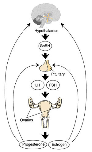
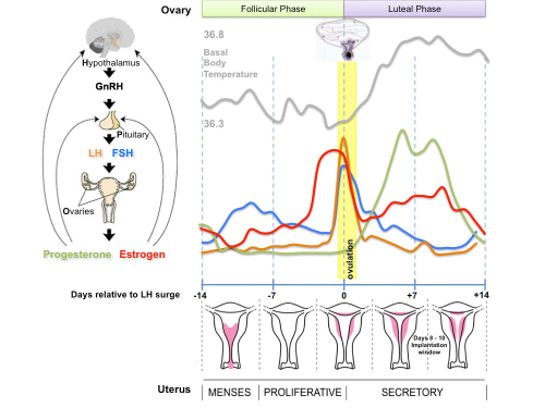
  • Menstrual Cycle a regular cycle of reproduction (28 days)
  • prenatal all oocytes produced
  • begins at puberty, release of 1 egg (oocyte) every cycle
  • Endocrine controlled (HPG axis) Hypothalamus - Pituitary - Gonad
  • postnatal continuous production of spermatozoa
  • begins at puberty, release millions of spermatozoa
  • Endocrine controlled (HPG axis) Hypothalamus - Pituitary - Gonad

XXhpgaxis.jpg Menstrual cycle.png

Gametogenesis

Male

The testes have two functions.

  1. produce the male gametes or spermatozoa
  2. produce male sexual hormone, testosterone (internal and external genitalia, sex characteristics)

Human spermatozoa take about 48 days from entering meiosis until morphologically mature spermatozoa.

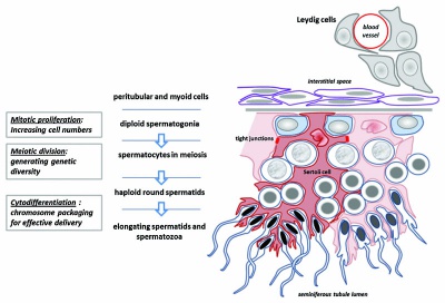
  • Spermatogonia - are the first cells of spermatogenesis
  • Primary spermatocytes - large, enter the prophase of the first meiotic division
  • Secondary spermatocytes - small, complete the second meiotic division
  • Spermatid - immature spermatozoa
  • Spermatozoa - differentiated gamete
Spermatozoa development: primordial germ cell - spermatogonia - primary spermatocyte - secondary spermatocytes - spermatid - spermatozoa

sertoli cells (support cells) Interstitial cells or Leydig cells (produce hormone testosterone)

Seminiferous tubule cartoon.jpg
Spermatozoa Development (expand to see terms)  

Spermatozoa Development

Note there are additional glossaries associated with genital, spermatozoa, oocyte and renal.

Spermatozoon
  • acroplaxome - structure forms the acrosome plate with intermediate filament bundles of the marginal ring at the leading edge of the acrosome. The sub-acrosomal layer located in the developing spermatozoa head perinuclear region, located between the inner acrosomal membrane and the nuclear envelope. The other part of the perinuclear region is the post-acrosomal sheath (PAS) at the post-acrosomal region.
  • acrosome - Cap-shaped cellular structure formed from the golgi apparatus and contains enzymes to dissolve the oocyte (egg) zona pellucida for fertilisation.
  • acrosome compaction - Acrosome reshaping process in final stages of spermatogenesis (spermatid to spermatozoa).
  • acrosome reaction - Chemical change within the spermatozoa following binding to the zona pellucida, only acrosome reacted spermatozoa have an ability to fuse with oocytes.
  • annulus - Cytoskeletal (septin) structure located between the midpiece and principal piece regions of the tail, thought to form a diffusion barrier between these two domains. PMID 20042538
  • asthenozoospermia - (asthenospermia) Term for reduced sperm motility and can be the cause of male infertility.
  • axoneme - (axonema) The basic structure in cilia and eukaryotic flagella and in the spermatozoa tail, consisting of parallel microtubules in a characteristic "9 + 2" pattern. This pattern describes 9 outer microtubule doublets (pairs) surrounding 2 central singlet microtubules, in humans 50 μm long. The motor protein dynenin move the outer microtubules with respect to the central pair, bending the cilia and generating motility. Note that prokaryotic bacteria have a similar process (flagellum) that uses an entirely different mechanism for motility.
  • capacitation - term describing the process by which spermaozoa become capable of fertilizing an oocyte, requires membrane changes, removal of surface glycoproteins and increased motility.
  • caput - proximal head of the epididymis, epithelium with stereocilia, involved in absorbing fluid to concentrate spermatozoa. Underlying smooth muscle aids movement. Epididymis three main parts : caput (head), corpus (body), cauda (tail).
  • CatSper - cationic (Ca2+) channel of spermatozoa, progesterone activated involved in hyperactivation, acrosome reaction, and possibly chemotaxis.
  • cauda - distal tail of the epididymis, region with a thin epithelium and the greatest quantity of smooth muscle. Epididymis three main parts : caput (head), corpus (body), cauda (tail).
  • centriole - a microtubule organising centre. First required for axoneme formation (distal centriole) that is lost and a second for pronuclei formation (proximal) following fertilisation. Rodents loose both and only have maternal centrioles.
  • connecting piece - linkage between the spermatozoa head and the midpiece of the tail. PMID 22767409
  • corpus - elongated body of the epididymis, This has an intermediate thickness of epithelium and thicker smooth muscle layer than caput. Epididymis three main parts : caput (head), corpus (body), cauda (tail).
  • cytoplasmic bridges - Transient cytoplasm connections between spermatids arising from one spermatogonium due to incomplete cytokinesis.
  • diploid - (Greek, di = double + ploion = vessel) Having two sets of chromosomes, the normal state for all cells other than the gametes.
  • end piece - Last portion of the spermatozoa tail region.
  • epididymis - testis tubular structure connecting the efferent ducts to the ductus deferent and functions for the storage and maturation of spermatozoa. Epididymis three main parts : caput (head), corpus (body), cauda (tail). PMID27307387
  • fibrous sheath - cytoskeletal structure surrounding the axoneme and outer dense fibers, defining the extent of the principal piece region.
  • haploid - (Greek, haploos = single) Having a single set of chromosomes as in mature germ/sex cells (oocyte, spermatozoa) following reductive cell division by meiosis. Normally cells are diploid, containing 2 sets of chromosomes.
  • interstitial cell - (Leydig cell) Male gonad (testis) cell which secrete the androgen testosterone, beginning in the fetus.
  • interstitium - testis developmental region (space between testis cords) that generates Leydig cells and other less well characterized cell types.
  • Johnsen score - a clinical score (1-10) for assessing spermatogenesis in a human testicular biopsy. Named after the author of the original article. PMID 5527187
  • Leydig cell - (interstitial cell) Male gonad (testis) cell that secrete the androgen testosterone, beginning in the fetus. Fetal Leydig cells develop from coelomic epithelium and undifferentiated perivascular cells in the gonad–mesonephros border region. Adult Leydig cells appear after birth from stem/progenitor cells among peritubular and peri-vascular cells. Leydig cells were first histologically identified in 1850 by Franz von Leydig (1821 - 1908) a German scientist.
  • meiosis - The cell division that occurs only in production of germ cells where there is a reduction in the number of chromosomes (diploid to haploid) which is the basis of sexual reproduction. All other non-germ cells in the body divide by mitosis.
  • midpiece - (middle piece) spermatozoa tail initial segment of axoneme surrounded outer dense fibres then by mitochondria. Next in the tail is the principal piece then finally the end piece.
  • mitosis - The normal division of all cells, except germ cells, where chromosome number is maintained (diploid). In germ cell division (oocyte, spermatozoa) meiosis is a modified form of this division resulting in reduction in genetic content (haploid). Mitosis, division of the nucleus, is followed by cytokinesis the division of the cell cytoplasm and the cytoplasmic contents. cytokinesis overlaps with telophase.
  • outer dense fibres - (ODF, outer dense fibers) cytoskeletal structures that surround the axoneme in the middle piece and principal piece of the spermatozoa tail.
  • primary spermatocyte - arranged in the seminiferous tubule wall deep (luminal) to the spermatogonia. These large cells enter the prophase of the first meiotic division. (More? meiosis)
  • principal piece - Spermatozoa tail segment containing the plasma membrane calcium channels (CatSper1 and CatSper2) required for hyperactivation of motility. Region is partially separated from the midpiece by a barrier called the annulus.
  • sertoli cells - (sustentacular cell) These cells are the spermatozoa supporting cells, nutritional and mechanical, as well as forming a blood-testis barrier. The cell cytoplasm spans all layers of the seminiferous tubule. The cells are named after Enrico Sertoli (1842 - 1910), and italian physiologist and histologist.
  • sperm annulus - (Jensen's ring; Latin, annulus = ring) A region of the mammalian sperm flagellum connecting the midpiece and the principal piece. The annulus is a septin-based structure formed from SEPT1, 4, 6, 7 and 12. Septins are polymerizing GTPases that can act as a scaffold forming hetero-oligomeric filaments required for cytokinesis and other cell cycle roles.
  • spermatogenesis - (Greek, genesis = origin, creation, generation) The term used to describe the process of diploid spermatagonia division and differentiation to form haploid spermatazoa within the testis (male gonad). The process includes the following cellular changes: meiosis, reoorganization of DNA, reduction in DNA content, reorganization of cellular organelles, morphological changes (cell shape). The final process of change in cell shape is also called spermiogenesis.
  • spermatogenesis - (Greek, genesis = origin, creation, generation) The maturation process of the already haploid spermatazoa into the mature sperm shape and organization. This process involves reorganization of cellular organelles (endoplasmic reticulum, golgi apparatus, mitochondria), cytoskeletal changes (microtubule organization) and morphological changes (cell shape, acrosome and tail formation).
  • spermatogonia - The cells located in the seminiferous tubule adjacent to the basal membrane that either divide and separate to renew the stem cell population, or they divide and stay together as a pair (Apr spermatogonia) connected by an intercellular cytoplasmic bridge to differentiate and eventually form spermatazoa.
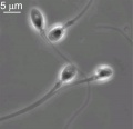
  • spermatozoa head - Following spermiogenesis, the first region of the spermatozoa containing the haploid nucleus and acrosome. In humans, it is a flattened structure (5 µm long by 3 µm wide) with the posterior part of nuclear membrane forming the basal plate region. The human spermatozoa is about 60 µm long, actively motile and divided into 3 main regions (head, neck and spermatozoa tail).
  • spermatozoa neck - Following spermiogenesis, the second region of the spermatozoa attached to basal plate, transverse oriented centriole, contains nine segmented columns of fibrous material, continue as outer dense fibres in tail. In humans, it forms a short structure (1 µm). The human spermatozoa is about 60 µm long, actively motile and divided into 3 main regions (head, neck and tail).
  • spermatozoa tail - Following spermiogenesis, the third region of the spermatozoa that has a head, neck and tail). The tail is also divided into 3 structural regions a middle piece, a principal piece and an end piece. In humans: the middle piece (5 µm long) is formed by axonema and dense fibres surrounded by mitochondria; the principal piece (45 µm long) fibrous sheath interconnected by regularly spaced circumferential hoops; the final end piece (5 µm long) has an axonema surrounded by small amount of cytoplasm and plasma membrane.
  • spermatogonial stem cells - (SSCs) The spermatagonia cells located beside the seminiferous tubule basal membrane that either divide and separate to renew the stem cell population, or they divide and stay together as a pair (|Apr spermatogonia) connected by an intercellular cytoplasmic bridge to differentiate and eventually form spermatazoa.
  • spermatozoon - singular form of of spermatozoa.
  • sperm protein 56 - A component of the spermatozoa acrosomal matrix released to the sperm surface during capacitation.
  • teratospermia - Clinical term for a spermatozoa with abnormal morphology (small, large, defects in the head, tail, and/or mid-piece) present in the semen or ejaculate.
  • testis cords - developmental structure that give rise to the adult seminiferous tubules, the other developmental region is the interstitium.
  • vasectomy - Clinical term for ligation of the scrotal portion of the ductus deferens.

See also: Spermatozoa Terms collapse table

Other Terms Lists  
Terms Lists: ART | Birth | Bone | Cardiovascular | Cell Division | Endocrine | Gastrointestinal | Genital | Genetic | Head | Hearing | Heart | Immune | Integumentary | Neonatal | Neural | Oocyte | Palate | Placenta | Radiation | Renal | Respiratory | Spermatozoa | Statistics | Tooth | Ultrasound | Vision | Historic | Drugs | Glossary

Female

The ovary has two main functions.

  1. produce the female gametes or oocytes
  2. produce female hormones, estrogen and progesterone (secondary sex characteristics, menstrual cycle)

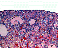
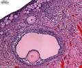
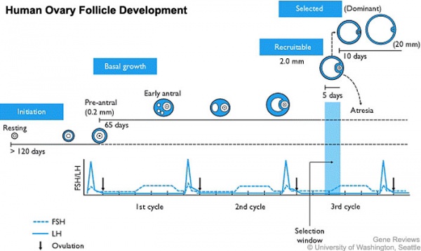
In an adult human female the development of a primordial follicle containing an oocyte to a preovulatory follicle takes in excess of 120 days.

Human ovary follicle development.jpg

Human ovary follicle development

Ovarian Follicle Stages: primordial follicle - primary follicle - secondary follicle - preovulatory follicle


Follicle cells (support cells) Theca cells (produce hormone)


Oocyte Development (expand to see terms)  

Oocyte Development

Note there are additional specific term glossaries available listed at bottom of this table.

  • antral follicle - (secondary) the stage following preantral in the decription of the sequence ovarian follicle development.
  • antrum - (L. a cave), cavity; a nearly-closed cavity or bulge. In the ovary this refers to the follicular fluid-filled space within the follicle.
  • atretic follicle - An ovarian follicle that fails to mature and degenerates. Also called "atresia" refering to the process of degeneration of the ovarian follicle. This process can occur at any stage of follicle development (folliculogenesis).
  • clomiphene citrate - drug taken orally to promote the process of follicle/egg maturation.
  • COCs - (cumulus-oocyte complexes) term used in Assisted Reproductive Technology to describe the ovulated Graafian follicle consisting of the oocyte surrounded by a packed layers of cumulus cells.
  • corona radiata - Layer of follicle cells of cumulus oophorus remaining directly attached to zona pellucida of the oocyte. These cells communicate with the oocyte through the zone pellucida, also called granulosa cells.
  • corpus albicans - (L. corpus = body, L. albicans = whitish); a degenerating corpus luteum in ovary.
  • corpus luteum - (L. corpus = body, L. luteum = yellow) The remains of the ovulating ovarian follicle after ovulation, that acts as the initial endocrine organ supporting pregnancy and preventing menstruation (loss of the endometrial lining). de Graaf first observed it in the ovary of a cow as a yellow structure.
  • cortical - (L. corticalis) at the outside (like the bark of a tree), usually combined with medulla meaning the core.
  • cumulus oophorus - (L. cumulus = a little mound G. oon = egg + phorus = bearing); part of the wall of an ovarian follicle surrounding and carrying the ovum (oocyte).
  • first polar body - a small cytoplasmic exclusion body contains the excess DNA from the oocyte meiosis formed during meiosis 1.
  • follicle - (L. folliculus = little bag,dim. of L. follis). A structure which develops in the ovary and contains a developing egg (oocyte).
  • follicle stimulating hormone - (FSH, gonadotropin) A glycoprotein hormone secreted by anterior pituitary (adenohypophysis gonadotrophs, a subgroup of basophilic cells) and acts on gametogenesis and other systems in both males and females. Females, FSH acts on the ovary to stimulate follicle development. Males, acts on the testis Sertoli cells to increase androgen-binding protein (ABP) that binds androgens and has a role in spermatogenesis. pituitary
  • follicular fluid - the fluid found in the antrum of a secondary follicle. Secreted by cells in the wall of the follicle. This fluid is released along with the oocyte at ovulation.
  • germinal epithelium - cellular component covering surface of ovary, it is continuous with mesothelium covering mesovarium. Note that it is a historical misnomer, as it is not the actual site of germ cell formation.
  • Graafian follicle - named after Regnier de Graaf (1641-1673), an historic Dutch physician embryologist who studied pregnancy using rabbits.
  • granulosa cells - the supporting cells that surround the developing egg within the follicle thecal layers.
  • homologs - maternal and paternal homologous chromosomes.
  • Izumo1 - a protein located on the equatorial segment of acrosome-reacted spermatozoa recognizes its receptor Juno, on the oocyte surface, for plasma membrane binding and fusion. Named for a Japanese shrine dedicated to marriage. OMIM609278
  • Juno - (folate receptor-δ; FOLR-δ) a glycophosphatidylinositol (GPI)-anchored, cysteine-rich glycoprotein on the oocyte surface for fertilisation that is the receptor of Izumo1 on the spermatozoa, for plasma membrane binding and fusion. OMIM615737
  • luteinizing hormone - (LH, gonadotropin, lutropin, Interstitial Cell Stimulating Hormone, ICSH) glycoprotein hormone releasd from anterior pituitary hormone that acts on the gonad and has a role in male and female reproduction. Female, LH triggers ovulation (release of the oocyte). Male, LH stimulates testis interstital cell (Leydig cell) production of testosterone. Have been used clinically in humans for the treatment of female infertility.
  • meiosis - oocyte reductive (diploid to haploid) cell division, with 1 round of DNA replication is followed by 2 rounds of chromosome segregation. The process beginning in the fetus and only completed at fertilization.
  • mesovarium - mesentry of the ovary formed from a fold of the broad ligament that attaches the ovary.
  • medullary - (L. medius = in the middle) relating to the medulla; pith, marrow, inner portion of an organ. Usually combined with cortex (cortical) meaning the outer layer.
  • oocyte - (Greek, oo = egg, ovum) The term used to describe the haploid egg or ovum formed within the ovary (female gonad) and released to enter the uterine tube and be transported to the uterus. The mature oocyte is the cell released from the ovary during ovulation.
  • oogenesis - (Greek, oo = egg + genesis = origin, creation, generation) process of diploid oogonia division and differentiation into an haploid oocyte (egg) within the ovary (female gonad). Mammalian meiosis will only be completed within the oocyte if fertilization occurs.
  • oogonia - (Greek, oo = egg) diploid germ cells within the ovary (female gonad) which provide the primary oocytes for oocyte (egg) formation. In humans, all oogonia form primary oocytes within the ovary before birth.
  • oolemma - (zona pellucida, vitelline membrane).
  • oophorus - (Greek, oo = egg + phorus = carrying, egg-bearing) cumulus oophorus, used to describe the granulosa cells within the follicle that tether or link the oocyte to the wall of the follicle.
  • ovarian reserve - Clinical term for the number of oocytes (non-growing follicles) available for possible fertilization at the different times during female reproductive life. A blood test for Anti-Mullerian Hormone (AMH) levels is used clinically as a measure of the ovarian reserve. human graph
  • ovastacin - an oocyte released enzyme following fertilization that cleaves ZP2 protein to prevent polyspermy.
  • ovulation - release of the oocyte from the mature follicle. In humans generally a single oocyte is released from a cohort of several maturing follicles.
  • ovum - oocyte, note that historically this same term was also used to describe the early stages following fertilisation.
  • polar body - small cytoplasmic exclusion body contains the excess DNA from the oocyte meiosis reductive division. The first polar body formed during meiosis 1, the second and sometimes third polar bodies are formed from meiosis 2 at fertilization.
  • polyspermy - abnormal fertilization by more that a single spermatozoa, may generate a hydatidiform mole.
  • preantral follicle - (primary) the stage following primordial in the description of the sequence ovarian follicle development.
  • primary follicle - (preantral) the stage following primordial in the description of the sequence ovarian follicle development.
  • primordial follicle - the first stage in the description of the sequence ovarian follicle development. Present in the ovary from birth, located in the stroma of the ovary cortex beneath the tunica albuginea. The primordial follicle is the oocyte and the surrounding follicular cells.
  • primordial germ cell - oocyte present in the primordial follicle ovary from birth, located in the stroma of the ovary cortex beneath the tunica albuginea. The primordial follicle is the oocyte and the surrounding follicular cells.
  • second polar body - a small cytoplasmic exclusion body contains the excess DNA from the oocyte formed during meiosis 2 at fertilization.
  • secondary follicles - the stage following primary in the description of the sequence ovarian follicle development.
  • stromal cells - in the ovary, cells surrounding the developing follicle that form a connective tissue sheath (theca folliculi). This layer then differentiates into 2 layers (theca interna, theca externa). This region is richly vascularized and involved in hormone secretion.
  • superovulation therapy - a fertility drug treatement (oral clomiphene citrate and/or injectable FSH with or without LH) aimed at stimulating development/release of more than one follicle during a single menstrual cycle.
  • tertiary follicle - (preovulatory, Graffian) the stage following secondary in the description of the sequence ovarian follicle development.
  • theca folliculi - stromal cells in the ovary, cells surrounding the developing follicle that form a connective tissue sheath. This layer then differentiates into 2 layers (theca interna, theca externa). This region is vascularized and involved in hormone secretion.
  • theca externa - stromal cells forming the outer layer of the theca folliculi surrounding the developing follicle. Consisting of connective tissue cells, smooth muscle and collagen fibers.
  • theca interna - stromal cells forming the inner layer of the theca folliculi surrounding the developing follicle. This vascularized layer of cells respond to LH (leutenizing hormone) synthesizing and secreting androgens which are processed into estrogen.
  • transzonal projection - (TZP) ovarian follicle term describing the cellular membraneous extension from the granulosa cell through the zona pellucida to the oocyte cell membrane where it forms gap junctions or adherens junctions allowing signalling and adhesion between the two cells.
  • tunica albuginea - dense connective tissue layer lying near the ovary surface, a layer of simple squamous to cuboidal epithelial covers this layer. A similar named structure is found in male genital system.
  • uterus - site of embryo implantation and development. Uterine wall has 3 major layers: endometrium, myometrium, and perimetrium. Endometrium can be further divided into the functional layer (shed/lost during menstruation) and basal layer (not lost during menstruation).
  • zinc sparks following fertilization the oocyte releases a burst of zinc atoms in brief bursts (zinc sparks) has a role in zonal pellucida induced structural changes (hardening) along with ovastacin cleavage of ZP2 protein.
  • zona hardening - following fertilization the structural changes that occur to the zona pellucida to prevents further spermatozoa binding acting as a block to polyspermy.
  • zona pellucida - extracellular layer lying directly around the oocyte underneath follicular cells. Has an important role in egg development, fertilization and blastocyst development. This thick extracellular matrix consists of glcosaminoglycans and 3 glycoproteins (ZP1, ZP2, ZP3). (More? Zona pellucida)
Other Terms Lists  
Terms Lists: ART | Birth | Bone | Cardiovascular | Cell Division | Endocrine | Gastrointestinal | Genital | Genetic | Head | Hearing | Heart | Immune | Integumentary | Neonatal | Neural | Oocyte | Palate | Placenta | Radiation | Renal | Respiratory | Spermatozoa | Statistics | Tooth | Ultrasound | Vision | Historic | Drugs | Glossary
Links: spermatozoa | oocyte | MBoC - Figure 20-18. Influence of Sry on gonad development | Endocrinology - Comparative anatomy of male and female reproductive tracts

Fertilization

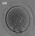
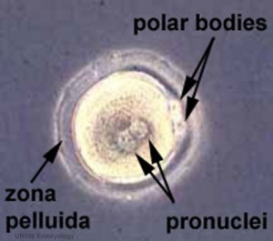
Early zygote showing polar bodies
  • oogenesis - 1 gamete produced/meiosis + 3 polar bodies, meiosis is slow, 1 egg produced and released at ovulation
  • spermatogenesis - 4 gametes produced/meiosis, meiosis is fast, 200-600 million sperm released at ejaculation


Ovulation icon.jpg Fertilization 001 icon.jpg

Fertilization Site

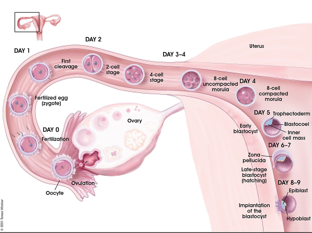
  • Fertilization usually occurs in first 1/3 of uterine tube (oviduct, Fallopian tube)
  • Fertilization can also occur outside uterine tube associated with Assisted Reproductive Technologies (ART, IVF, GIFT, ZIFT...) and ectopic pregnancy
  • The majority of fertilized eggs do not go on to form an embryo

Fertilization - Spermatozoa


Approximate Timing of Early Human Events (in vitro)
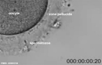
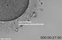
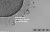
Human fertilization movie 2 frame 01.jpg Human fertilization movie 2 frame 02.jpg Human fertilization movie 2 frame 03.jpg
20 min - components 28 min - spermatozoa penetrates zone pellucida 31 min - spermatozoa penetrates oocyte - fertilization
See also clock in lower righthand corner for the approximate timing of events.
Links: Human Fertilization Detail Movie | Human Fertilization Movie
Reference: PMID 22695746 J Assist Reprod Genet.

Fertilization - Oocyte

Embryo mitosis icon.jpg Week1 001 icon.jpg

Week 1 and 2

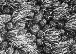
Human uterine tube ciliated epithelium (SEM)

Week1 summary.jpg

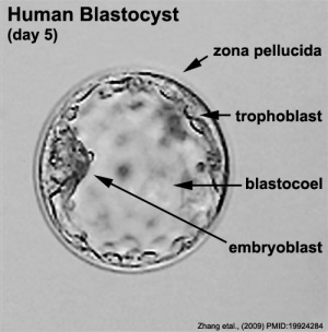
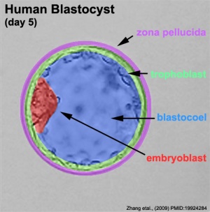
Human embryo day 5 label.jpg Human embryo day 5 label2.jpg

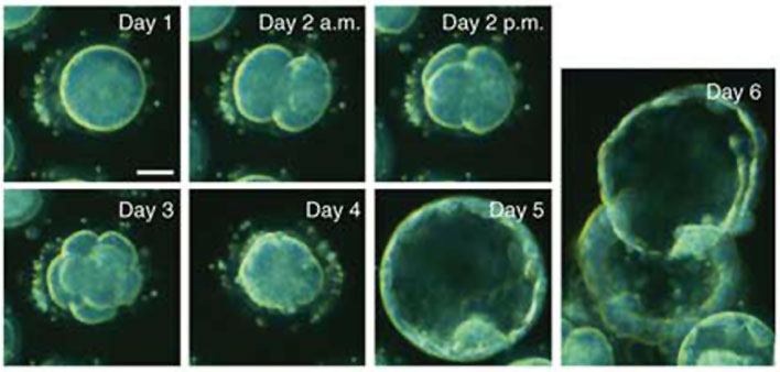
Human blastocyst day 1-6.jpg

Week 2 Implantation


Week2 001 icon.jpg Chorion 001 icon.jpg

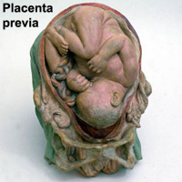
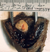
Uterine Implantation Ectopic Implantation
  • Uterine body
    • posterior, anterior, superior, lateral (most common posterior)
    • Placenta Previa inferior implantation, placenta overlies internal os of uterus
  • Outside Uterine body
    • external surface of uterus, ovary, bowel, gastrointestinal tract, mesentery, peritoneal wall
    • Tubal pregnancy - (uterine tube) most common ectopic
Galletti1770 placenta previa.jpg Tubal pregnancy.gif

Early Placenta

  • interaction between implanting conceptus and uterine wall (endometrium)
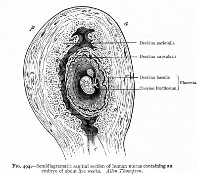
  • The uterine lining following implantation (Decidua)
    • forms 3 distinct regions, at approx 3 weeks
    • Decidua Basalis - implantation site
    • Decidua Capsularis - enclosing the conceptus
    • Decidua Parietalis - remainder of uterus
  • uterine cavity is lost by 12 weeks
Bailey494.jpg

Week 3 Gastrulation

Stage7-sem2.jpg Stage7 primitive streak Chicken-gastrulation3.jpg
Embryonic disc Primitive Streak Gastrulation
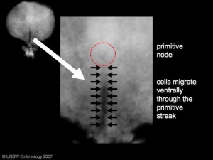
  • Primitive node - region in the middle of the early embryonic disc epiblast from which the primitive streak extends caudally (tail)
    • nodal cilia establish the embryo left/right axis
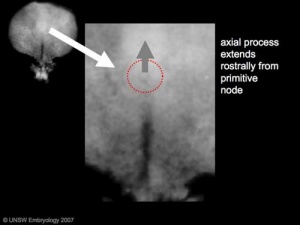
    • axial process extends from the nodal epiblast
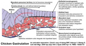
  • Primitive streak - region of cell migration (gastrulation) from the epiblast layer forming sequentially the two germ cell layers (endoderm and mesoderm)
Gastrulation, (Greek = belly)

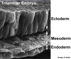
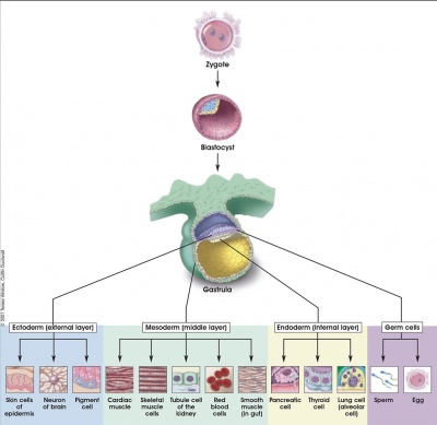
Means the formation of gut, but has been used in a more looser sense to to describe the formation of the trilaminar embryo. The epiblast layer, consisting of totipotential cells, derives all 3 embryo layers:

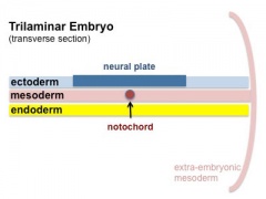
  1. ectoderm
  2. mesoderm
  3. endoderm

The primitive streak is the visible feature which represents the site of cell migration to form the additional layers. Historically, gastrulation was one of the earliest observable morphological event occurring in the frog embryo.

Mesoderm 001 icon.jpg
 ‎‎Week 3 Mesoderm
Page | Play
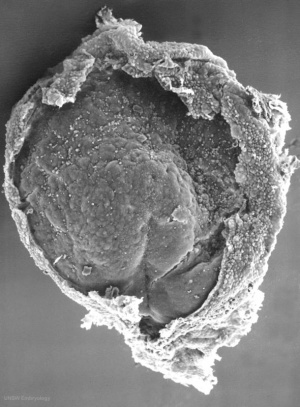
Trilaminar embryo.jpg

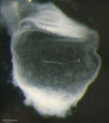
Trilaminar embryo (SEM)

Trilaminar embryo

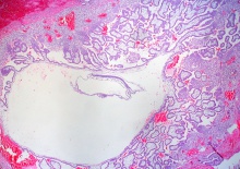
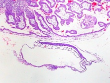
Virtual Slides - Human Embryo (stage 7) 
Stage 7 - Implanted Conceptus

Stage7 bf5.jpg

 ‎‎Mobile | Desktop | Original

Stage 7 | Embryo Slides
Stage 7 - Embryonic Disc

Stage7 bf51.jpg

 ‎‎Mobile | Desktop | Original

Stage 7 | Embryo Slides
Stage 7 - Vesicle (Kyoto 15458)

Kyoto15458-01-icon.jpg

 ‎‎Mobile | Desktop | Original

Stage 7 | Embryo Slides
Stage 7 - Conceptus

Stage7-bf1.jpg

 ‎‎Mobile | Desktop | Original

Stage 7 | Embryo Slides

Notochord

Stage7 axial process

The notochord is a structure which has an early mechanical role in embryonic disc folding and a major signaling role in patterning surrounding embryonic tissue development. This signaling role patterns many different tissues (neural plate, neural tube, somites, endodermal organs). It has its own sequence of development from a primitive axial process and is a developmental feature not present in the adult anatomy.

Notochord 02 icon.jpg

Page

  • axial process an initial epiblast hollow epithelial tube which extends in the midline from the primitive pit, cranially in the embryonic disc (toward the oral membrane).
    • neuroenteric canal is a transient communication between the amnionic cavity and the yolk sac cavity formed by the axial process.
  • notochordal plate forms from the axial process merging with the endoderm layer.
  • notochord forms from the notochordal plate which then separates back into the mesoderm layer as a solid column of cells lying in the midline of the embryonic disc and running rostro-caudally (head to tail).
    • An alternate name for the notochord is "axial mesoderm".

Somitogenesis

stage 9 Embryo
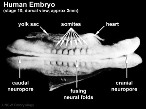
stage 10 Embryo

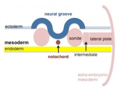
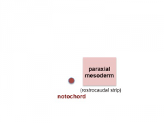
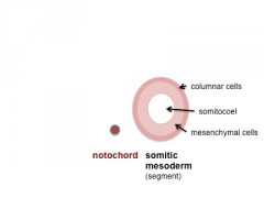
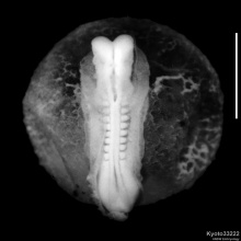
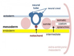
Mesoderm means the "middle layer" and it is from this layer that nearly all the bodies connective tissues are derived. In early mesoderm development a number of transient structures will form and then be lost as tissue structure is patterned and organised. Humans are vertebrates, with a "backbone", and the first mesoderm structure we will see form after the notochord will be somites.

Coelom, meaning "cavity", and major fluid-filled cavities can be seen to form both within the embryo (|intraembryonic coelom) and outside the embryo (extraembryonic coelom).

The intraembryonic coelom is the single primitive cavity that lies within the mesoderm layer that will eventually form the 3 major anatomical body cavities (pericardial, pleural, peritoneal).


stage 11 Embryo

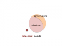
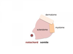
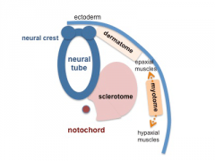
Somite initially forms 2 main components

  • sclerotome - (ventromedial) forms axial skeleton - vertebral body and intervertebral disc
  • dermomyotome - (dorsolateral) forms dermis and skeletal muscle


Neural

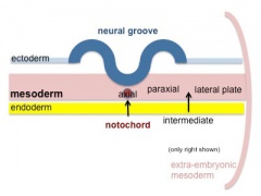
Week 3 ectoderm - 2 parts

  • midline - neural plate (columnar cells)
    • central nervous system (CNS)
  • edge of neural plate - neural crest
    • peripheral nervous system (PNS), and many other structures.
  • lateral - surface ectoderm (cuboidal cells)
    • epidermis of skin, hair, glands, anterior pituitary, teeth enamel
    • head region - sensory placodes
  • extends from buccopharyngeal membrane to primitive node
  • forms above notochord and paraxial mesoderm
  • neuroectodermal cells
    • broad brain plate
    • narrower spinal cord
  • 3 components form: floor plate,neural plate, neural crest
Neuralplate 001 icon.jpg
 ‎‎Neural Plate
Page | Play
Neuraltube 001 icon.jpg
 ‎‎Neural Tube
Page | Play
Mouse neural tube 01 movie icon.jpg
 ‎‎Neural Tube Close
Page | Play
Week 4 Embryo (dorsal view)  
Stage 10 - Dorsal View

Stage10 bf10.jpg

 ‎‎Mobile | Desktop | Original

Stage 10 | Embryo Slides

Embryo Virtual Slides

Neural Tube Defects
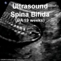
US Spina bifida GA19week.jpg
 ‎‎Spina Bifida
Page | Play
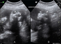
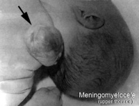
Anencephaly ultrasound.jpg Neural tube defect meningomyelocele.jpg
Spina-Bifida Anencephaly Meningomyelocele


Links: Neural System - Abnormalities | Folic Acid and Neural Tube Defects

Cardiogenesis

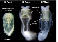
Heart Tube Fusion
Heart Tube Fusion

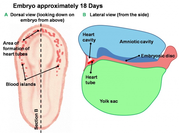
Early Development of Heart Tube.jpg

Early Development of Heart Tube

Human heart SEM1.jpg

The Human Heart from day 10 to 25 (scanning electron micrograph)

  • forms initially in splanchnic mesoderm of prechordal plate region - cardiogenic region
    • growth and folding of the embryo moves heart ventrallly and downward into anatomical position
  • week 3 begins as paired heart tubes that fuse to form single heart tube
  • begins to beat in Humans- day 22-23
Week3 folding icon.jpg

Blood Islands

fetal blood
  • 2 populations of cells
  • all vessels (arteries and veins) appear initially the same

Blood Formation

Mouse hematopoietic stem cell location
  • blood formation from stem cells occurs initially in the extra-embryonic mesoderm of the yolk sac
  • then later (week 5) throughout embryonic mesenchyme
  • blood stem cells then migrate into the liver
    • then spleen, bone marrow, lymph nodes

Red Blood Cells

The only cells in the blood are initially entirely fetal red blood cells (RBC).

These red blood cells differ from adult red blood cells in:

  • may remaining nucleated
  • contain fetal haemoglobin - has different oxygen and carbon dioxide binding characteristics


Links: Basic Cardiac Embryology

Early Placentation

Week2 001 icon.jpg

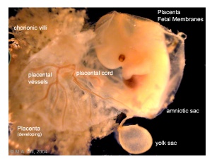
Placenta and placental membranes
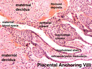
Placenta anchoring villi

The trophoblast layer has now differentiated into two morphologically distinct cellular layers.

  • Syncitiotrophoblasts - form a multinucleated cytoplasmic mass by cytotrophoblast cell fusion and both invade the decidua and secrete hCG
  • Cytotrophoblasts - form a cellular layer around the blastocyst, proliferates and extends behind syncitiotrophoblasts

Early Utero-Placental exchange - transfer of nutrition from maternal lacunae filled with secretions from uterine glands and maternal blood from blood vessels. The development of trophoblast villi extending into the uterine decidua.

There are three stages of villi development:

  1. Primary Villi - cytotrophoblast
  2. Secondary Villi - cytotrophoblast + extraembryonic mesoderm
  3. Tertiary Villi - cytotrophoblast + extraembryonic mesoderm + blood vessels

There are two main types of early villi:

  • Anchoring villi - attached to decidua
  • Floating villi - not attached to decidua, floating in maternal lacunae.

Abnormalities

Critical periods, Genetic and Environmental factors leading to abnormal development will be covered in the associated practical classes.


BGDsmall.jpg

BGDA: Lecture 1 | Lecture 2 | Practical 3 | Practical 6 | Practical 12 | Lecture Neural | Practical 14 | Histology Support - Female | Male | Tutorial

Glossary Links

Glossary: A | B | C | D | E | F | G | H | I | J | K | L | M | N | O | P | Q | R | S | T | U | V | W | X | Y | Z | Numbers | Symbols | Term Link

Cite this page: Hill, M.A. (2024, March 19) Embryology BGDA Lecture - Development of the Embryo/Fetus 1. Retrieved from https://embryology.med.unsw.edu.au/embryology/index.php/BGDA_Lecture_-_Development_of_the_Embryo/Fetus_1

What Links Here?
© Dr Mark Hill 2024, UNSW Embryology ISBN: 978 0 7334 2609 4 - UNSW CRICOS Provider Code No. 00098G