File:Human fetal temporal bone and mandible 01.jpg
From Embryology
Size of this preview: 800 × 537 pixels. Other resolution: 1,200 × 805 pixels.
Original file (1,200 × 805 pixels, file size: 170 KB, MIME type: image/jpeg)
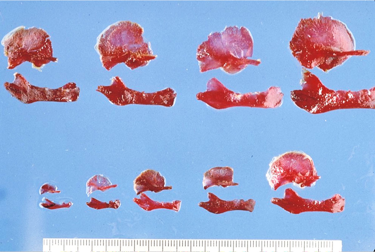
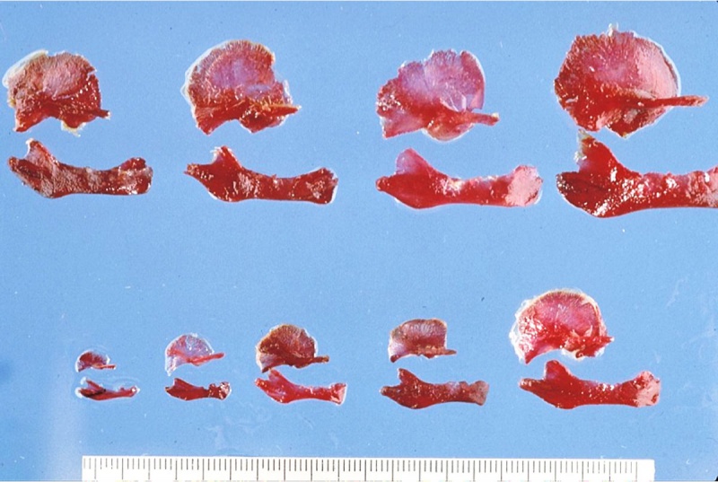
Human Temporal Bone and Mandible
Image shows growth of both bones from the end of the embryonic period (week 8, GA week 10) through the fetal period of development (to 9 months).
Temporal Bone
Temporal styloid process growth can also be noted.
- pointed piece of bone extending from the temporal bone just below the ear.
- anchor point for several muscles associated with the tongue and larynx.
Mandible
- develops beside the Meckel's cartilage template.
- bone formation by intramembranous ossification.
- at birth not completely ossified.
- there is a sex difference in overall growth.
- Links: Skull Development
Reference
Image Source: Prof Virginia Diewert
Cite this page: Hill, M.A. (2024, April 26) Embryology Human fetal temporal bone and mandible 01.jpg. Retrieved from https://embryology.med.unsw.edu.au/embryology/index.php/File:Human_fetal_temporal_bone_and_mandible_01.jpg
- © Dr Mark Hill 2024, UNSW Embryology ISBN: 978 0 7334 2609 4 - UNSW CRICOS Provider Code No. 00098G
File history
Click on a date/time to view the file as it appeared at that time.
| Date/Time | Thumbnail | Dimensions | User | Comment | |
|---|---|---|---|---|---|
| current | 14:42, 15 May 2013 | | 1,200 × 805 (170 KB) | Z8600021 (talk | contribs) | ==Human Temporal Bone and Mandible== Image shows growth of both bones from the end of the embryonic period (week 8) through the fetal period of development (to 9 months). ---- Image Source: Prof Virginia Diewert {{Template:Footer}} [[Category:Human... |
You cannot overwrite this file.
File usage
The following page uses this file: