File:Heart outflow tract stage 14 02.jpg

Original file (996 × 996 pixels, file size: 139 KB, MIME type: image/jpeg)
Heart Outflow Tract (Carnegie Stage 14) EFIC
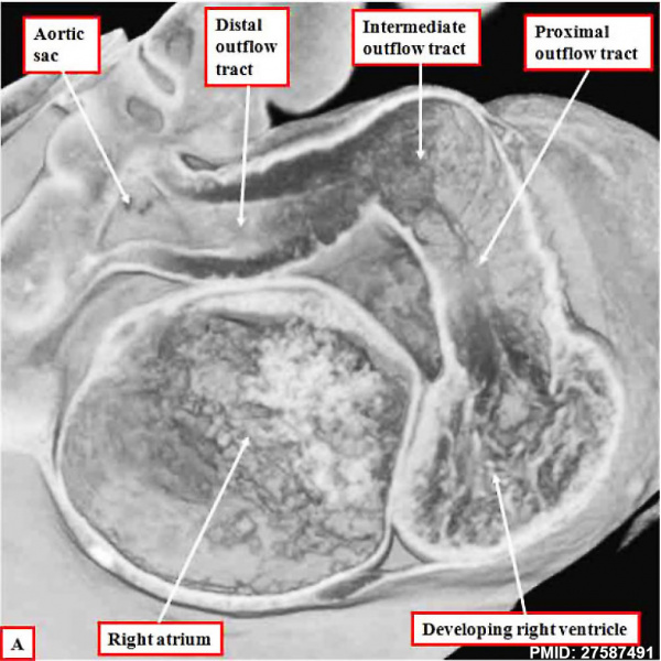
EFIC images are from an episcopic data set prepared from a human embryo at Carnegie stage 14, representing the end of the fifth week of development.
A shows the dog-leg bend in the outflow tract, which at this early stage is supported exclusively from the developing right ventricle.
- Links: Image EFIC outflow tract frontal | Image EFIC - OFT RA RV | Image EFIC - OFT RA LA | Carnegie stage 14 | Week 5 | EFIC
Reference
<pubmed>27587491</pubmed>
https://www.ncbi.nlm.nih.gov/pmc/articles/PMC5011314/
http://journals.sagepub.com/doi/abs/10.1177/2150135116651114
PMID 27587491
Copyright
© The Author(s) 2016
https://creativecommons.org/licenses/by/3.0/
Figure 2. cropped and resized.
Cite this page: Hill, M.A. (2024, May 11) Embryology Heart outflow tract stage 14 02.jpg. Retrieved from https://embryology.med.unsw.edu.au/embryology/index.php/File:Heart_outflow_tract_stage_14_02.jpg
- © Dr Mark Hill 2024, UNSW Embryology ISBN: 978 0 7334 2609 4 - UNSW CRICOS Provider Code No. 00098G
File history
Click on a date/time to view the file as it appeared at that time.
| Date/Time | Thumbnail | Dimensions | User | Comment | |
|---|---|---|---|---|---|
| current | 11:59, 29 January 2017 | | 996 × 996 (139 KB) | Z8600021 (talk | contribs) | |
| 11:59, 29 January 2017 | 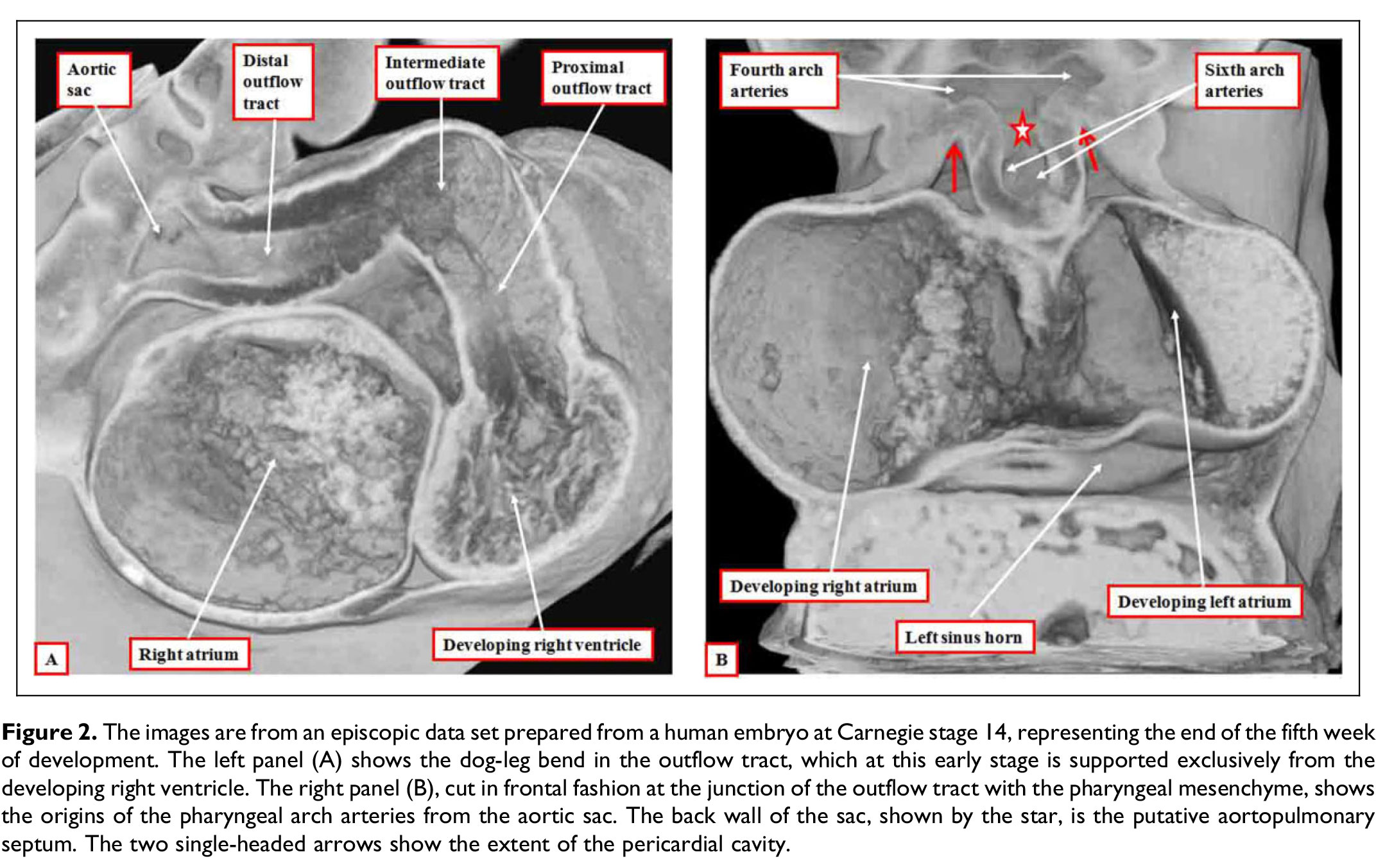 | 2,144 × 1,353 (421 KB) | Z8600021 (talk | contribs) |
You cannot overwrite this file.
File usage
The following 2 pages use this file: