File:Hamster oocyte and spermatozoa.jpg
Original file (883 × 836 pixels, file size: 266 KB, MIME type: image/jpeg)
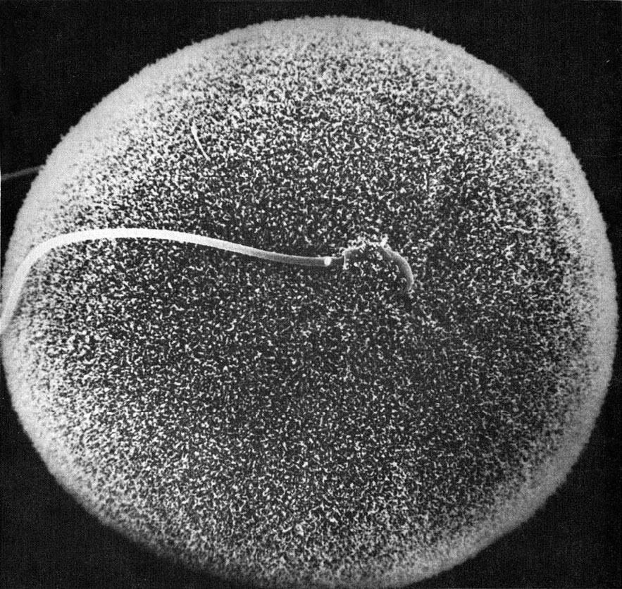
Hamster oocyte and spermatozoa
Figure 32 from Chapter 2 (Specializations of the Free Surface) of 'The Cell, 2nd Ed.' by Don W. Fawcett M.D. Scanning electron micrograph showing in vitro fertilization of a hamster oocyte.
The sperm head, near the center of the field of view, is engulfed by the microvilli on the surface of the oocyte membrane; the sperm tail extends to the left. Image by David Phillips.
NCBI Organism Classification: Phodopus
Cell Type:oocyte, sperm
Cellular Component: microvillus membrane
- Links: Hamster oocyte and spermatozoa | Hamster fused oocyte and spermatozoa | Oocyte Development | Spermatozoa Development
Original image name: 11094.jpg http://www.cellimagelibrary.org/images/11094
Licensing: Attribution Non-Commercial; No Derivatives:This image is licensed under a Creative Commons Attribution, Non-Commercial, No Derivatives License.
File history
Click on a date/time to view the file as it appeared at that time.
| Date/Time | Thumbnail | Dimensions | User | Comment | |
|---|---|---|---|---|---|
| current | 07:30, 26 April 2011 | | 883 × 836 (266 KB) | S8600021 (talk | contribs) | ==Hamster oocyte and spermatozoa== Figure 32 from Chapter 2 (Specializations of the Free Surface) of 'The Cell, 2nd Ed.' by Don W. Fawcett M.D. Scanning electron micrograph showing in vitro fertilization of a hamster oocyte. The sperm head, near the ce |
You cannot overwrite this file.
File usage
The following 2 pages use this file: