File:Hamster fused oocyte and spermatozoa.jpg
Original file (888 × 405 pixels, file size: 98 KB, MIME type: image/jpeg)
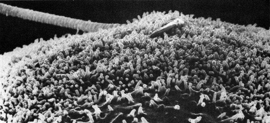
Hamster fused oocyte and spermatozoa
Figure 33 from Chapter 2 (Specializations of the Free Surface) of 'The Cell, 2nd Ed.' by Don W. Fawcett M.D. Scanning electron micrograph showing a lateral view of sperm penetration of a hamster oocyte during vitro fertilization. As membrane of the sperm head fuses with that of the oocyte, the microvilli on the oocyte reform over the hybrid membrane. Image by David Phillips.
- Links: Hamster oocyte and spermatozoa | Hamster fused oocyte and spermatozoa | Oocyte Development | Spermatozoa Development
NCBI Organism Classification: Phodopus
Cell Type:oocyte, sperm
Cellular Component: microvillus membrane
Original Image name: 11096.jpg http://www.cellimagelibrary.org/images/11096
Licensing: Attribution Non-Commercial; No Derivatives:This image is licensed under a Creative Commons Attribution, Non-Commercial, No Derivatives License.
File history
Click on a date/time to view the file as it appeared at that time.
| Date/Time | Thumbnail | Dimensions | User | Comment | |
|---|---|---|---|---|---|
| current | 07:36, 26 April 2011 | | 888 × 405 (98 KB) | S8600021 (talk | contribs) | ==Hamster fused oocyte and spermatozoa== Figure 33 from Chapter 2 (Specializations of the Free Surface) of 'The Cell, 2nd Ed.' by Don W. Fawcett M.D. Scanning electron micrograph showing a lateral view of sperm penetration of a hamster oocyte during vitr |
You cannot overwrite this file.
File usage
The following page uses this file: