File:Gray1014.jpg
Gray1014.jpg (619 × 600 pixels, file size: 95 KB, MIME type: image/jpeg)
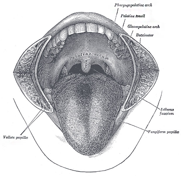
Fig. 1014. The Mouth Cavity
The cheeks have been slit transversely and the tongue pulled forward.
The Mouth Cavity Proper (cavum oris proprium) (Fig. 1014) is bounded laterally and in front by the alveolar arches with their contained teeth; behind, it communicates with the pharynx by a constricted aperture termed the isthmus faucium. It is roofed in by the hard and soft palates, while the greater part of the floor is formed by the tongue, the remainder by the reflection of the mucous membrane from the sides and under surface of the tongue to the gum lining the inner aspect of the mandible. It receives the secretion from the submaxillary and sublingual salivary glands.
The tongue (lingua) is the principal organ of the sense of taste, and an important organ of speech; it also assists in the mastication and deglutition of the food. It is situated in the floor of the mouth, within the curve of the body of the mandible.
- Links: Tongue Development | Salivary Gland Development | Head Development | Musculoskeletal System Development |
- Gray's Images: Development | Lymphatic | Neural | Vision | Hearing | Somatosensory | Integumentary | Respiratory | Gastrointestinal | Urogenital | Endocrine | Surface Anatomy | iBook | Historic Disclaimer
| Historic Disclaimer - information about historic embryology pages |
|---|
 |
| iBook - Gray's Embryology | |
|---|---|

|
|
Reference
Gray H. Anatomy of the human body. (1918) Philadelphia: Lea & Febiger.
Cite this page: Hill, M.A. (2024, April 26) Embryology Gray1014.jpg. Retrieved from https://embryology.med.unsw.edu.au/embryology/index.php/File:Gray1014.jpg
- © Dr Mark Hill 2024, UNSW Embryology ISBN: 978 0 7334 2609 4 - UNSW CRICOS Provider Code No. 00098G
File history
Click on a date/time to view the file as it appeared at that time.
| Date/Time | Thumbnail | Dimensions | User | Comment | |
|---|---|---|---|---|---|
| current | 08:06, 11 May 2014 | | 619 × 600 (95 KB) | Z8600021 (talk | contribs) | ==Fig. 1014. The Mouth Cavity== The cheeks have been slit transversely and the tongue pulled forward. The Mouth Cavity Proper (cavum oris proprium) (Fig. 1014) is bounded laterally and in front by the alveolar arches with their contained teeth; behin... |
You cannot overwrite this file.
File usage
The following 3 pages use this file: