BGDB Practical - Upper Gastrointestinal Tract Histology
Introduction


This current page provides background support information for Medicine phase 1 BGD Histology Practical Virtual Slides and is not part of the BGDB practical class virtual slides.
Practical 4: Upper Gastrointestinal Tract Histology Principal Teacher: Patrick de Permentier

|
Moodle Lab Slides
Note - Moodle icon links appearing below on this page go directly to Lab Slide. |
- Related Content: GIT Development Lecture | GIT Development Practical | Salivary Gland Development
Aim
To introduce the histology of the upper GIT (gastro-intestinal tract) Specific Objectives:
- To describe the histology of the lip and the tongue, including epithelia, muscles, glands, papillae and taste buds.
- To appreciate the histological features of the three major salivary glands.
- To describe the general architecture of the wall of the alimentary canal and the functions of the mucosa, submucosa, muscle layer and serosal or adventitial layer in the various anatomical divisions of the canal.
- To describe the main features of the structure of the wall of the oesophagus, cardio-oesophageal junction, stomach, and duodenum.
- To identify the cells of gastric epithelium and distinguish the histological features of the cardiac, body and pyloric regions of stomach. To appreciate the role of mucus on the surface of gastric epithelium.
- To describe the histological features of the duodenum including villi, intestinal mucosal glands (crypts of Lieberkühn), lymphatic tissue and nerve plexuses.
Learning Activities
Lip and Infant lip
|
|
In these 2 slides, identify the mucosal surface and skin surface on opposite sides of the lip.
Note the mucocutaneous transition zone (also called "red margin" or vermillion border). Observe the orbicularis oris skeletal muscle; labial salivary glands (mucoserous glands; mostly serous in infant lip); salivary ducts with stratified epithelium; nerve fascicles.

Soft Palate
| Slide - Soft Palate |
Note palatal salivary glands secreting mucus; ducts lined by stratified cuboidal epithelium; skeletal muscle; palatine tonsil.
- What type of epithelium covers the respiratory and oral surface respectively?
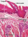
Root of Tongue
| Slide - Lingual tonsil, tongue |
| Slide - Tongue circumvallate papillae |
It shows interlacing striated muscle fibres, lingual tonsils with lymphatic nodules, crypts, mucoserous glands, and ducts of the glands (some mucous acini have serous demilunes).
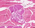
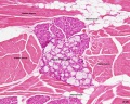
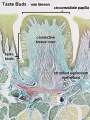
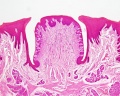
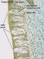
Tongue: circumvallate papillae Taste buds in the lateral wall of circumvallate papillae; von Ebner's serous glands and ducts; striated muscle fascicles; blood vessels; nerve fascicles; lymph aggregations.

Circumvallate Papilla


Filiform papillae

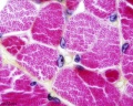
Human Tongue ( lingual salivary gland, white adipose tissue, skeletal muscle)
von Ebner's gland

Tongue (rabbit)
| Slide - Tongue (taste buds) |
Foliate papillae with taste buds in walls; serous glands; LS and TS of striated muscle fibres.

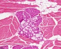
Submandibular gland
| Slide - Salivary gland (submandibular) |
Mixed salivary gland; predominantly serous acini; some mucous acini with serous demilunes; interlobular ducts with stratified cuboidal or stratified columnar epithelium; connective tissue; striated ducts with simple cuboidal lining epithelium; short intercalated ducts.

Sublingual gland
| Slide - Salivary gland (sublingual) |
Predominantly mucous acini; some serous demilunes; interlobular ducts with stratified cuboidal/columnar epithelium; connective tissue; striated ducts with simple columnar lining epithelium; short intercalated ducts.


Parotid gland
| Slide - Salivary gland (parotid) |
Serous salivary gland; lobules; connective tissue septa; serous acini, zymogen granules; striated ducts; interlobular ducts with stratified epithelium; intercalated ducts; lymph node with capsule.


Oesophagus

| Slide - Oesophagus |
Mucosa; submucosa; muscularis externa.
- What type of epithelium lines the lumen?
- What types of muscle fibres can you recognize in the 2 sublayers of the muscularis externa?


Cardio-oesophageal junction

| Slide - Cardio-oesophageal junction |
Oesophageal epithelium; sharp transition to gastric epithelium; cardiac glands (of stomach); smooth muscle fascicles.
- What are the 2 sublayers of the muscularis externa in the oesophagus?

Stomach fundus
| Slide - Stomach fundus |
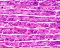
Surface secretory sheet gland (secreting mucus) folded macroscopically to form gastric rugae; gastric pits; gastric simple branched tubular glands; mucous neck cells; parietal cells (pale staining); zymogen (chief) cells (dark staining) in the deeper regions of glands; muscularis mucosae; extensive submucosa; muscularis externa (part).
| parietal cells - chief cells | mucus neck - parietal cells - chief cells |
|---|---|

|

|
stomach mucosa
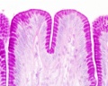
mucosa - secretory epithelial sheath - goblet cell
gastric glands - parietal cells - chief cells
stomach overview


- Stomach Histology Links: stomach labeled overview | parietal cells - chief cells | mucus neck - parietal cells - chief cells | stomach overview | stomach mucosa | mucosa - secretory epithelial sheath - goblet cell | gastric glands - parietal cells - chief cells | stomach overview | Stomach Histology | Stomach Development | Gastrointestinal Tract Development
Pyloro-duodenal junction
| Slide - Pyloro-duodenal junction |
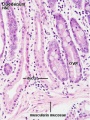
In the pyloric region of stomach, note the deep pyloric pits and shorter, straighter pyloric glands lined mainly by mucus-secreting cells (the specimen shows some post-mortem loss of mucins). Note the pyloric sphincter (smooth muscle) and the transition of epithelium at the duodenum with its villi and intestinal crypts (of Lieberkühn). A striking feature of the first part of the duodenum is the presence of Brunner's submucosal glands.
- What is the secretion from Brunner’s glands?
- What structure defines the location of Brunner’s glands as being part of the submucosa?

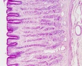
In the duodenum of the small intestine, surface epithelium of columnar absorptive cells; goblet cells; villi; intestinal crypts (tubular glands); duodenal (Brunner's) glands of submucosa; submucosa; muscularis externa with two sublayers; lacteals. This part of the duodenum is retroperitoneal and so it is covered by a tunica adventitia and not a tunica serosa. (Paneth cells in the bases of the crypts of Lieberkühn are not stained in this preparation.)
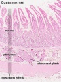
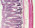
Duodenum overview
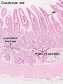
Duodenum villi and crypts
Duodenum
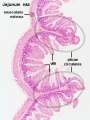
Jejunum overview
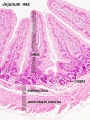
Jejunum villus
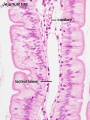
Jejunum labeled
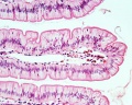
Jejunum unlabeled


- Intestine Histology Links: Duodenum overview | Duodenum villi and crypts | Duodenum | Jejunum overview | Jejunum villus | Jejunum labeled | Jejunum unlabeled | Gastrointestinal Tract Histology | Intestine Development
Terms
- Histology Glossary: A | B | C | D | E | F | G | H | I | J | K | L | M | N | O | P | Q | R | S | T | U | V | W | X | Y | Z | ANAT2241 Support | Histology | Histology Stains | Embryology Glossary
Abbreviations: ( ) plural form in brackets, A. Arabic, abb. abbreviation, c. circa =about, F. French adj. adjective, G. Greek, Ge. German, cf. compare, L. Latin, dim. diminutive, NA. Nomina anatomica, q.v. which see, OF. Old French
- acinus (-i) - L. = a juicy berry, a grape; applied to small, rounded terminal secretory units of compound exocrine glands that have a small lumen (adj. acinar).
- adventitia - layer of loose connective tissue, separates alimentary canal from other tissues.
- Auerbach plexus - Auerbach, Leopold (1828-1897) Breslau neuropathologist & anatomist; Auerbach's nerve plexus in external muscle layer of intestinal wall (1862).
- Bartholin’s duct - see sublingual duct.
- Brunner gland - Brunner, Johann Konrad (1653-1727) Professor of Medince at Heidelberg; B's glands = compound mucus-secreting glands of duodenal submucosa (1687).
- buccal - L. bucca = cheek; related to cheek or mouth.
- chief cell - (peptic cell, gastric zymogenic cell) gastric cells that release pepsinogen, gastric lipase and chymosin.
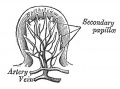
- circumvallate papillae - (vallate papillae) tongue largest and least numerous papillae (human 8 to 12) occur in depressions of the surface of the tongue and are surrounded with a trench formed by the infolding of the epithelium. Taste buds are numerous on the lateral surfaces of these papillae and the excretory ducts of serous glands (glands of von Ebner) open into the trenches surrounding the papillae.
- demilune - L. dimidius = half + luna = moon; crescent-shaped cap of serous cells over mucous alveolus in some salivary glands.
- duodenum - L. duodenum digitorum = "twelve fingers' breadth" (25 cm long 5 cm diameter) the first part of the small intestine lying between stomach and jejunum. Food mixes with bile (gallbladder) and digestive juices (pancreas) and absorption of vitamins, minerals, and other nutrients begins here.
- ductus (-us) - L. = passage from L. ducere = to lead; tube lined by epithelium for exocrine glandular secretions to reach surface.
- fundus - L. = bottom, base (as in fundamental); refers to region of organ (e.g., stomach, uterus, eye), gland (e.g., gastric glands).
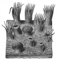
- filiform papillae - tongue smallest and most numerous papillae, provide a rough surface to aid in the manipulation and processing of foods.
- fungiform - L. fungus = mushroom + forma = a shape; of lingual papillae.
- fungiform papillae - tongue single evenly spaced between the filiform papillae. Epithelium is slightly thinner than on the remaining surface of the tongue and connective tissue core is richly vascularised.
- gastric - adj. L. gastricus , from G. gaster = stomach, belly; relating to the stomach.
- gland - L. glandula , dim of L. glans = an acorn, a pellet; term used to describe mesenteric lymph nodes (Herophilus, c. 300 BC).
- intercalated - L. inter = between + calare = to proclaim, calatus = inserted; of a duct inserted between the end of the gland (acinus, or alveolus) and a larger duct.
- intercalated duct - cuboidal epithelium, modify saliva, add bicarbonate ions (buffering) and absorb chloride.
- intramural - L. intra = within + murus = wall; within the wall of an organ.
- labial - adj. L. labialis = of the lips, L. labium = lip, rim of a vessel.
- labial salivary gland - (glandulœ labiales) several glands located between the mucous membrane and the Orbicularis oris, around the orifice of the mouth. They are circular in form (about the size of small peas) their ducts open by minute orifices onto the mucous membrane. In structure they resemble the salivary glands.
- lacteal - L. lac = milk ( lacteus = of milk, lactare = to suckle); intestinal lymphatic, containing chyle after a fatty meal.
- lining epithelium - non-keratinised stratified squamous epithelium, covers the remaining (non-masticatory epithelium) surfaces of the oral cavity.
- lingual - adj. L. lingua = tongue.
- lips - outside (lined by skin) and inside (oral mucosa), vermilion border (prolabium) transition from the skin to the oral mucosa, forms only small part of the anatomical lips.
- lumen - L. = light; space enclosed by tubular or vesicular structure; hence luminal.
- masticatory epithelium - covers the tongue, gingivae and hard palate. Keratinised epithelium to different degrees depending on the extent of physical forces exerted on the epithelium.
- microvilli - epithelial cell apical surface specialisation seen in the small intestinal. These microfilament filled structures increase the surface area for absorption and secretion.
- mucin - L. mucus from G. muxa = snot, slime; protein constituent of all mucus; occurs as granules in secretory cells.
- mucosa - (-ae) L. (mucous membrane) - the moist tissue that lines parts of the inside of the body (nose, mouth, digestive tract, lungs, and urinary). Glands in this tissue release a thick fluid called mucus. The underlying connective tissue is described as the submucosa.
- mucous membrane - see mucosa.
- muscularis externa - in oesophagus contains striated muscle (upper third), a mixture of striated muscle and smooth muscle (middle third) and smooth muscle (lower third).
- mucus - L. = slime (adj. mucous).
- myenteric - G. mys = muscle + enteron = intestine (cf. Auerbach).
- oesophagus - G. oiso = future of phero = I carry + pahgein = to eat; G. oisophagos = gullet, or tube carrying food from pharynx to stomach.
- oesophageal glands - located in the submucosa produce a mucous secretion, which lubricates the epithelium and aids the passage of food. Oesophagus closest to the stomach may also be mucosal mucus-producing glands, similar to the glands in the adjacent mucosa of the stomach.
- oral - adj. L. os, oris = mouth.
- oxyntic cell - see parietal cell.
- palate - L. palatum = roof of mouth.
- palisade - L. palus = stake; like a fence of stakes.
- Paneth cell - Paneth, Josef (1857-1890) Breslau & Vienna physiologist; P. cells (1887) = eosinophilic cells at base of intestinal crypts of Lieberkühn.
- papilla - (-ae) L. = a teat, a nipple; a nipple-like projection, e.g., on the tonge (Malpighi, c. 1670; cf. circumvallate, filiform, foliate, fungiform, vallate); duodenal papilla (containing duodenal ampulla).
- parietal cell - (oxyntic cell, delomorphous cell), gastric epithelial cell that secretes gastric acid (HCl) and intrinsic factor.
- parotid - G. para = beside + otos = of the ear; a salivary gland.
- pepsinogen - G. pepis = digestion + gennan = to produce; a precusor of pepsin = enzyme that aids digestion.
- peptic cell - see chief cell.
- plica - (-ae) a corruption from L. plicare = to fold; in13th century a scalp infection endemic in Poland was called plica polonica (Polish plait); any kind of fold.
- plicae circulares - L. = circular folds; actually transverse folds that are not circular in small intestine = valves of Kerckring, q.v.
- ruga - (-ae) L. = a fold or wrinkle, e.g., in stomach.
- sac - L. saccus = sack, bag, from G. sakkos .
- salivary - L. saliva = spittle.
- secretion - L. secretus = separated; production of materials by glandular activity.
- serous - adj. L. = having nature of serum, watery fluid.
- Stenson's duct - (parotid duct) Historic term for the major duct of the parotid gland that allows salivary gland secretions to empty into the oral cavity. Named after Niels Stensen (1638 - 1686) a Danish anatomist, natural scientist, and theologist.
- stomach - G. stomachos = gullet or oesophagus, from G. stoma = a mouth + cheo = I pour; lower end of the gullet; organ attached to lower end. NB. Greeks used gaster for stomach.
- striated border - L. striatus = striped; light microscopic term for the fine microvilli on intestinal absorptive cells.
- striated ducts - modifies saliva (secretion of potassium and the absorption of sodium), columnar cells with the nucleus of located approximately midways between the apical and basal cell surfaces. Striations are found in the basal part of the cytoplasm of the cells, where numerous mitochondria are found between infoldings of the basal cell membrane. These cells can also take up a secretable form of antibodies and release them into the saliva.
- sublingual caruncle - location on either side of the frenulum linguae on the sublingual surface of the tongue.
- sublingual duct - (major sublingual duct, Bartholin’s duct) from the sublingual gland that opens in the sublingual caruncle. There are also numerous minor sublingual ducts opening on the sublingual fold.
- submandibular - adj. L. " + mandibula = jaw.
- submandibular duct - (Wharton’s duct) from the submandibular gland, runs forward along the lingual nerve in the sublingual space and opens in the sublingual caruncle.
- submucosa - the connective tissue underlying the mucosa.
- succus entericus - L. = juice + of the intestine.
- succus gastricus - L. = juice + of the stomach.
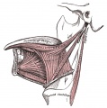
- tongue muscles - skeletal muscle organized into strands oriented more or less perpendicular to each other.
- tongue nerves - movement (XII, hypoglossal nerve - motor) and sensory information. (V, trigeminal nerve - sensory - anterior two-thirds; VII, facial nerve - taste; IX, glossopharyngeal nerve - sensory/taste - posterior one-third).
- tonsil - L. tonsilla (origin obscure); mass of lymphocytes close to an epithelium, e.g., lingual tonsil, palatine tonsil (the "tonsil"), pharyngeal tonsil (adenoid, tonsil of Luschka, q.v.), tubal tonsil (of auditory tube).
- tubuloacinar gland - secretory acini with the first part of the duct system also participating in the secretory process.
- uvula - L. = a little grape; pendulous posterior end of soft palate used to produce gutteral consonants (1695).
- vallate papillae - see circumvallate papillae.
- vermilion border - (mucocutaneous transition zone, "red margin") A small region of the lip that occurs between the lip and the adjacent normal skin, It has no sebaceous glands, sweat glands, or hair. Change in the epidermis from external skin (highly keratinised) to internal skin (less keratinised).
- von Ebner's glands - serous glands associated with circumvallate papillae, their ducts open into the trenches surrounding the papillae ("rinsing glands").
- Waldeyer's ring - Waldeyer, Heinrich Wilhelm Gottfried (1836-1921) Brelau & Berlin anatomist; ring of lymphatic tissue at junction of oro- and nasopharynx (1884).
- Wharton’s duct - see submandibular duct.
- zymogenic - adj. G. zyme = leaven, ferment, sour dough + gennan = to produce; of the granules in glandular cells producing enzymes.
- zymogenic cell - see chief cell.
Images
Salivary gland sk muscle HE
Salivary gland sk muscle HE
Filiform papillae HE
circumvallate papilla VG
circumvallate papilla HE
Taste buds VG
Salivary gland sk muscle HE x10 transverse
Tongue x100


Historic Images
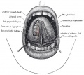
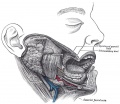
1013 The mouth cavity
1014 The mouth cavity
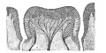
1015 Circumvallate papilla
1016 Filiform papilla
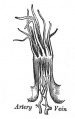
1017 Fungiform papilla
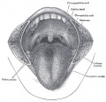
1018 Mucous membrane of the tongue
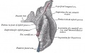
1019 Extrinsic muscles of the tongue
1020 Intrinsic muscles of the tongue
1022 Parotid
1023 Parotid
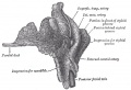
1024 Salivary Glands
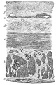
1033 Section of the Human Esophagus
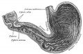
1050 Interior of stomach


Cite this page: Hill, M.A. (2024, April 24) Embryology BGDB Practical - Upper Gastrointestinal Tract Histology. Retrieved from https://embryology.med.unsw.edu.au/embryology/index.php/BGDB_Practical_-_Upper_Gastrointestinal_Tract_Histology
- © Dr Mark Hill 2024, UNSW Embryology ISBN: 978 0 7334 2609 4 - UNSW CRICOS Provider Code No. 00098G