File:Fetal ductus venosus cartoon.jpg
From Embryology
Size of this preview: 451 × 599 pixels. Other resolution: 1,000 × 1,329 pixels.
Original file (1,000 × 1,329 pixels, file size: 255 KB, MIME type: image/jpeg)
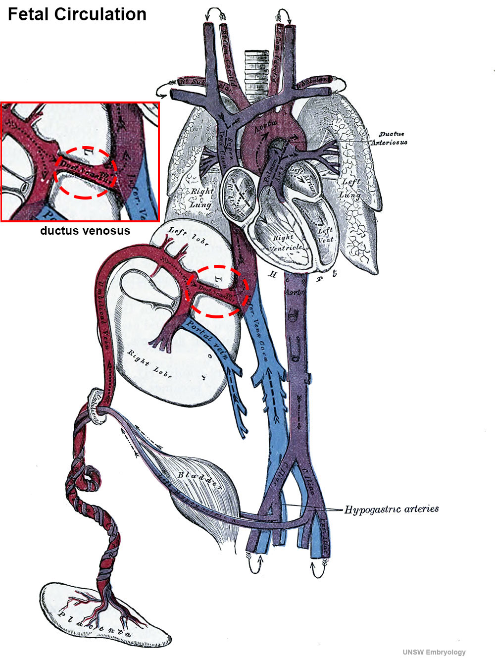
Fetal Ductus Venosus
Cartoon showing the position and function significance of the ductus venous to fetal circulation.
Fetal blood is returned from the placenta to the fetus by the placental or umbilical vein.
- Placental vein enters the abdomen at the umbilicus, and passes upward along the free margin of the falciform ligament of the liver to the under surface of that organ, where it gives off two or three branches:
- 1 large to the left lobe.
- 1-2 smaller to the lobus quadratus and lobus caudatus.
- Placental vein at the porta hepatis (transverse fissure of the liver) divides into two branches:
- the larger is joined by the portal vein and enters the right lobe
- the smaller ductus venosus continues upward and joins the inferior vena cava.
- Links: image - cartoon excerpt | Ductus Venosus | Gray Fig. 502
Cartoon modified from Gray's Anatomy Fig. 502.
Cite this page: Hill, M.A. (2024, May 4) Embryology Fetal ductus venosus cartoon.jpg. Retrieved from https://embryology.med.unsw.edu.au/embryology/index.php/File:Fetal_ductus_venosus_cartoon.jpg
- © Dr Mark Hill 2024, UNSW Embryology ISBN: 978 0 7334 2609 4 - UNSW CRICOS Provider Code No. 00098G
File history
Click on a date/time to view the file as it appeared at that time.
| Date/Time | Thumbnail | Dimensions | User | Comment | |
|---|---|---|---|---|---|
| current | 13:13, 21 February 2015 | | 1,000 × 1,329 (255 KB) | Z8600021 (talk | contribs) | |
| 12:58, 21 February 2015 |  | 1,000 × 1,329 (254 KB) | Z8600021 (talk | contribs) | ==Fetal Ductus Venosus== Cartoon showing the position and function significance of the ductus venous to fetal circulation. Cartoon modified from Gray's Anatomy Fig. 502. Category:Cartoon |
You cannot overwrite this file.
File usage
The following page uses this file: