File:Ectopic pregnancy CT 02.jpg
Original file (800 × 676 pixels, file size: 59 KB, MIME type: image/jpeg)
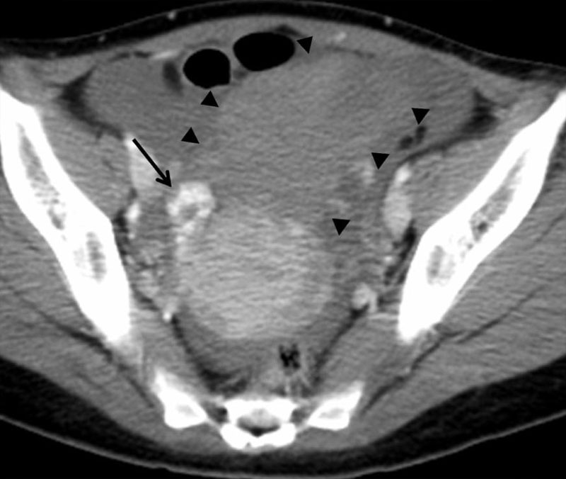
Ectopic Tubal Pregnancy Computed Tomography
Imaging findings are presented for 37-year-old woman with interstitial pregnancy.
- Follow-up CT image obtained after four days shows amorphous hematoma (arrowheads) with massive hemoperitoneum caused by ruptured interstitial pregnancy.
- Previous strong enhancing ring-like mass in right pelvis is now seen as heterogeneous enhancing mass (arrow) after rupture of interstitial pregnancy.
- Links: Ectopic pregnancy Initial CT | Ectopic pregnancy Follow-up CT | Ectopic pregnancy CT | Ectopic Implantation | Computed Tomography
Reference
Shin BS & Park MH. (2010). Incidental detection of interstitial pregnancy on CT imaging. Korean J Radiol , 11, 123-5. PMID: 20046504 DOI.
Copyright
This is an Open Access article distributed under the terms of the Creative Commons Attribution Non-Commercial License (http://creativecommons.org/licenses/by-nc/3.0) which permits unrestricted non-commercial use, distribution, and reproduction in any medium, provided the original work is properly cited.
Original file name: Fig. 1 http://www.ncbi.nlm.nih.gov/pmc/articles/PMC2799642/figure/F1/
Cite this page: Hill, M.A. (2024, April 26) Embryology Ectopic pregnancy CT 02.jpg. Retrieved from https://embryology.med.unsw.edu.au/embryology/index.php/File:Ectopic_pregnancy_CT_02.jpg
- © Dr Mark Hill 2024, UNSW Embryology ISBN: 978 0 7334 2609 4 - UNSW CRICOS Provider Code No. 00098G
File history
Click on a date/time to view the file as it appeared at that time.
| Date/Time | Thumbnail | Dimensions | User | Comment | |
|---|---|---|---|---|---|
| current | 15:02, 3 September 2011 | | 800 × 676 (59 KB) | S8600021 (talk | contribs) | Ectopic---PMC2799642-B.jpg |
You cannot overwrite this file.
File usage
The following 2 pages use this file: