File:Day 16 Reposition of umbilical hernia.JPG

Day_16_Reposition_of_umbilical_hernia.JPG (640 × 532 pixels, file size: 34 KB, MIME type: image/jpeg)
Mouse Embryo E16
Day 16: the umbilical hernia repositions. superficial veins are visible through the transparet skin of the embryo.
Vc= vena cephalica, Vth= Vena thoracica lateralis, Vt= Vena temporalis superficialis, Vu= Vena umbilica, Vv= Vena vitellina, P= Placenta, Vm+ Vena metatarseae dorsales, Nh= umbilical hernia
- Links: Mouse Development | Mouse Stages | Category:Mouse E16.0 | Student Project
Reference
Illustration by z3252340, based upon Fig 345 and Fig 246, in Dr Karl Theiler’s ‘The House Mouse; Atlas of Embryonic Development’, Springer - Verlag New York Inc, New York, 1989
Copyright
Beginning six months after publication, I, z3252340, grant the public the non-exclusive right to copy, distribute, or display the Work under a Creative Commons Attribution-Noncommercial-Share Alike 3.0 Unported license, as described at http://creativecommons.org/licenses/by-nc-sa/3.0/ and http://creativecommons.org/licenses/by-nc-sa/3.0/legalcode.
- Note - This image was originally uploaded as part of an undergraduate science student project and may contain inaccuracies in either description or acknowledgements. Students have been advised in writing concerning the reuse of content and may accidentally have misunderstood the original terms of use. If image reuse on this non-commercial educational site infringes your existing copyright, please contact the site editor for immediate removal.
File history
Click on a date/time to view the file as it appeared at that time.
| Date/Time | Thumbnail | Dimensions | User | Comment | |
|---|---|---|---|---|---|
| current | 14:07, 19 September 2009 | | 640 × 532 (34 KB) | Z3252340 (talk | contribs) | Reverted to version as of 04:06, 19 September 2009 |
| 14:06, 19 September 2009 |  | 640 × 532 (34 KB) | Z3252340 (talk | contribs) | Reverted to version as of 04:05, 19 September 2009 | |
| 14:06, 19 September 2009 | 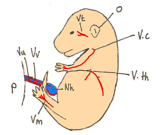 | 640 × 532 (34 KB) | Z3252340 (talk | contribs) | ||
| 14:05, 19 September 2009 |  | 640 × 532 (34 KB) | Z3252340 (talk | contribs) | colour | |
| 10:44, 16 September 2009 |  | 640 × 532 (33 KB) | Z3252340 (talk | contribs) | Day 16: the umbilical hernia repositions. superficial veins are visible through the transparet skin of the embryo. Vc= vena cephalica, Vth= Vena thoracica lateralis, Vt= Vena temporalis superficialis, Vu= Vena umbilica, Vv= Vena vitellina, P= Placenta, |
You cannot overwrite this file.
File usage
The following 3 pages use this file: