File:Cleidocranial dysplasia 01.jpg
Original file (518 × 700 pixels, file size: 65 KB, MIME type: image/jpeg)
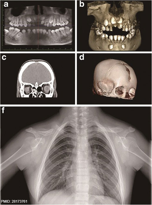
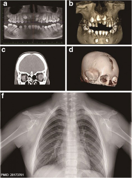
Cleidocranial Dysplasia
Radiological findings for a patient with cleidocranial dysplasia.
a, b Cone-beam computed tomography results showing detailed dental abnormalities, including impacted supernumerary teeth, the retention of primary teeth, eruption failure of the permanent teeth, and impaired root development.
c, d A skull CT scan showed the presence of open fontanelles.
e Radiographs revealed hypoplastic or aplastic distal ends of clavicles and structural abnormalities occurring in the right shoulder peak joint.
Reference
<pubmed>28173761</pubmed>
doi: 10.1186/s12881-017-0375-x.
Copyright
© Wen’an Xu, Qiuyue Chen, Cuixian Liu, Jiajing Chen, Fu Xiong, Buling Wu. 2017
Original file - 12881_2017_375_Fig1_HTML.jpg resized and PMID added to original image.
Cite this page: Hill, M.A. (2024, May 21) Embryology Cleidocranial dysplasia 01.jpg. Retrieved from https://embryology.med.unsw.edu.au/embryology/index.php/File:Cleidocranial_dysplasia_01.jpg
- © Dr Mark Hill 2024, UNSW Embryology ISBN: 978 0 7334 2609 4 - UNSW CRICOS Provider Code No. 00098G
File history
Click on a date/time to view the file as it appeared at that time.
| Date/Time | Thumbnail | Dimensions | User | Comment | |
|---|---|---|---|---|---|
| current | 14:18, 13 February 2017 | | 518 × 700 (65 KB) | Z8600021 (talk | contribs) | A novel, complex RUNX2 gene mutation causes cleidocranial dysplasia. Xu W, Chen Q, Liu C, Chen J, Xiong F, Wu B. BMC Med Genet. 2017 Feb 7;18(1):13. doi: 10.1186/s12881-017-0375-x. PMID 28173761 12881_2017_375_Fig1_HTML.jpg |
You cannot overwrite this file.
File usage
The following page uses this file: