File:Choanal atresia computed tomography 01.jpg
From Embryology
No higher resolution available.
Choanal_atresia_computed_tomography_01.jpg (598 × 477 pixels, file size: 35 KB, MIME type: image/jpeg)
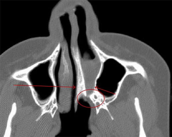
Choanal Atresia
Axial computed tomography
- long arrow - widening of the vomer
- short arrow - bowing of the posteromedial maxilla
- ring - narrowing of the choana anterior to the pterygoid
Original image name: Figure 1 IJPED2011-280763.001.jpg
Reference
Al-Noury K & Lotfy A. (2011). Role of multislice computed tomography and local contrast in the diagnosis and characterization of choanal atresia. Int J Pediatr , 2011, 280763. PMID: 21772853 DOI.
This is an open access article distributed under the Creative Commons Attribution License, which permits unrestricted use, distribution, and reproduction in any medium, provided the original work is properly cited.
- Note - This image was originally uploaded as part of an undergraduate science student project and may contain inaccuracies in either description or acknowledgements. Students have been advised in writing concerning the reuse of content and may accidentally have misunderstood the original terms of use. If image reuse on this non-commercial educational site infringes your existing copyright, please contact the site editor for immediate removal.
File history
Click on a date/time to view the file as it appeared at that time.
| Date/Time | Thumbnail | Dimensions | User | Comment | |
|---|---|---|---|---|---|
| current | 11:48, 17 August 2011 | | 598 × 477 (35 KB) | S8600021 (talk | contribs) | ==Choanal Atresia== Axial computed tomography widening of the vomer (long arrow), bowing of the posteromedial maxilla (short arrow), and narrowing of the choana anterior to the pterygoid. Original image name: Figure 1 IJPED2011-280763.001.jpg ===Refere |
You cannot overwrite this file.