File:AV Valves.jpg
From Embryology
Size of this preview: 654 × 600 pixels. Other resolution: 1,183 × 1,085 pixels.
Original file (1,183 × 1,085 pixels, file size: 129 KB, MIME type: image/jpeg)
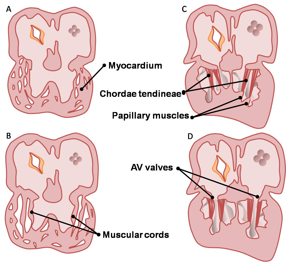
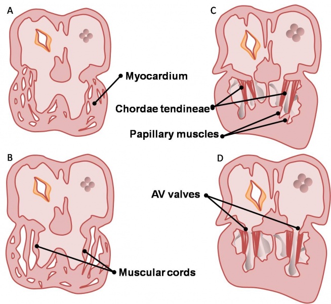
Sequence of events in the development of the atrioventricular valves.
The structures of the valves i.e. the papillary muscles, chordae tendineae and cusps are sculpted from the muscular ventricular walls.
| Begin Advanced | Heart Fields | Heart Tubes | Cardiac Looping | Cardiac Septation | Outflow Tract | Valve Development | Cardiac Conduction | Cardiac Abnormalities | Molecular Development |
| Cardiac Embryology | Begin Basic | Begin Intermediate | Begin Advanced |
File history
Click on a date/time to view the file as it appeared at that time.
| Date/Time | Thumbnail | Dimensions | User | Comment | |
|---|---|---|---|---|---|
| current | 11:15, 14 March 2010 | | 1,183 × 1,085 (129 KB) | Z3212774 (talk | contribs) | category:Heart ILP Sequence of events in the development of the atrioventricular valves. The structures of the valves i.e. the papillary muscles, chordae tendineae and cusps are sculpted from the muscular ventricular walls. |
You cannot overwrite this file.
File usage
The following file is a duplicate of this file (more details):