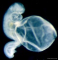
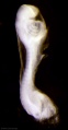
Category:Scanning EM
From Embryology
This Embryology category shows pages and media related to the research imaging technique of scanning electron micrographs (SEM) in development. Note:
- images in this category may also include some of the associated bright field images taken before SEM fixation and imaging.
- there is a separate Category:Electron Micrograph.
Pages in category 'Scanning EM'
The following 16 pages are in this category, out of 16 total.
C
Media in category 'Scanning EM'
The following 200 files are in this category, out of 426 total.
(previous page) (next page) Anderson2016-fig01.jpg 800 × 800; 93 KB
 Anderson2016-fig02.jpg 800 × 788; 75 KB
 Anderson2016-fig05.jpg 796 × 573; 66 KB
 Cat oocyte zona pellucida 01.jpg 832 × 817; 102 KB
 Cat oocyte zona pellucida 02.jpg 1,000 × 991; 146 KB
 Cat spermatozoa bound to oocyte zona pellucida.jpg 1,000 × 917; 161 KB
 Chicken- PGC grown in vitro 01.jpg 1,174 × 577; 122 KB
 Chicken- PGC grown in vitro 02.jpg 582 × 577; 60 KB
 Chicken- PGC grown in vitro 03.jpg 582 × 577; 61 KB
 Divisions of Early Heart Tube.jpg 1,105 × 978; 85 KB
 Early Heart Tube (Dorsal).jpg 1,282 × 1,124; 111 KB
 Early Heart Tube (Lateral).jpg 1,504 × 972; 110 KB
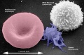
 Erythrocyte and lymphocyte SEM01.jpg 800 × 522; 74 KB
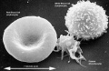
 Erythrocyte and lymphocyte SEM02.jpg 800 × 522; 78 KB
 Erythrocyte and lymphocyte SEM03.jpg 800 × 522; 80 KB
 Fetal temporomandibular joint 05.jpg 600 × 392; 71 KB
 Fetal temporomandibular joint 06.jpg 600 × 389; 50 KB
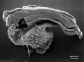
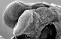
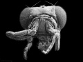 Fly antennapedia head.jpg 900 × 675; 71 KB
 Fly Hippo-type dorsal view head thorax SEM.jpg 555 × 796; 103 KB
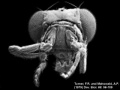
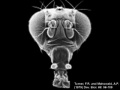
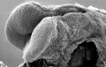
 Fly wild-type head.jpg 900 × 675; 54 KB
 Fly WT dorsal view head thorax SEM.jpg 555 × 796; 95 KB
 Fly-antennapedia head.jpg 320 × 240; 12 KB
 Fly-wild type head.jpg 320 × 240; 10 KB
 Hamster fused oocyte and spermatozoa.jpg 888 × 405; 98 KB
 Hamster oocyte and spermatozoa.jpg 883 × 836; 266 KB
 Hamster oocyte zona pellucida SEM.jpg 800 × 626; 101 KB
 Heart Looping Sequence (SEMs).jpg 1,928 × 776; 212 KB
 Heart Tube Fusion.jpg 1,551 × 1,139; 125 KB
 Heart Tube Segments.jpg 1,082 × 771; 63 KB
 HeartILP draft HeartTubeDorsal.jpg 1,282 × 1,124; 111 KB
 HeartILP draft hearttubesegments.jpg 1,082 × 771; 63 KB
 HeartILP001.jpg 1,507 × 898; 125 KB
 Human heart SEM1.jpg 1,200 × 330; 47 KB
 Human sperm pathology EM02.jpg 800 × 256; 22 KB
 Human uterine tube ciliated epithelium SEM.jpg 1,200 × 855; 248 KB
 Liver SEM01.jpg 2,000 × 1,333; 350 KB
 Lung epithelium sem11.jpg 1,000 × 999; 240 KB
 Lymphocyte rosettes EM01-06.jpg 1,364 × 2,100; 334 KB
 Lymphocyte rosettes EM01.jpg 661 × 665; 58 KB
 Lymphocyte rosettes EM012.jpg 618 × 661; 59 KB
 Lymphocyte rosettes EM02.jpg 661 × 665; 62 KB
 Lymphocyte rosettes EM03.jpg 661 × 665; 51 KB
 Lymphocyte rosettes EM04.jpg 661 × 665; 53 KB
 Lymphocyte rosettes EM05.jpg 661 × 665; 55 KB
 Lymphocyte rosettes EM06.jpg 661 × 665; 66 KB
 ME16 001.jpg 1,740 × 2,500; 557 KB
 ME16 002.jpg 1,037 × 1,500; 272 KB
 ME18 001.jpg 773 × 1,200; 176 KB
 ME34 002.jpg 1,280 × 871; 216 KB
 ME45 002.jpg 2,000 × 1,412; 373 KB
 Microscopy LM and SEM cartoon.jpg 720 × 500; 53 KB
 Mouse circumvallate papilla 01.jpg 712 × 955; 114 KB
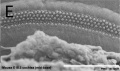
 Mouse E18.5 cochlea sem01.jpg 902 × 774; 150 KB
 Mouse E18.5 cochlea sem02.jpg 901 × 489; 75 KB
 Mouse E18.5 cochlea sem03.jpg 905 × 1,048; 183 KB
 Mouse E18.5 cochlea sem04.jpg 905 × 535; 85 KB
 Mouse E18.5 cochlea sem05.jpg 807 × 533; 78 KB
 Mouse in vitro follicle 04.jpg 799 × 537; 81 KB
 Mouse in vitro follicle 05.jpg 800 × 639; 82 KB
 Mouse in vitro follicle 06.jpg 800 × 639; 120 KB
 Mouse primitive node cilia.jpg 592 × 981; 130 KB
 Mouse renal podocyte EM01.jpg 1,000 × 1,338; 366 KB
 Mycobacterium-tuberculosis.jpg 320 × 240; 18 KB
 RBC with hereditary persistence of fetal haemoglobin SEM01.jpg 1,200 × 816; 220 KB
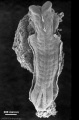
 Sea urchin SEM01.jpg 1,000 × 712; 95 KB
 Sea urchin SEM02.jpg 1,000 × 712; 120 KB
 Sea urchin SEM03.jpg 1,000 × 712; 154 KB
 Sickle cell RBC SEM01.jpg 1,000 × 750; 219 KB
 Stage 9 SEM1.jpg 347 × 450; 42 KB
 Stage10 bf1.jpg 1,000 × 748; 39 KB
 Stage10 bf1a.jpg 800 × 598; 29 KB
 Stage10 bf1b.jpg 600 × 449; 19 KB
 Stage10 bf1c.jpg 400 × 299; 10 KB
 Stage10 bf2.jpg 1,000 × 809; 29 KB
 Stage10 bf2c.jpg 400 × 323; 8 KB
 Stage10 bf3.jpg 836 × 1,000; 35 KB
 Stage10 SEM1.jpg 277 × 450; 28 KB
 Stage10 sem1.jpg 1,000 × 484; 46 KB
 Stage10 sem10.jpg 1,000 × 740; 68 KB
 Stage10 sem10a.jpg 800 × 592; 48 KB
 Stage10 sem10b.jpg 600 × 444; 32 KB
 Stage10 sem10c.jpg 400 × 296; 16 KB
 Stage10 sem11.jpg 1,000 × 636; 76 KB
 Stage10 sem11a.jpg 800 × 509; 59 KB
 Stage10 sem11b.jpg 600 × 382; 38 KB
 Stage10 sem11c.jpg 400 × 255; 20 KB
 Stage10 sem12.jpg 657 × 1,000; 57 KB
 Stage10 sem12a.jpg 526 × 800; 40 KB
 Stage10 sem12b.jpg 395 × 600; 25 KB
 Stage10 sem12c.jpg 263 × 400; 13 KB
 Stage10 sem13.jpg 672 × 1,000; 90 KB
 Stage10 sem13a.jpg 538 × 800; 65 KB
 Stage10 sem13b.jpg 404 × 600; 42 KB
 Stage10 sem13c.jpg 269 × 400; 21 KB
 Stage10 sem14.jpg 880 × 1,000; 100 KB
 Stage10 sem14a.jpg 704 × 800; 74 KB
 Stage10 sem14b.jpg 528 × 600; 50 KB
 Stage10 sem14c.jpg 352 × 400; 28 KB
 Stage10 sem15.jpg 1,000 × 750; 79 KB
 Stage10 sem15a.jpg 800 × 600; 58 KB
 Stage10 sem15b.jpg 600 × 450; 39 KB
 Stage10 sem15c.jpg 400 × 300; 21 KB
 Stage10 sem2.jpg 1,000 × 590; 60 KB
 Stage10 sem3.jpg 634 × 1,000; 55 KB
 Stage10 sem4.jpg 1,000 × 814; 83 KB
 Stage10 sem5.jpg 845 × 1,000; 83 KB
 Stage10 sem6 annotated.jpg 720 × 960; 122 KB
 Stage10 sem6.jpg 614 × 1,000; 57 KB
 Stage10 sem7.jpg 1,243 × 1,000; 116 KB
 Stage10 sem8.jpg 648 × 1,000; 84 KB
 Stage10 sem9.jpg 740 × 1,000; 72 KB
 Stage10 sem9a.jpg 592 × 800; 51 KB
 Stage10 sem9b.jpg 444 × 600; 32 KB
 Stage10 sem9c.jpg 296 × 400; 16 KB
 Stage11 bf1.jpg 429 × 1,000; 42 KB
 Stage11 bf10.jpg 1,762 × 2,304; 268 KB
 Stage11 bf10a.jpg 765 × 1,000; 86 KB
 Stage11 bf10b.jpg 612 × 800; 63 KB
 Stage11 bf10c.jpg 459 × 600; 42 KB
 Stage11 bf11.jpg 1,400 × 1,563; 128 KB
 Stage11 bf11a.jpg 896 × 1,000; 69 KB
 Stage11 bf11b.jpg 717 × 800; 51 KB
 Stage11 bf11c.jpg 538 × 600; 34 KB
 Stage11 bf12.jpg 1,924 × 2,000; 227 KB
 Stage11 bf12a.jpg 962 × 1,000; 87 KB
 Stage11 bf12b.jpg 770 × 800; 64 KB
 Stage11 bf12c.jpg 578 × 600; 43 KB
 Stage11 bf1a.jpg 343 × 800; 30 KB
 Stage11 bf1b.jpg 257 × 600; 20 KB
 Stage11 bf1c.jpg 171 × 400; 5 KB
 Stage11 bf2.jpg 521 × 1,000; 43 KB
 Stage11 bf2a.jpg 417 × 800; 31 KB
 Stage11 bf2b.jpg 313 × 600; 20 KB
 Stage11 bf2c.jpg 209 × 400; 11 KB
 Stage11 bf3.jpg 464 × 1,000; 45 KB
 Stage11 bf3a.jpg 371 × 800; 34 KB
 Stage11 bf3b.jpg 278 × 600; 22 KB
 Stage11 bf3c.jpg 185 × 400; 13 KB
 Stage11 bf4.jpg 533 × 1,000; 56 KB
 Stage11 bf4a.jpg 426 × 800; 41 KB
 Stage11 bf4b.jpg 320 × 600; 26 KB
 Stage11 bf4c.jpg 213 × 400; 14 KB
 Stage11 bf5.jpg 474 × 1,000; 62 KB
 Stage11 bf5a.jpg 379 × 800; 43 KB
 Stage11 bf5b.jpg 284 × 600; 26 KB
 Stage11 bf5c.jpg 189 × 400; 5 KB
 Stage11 bf6.jpg 525 × 1,000; 19 KB
 Stage11 bf6a.jpg 420 × 800; 14 KB
 Stage11 bf6b.jpg 315 × 600; 10 KB
 Stage11 bf6c.jpg 210 × 400; 6 KB
 Stage11 bf8.jpg 1,312 × 1,603; 124 KB
 Stage11 bf9.jpg 1,797 × 2,247; 289 KB
 Stage11 bf9a.jpg 800 × 1,000; 94 KB
 Stage11 bf9b.jpg 640 × 800; 67 KB
 Stage11 bf9c.jpg 480 × 600; 42 KB
 Stage11 histology-neural tube roof plate 1.jpg 800 × 594; 134 KB
 Stage11 histology-neural tube roof plate 2.jpg 800 × 552; 145 KB
 Stage11 histology-optic pit.jpg 800 × 510; 157 KB
 Stage11 histology-optic vesicle-hindbrain.jpg 800 × 536; 158 KB
 Stage11 sem10.jpg 1,000 × 898; 162 KB
 Stage11 sem100.jpg 1,000 × 898; 109 KB
 Stage11 sem100a.jpg 800 × 718; 82 KB
 Stage11 sem100b.jpg 600 × 539; 56 KB
 Stage11 sem100c.jpg 400 × 359; 30 KB
 Stage11 sem101.jpg 1,000 × 898; 175 KB
 Stage11 sem10a.jpg 800 × 718; 120 KB
 Stage11 sem10b.jpg 600 × 539; 81 KB
 Stage11 sem10c.jpg 400 × 359; 44 KB
 Stage11 sem11.jpg 778 × 1,000; 144 KB
 Stage11 sem11a.jpg 622 × 800; 102 KB
 Stage11 sem11b.jpg 467 × 600; 64 KB
 Stage11 sem11c.jpg 311 × 400; 32 KB
 Stage11 sem13.jpg 620 × 1,000; 84 KB
 Stage11 sem13a.jpg 496 × 800; 58 KB
 Stage11 sem13b.jpg 372 × 600; 36 KB
 Stage11 sem13c.jpg 248 × 400; 17 KB
 Stage11 sem2.jpg 976 × 1,000; 188 KB
 Stage11 sem20.jpg 668 × 1,000; 132 KB
 Stage11 sem20a.jpg 534 × 800; 92 KB
 Stage11 sem20b.jpg 401 × 600; 58 KB
 Stage11 sem20c.jpg 267 × 400; 29 KB
 Stage11 sem21.jpg 600 × 447; 79 KB
 Stage11 sem2a.jpg 781 × 800; 81 KB
 Stage11 sem2b.jpg 586 × 600; 54 KB
 Stage11 sem2c.jpg 391 × 400; 29 KB
 Stage11 sem3.jpg 936 × 1,000; 68 KB
 Stage11 sem3a.jpg 749 × 800; 44 KB
 Stage11 sem3b.gif 468 × 500; 182 KB
 Stage11 sem3b.jpg 468 × 500; 45 KB
 Stage11 sem3c.jpg 375 × 400; 16 KB
 Stage11 sem4.jpg 808 × 1,000; 129 KB
 Stage11 sem4a.jpg 646 × 800; 93 KB
 Stage11 sem4b.jpg 485 × 600; 60 KB
 Stage11 sem4c.jpg 323 × 400; 17 KB
 Stage11 sem5.jpg 612 × 1,000; 59 KB
 Stage11 sem5a.jpg 490 × 800; 42 KB
 Stage11 sem5b.jpg 368 × 600; 27 KB
 Stage11 sem5c.jpg 245 × 400; 14 KB
 Stage11 sem6.jpg 612 × 1,000; 57 KB
 Stage11 sem7.jpg 1,437 × 1,972; 377 KB


.jpg)
.jpg)


.jpg)