Category:Ovary
From Embryology
This Embryology category shows media and pages related to the ovary and its development.
This category may also include ovary related topics such as female genital development, oogenesis, folliculogenesis, corpus luteum, menstrual cycle, and ovary histology.
Subcategories
This category has the following 5 subcategories, out of 5 total.
Pages in category 'Ovary'
The following 133 pages are in this category, out of 133 total.
B
C
E
F
G
M
O
- Template:Oestrogen
- Template:Oestrogens
- Template:Oogenesis
- Template:Ova
- Template:Ovarian reserve
- Template:Ovary
- Template:Ovary abnormalities
- Ovary Development
- Ovary Development Movie
- Template:Ovary timeline
- Template:Ovary timeline table
- Template:Ovary Vignette
- Template:Ovulation
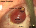
- Ovulation in the human ovary - Its mechanism and anomalies
- Ovulation Movie
P
- Paper - Cyclic changes in the ovaries and uterus of swine and their relations to the mechanism of implantation (1921)
- Paper - Development of the human ovary from birth to sexual maturity
- Paper - Human ova from large follicles
- Paper - Human ova from large follicles - including a search for maturation divisions and observations on atresia
- Paper - Human ova from large follicles - including a search for maturation divisions and observations on atresia (1930)
- Paper - Notes on the formation, structure and physiology of the corpus luteum of man, the pig, and the duck-billed platypus (1924)
- Paper - Selective elimination of ova in the adult ovary
- Paper - Selective elimination of ova in the adult ovary (1925)
- Paper - The corpus luteum in the ovary of the chicken (1918)
- Paper - The events of the primate ovarian cycle
- Paper - The fate of the graafian follicle in the human ovary
- Paper - The hormone of the corpus luteum
- Paper - The morphogenesis of the mammalian ovary (1913)
- Paper - The occurrence of polyovular graafian follicles (1924)
- Paper - The origin of the lutein cells of the corpus luteum
- Paper - The physiological descent of the ovaries in the human foetus
- Template:PCOS
- Template:Polycystic ovarian syndrome
- Template:Polycystic ovary syndrome
- Template:Preovulatory follicle
- Template:Primary follicle
- Template:Primordial follicle
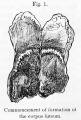
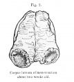
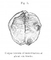
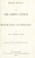
- Prize Essay on the Corpus Luteum (1851) 1
- Prize Essay on the Corpus Luteum (1851) 2
- Prize Essay on the Corpus Luteum (1851) 3
- Prize Essay on the Corpus Luteum (1851) Plates
- Template:Progesterone
R
- Rabbit Ovulation Movie
- Template:Ref-AdamsHertig1969a
- Template:Ref-AdamsHertig1969b
- Template:Ref-Allen1904
- Template:Ref-Allen1925
- Template:Ref-Allen1930
- Template:Ref-Allen1930b
- Template:Ref-Arai1920
- Template:Ref-Arnold1912
- Template:Ref-Brambell1927a
- Template:Ref-Brambell1928
- Template:Ref-Catchpole1949
- Template:Ref-Clark1899b
- Template:Ref-ClarkGJ1900
- Template:Ref-Corner1919
- Template:Ref-Corner1923
- Template:Ref-Corner1937
- Template:Ref-Corner1937b
- Template:Ref-Corner1952
- Template:Ref-Falkiner1933
- Template:Ref-Guttmacher1921
- Template:Ref-Hart1909d
- Template:Ref-Kampmeier1929
- Template:Ref-Kennedy1924
- Template:Ref-Kingsbury1913
- Template:Ref-Kingsbury1914
- Template:Ref-Kingsbury1914a
- Template:Ref-PearlBoring1918
- Template:Ref-Shaw1925
- Template:Ref-Shaw1926
- Template:Ref-Simkins1932
- Template:Ref-Solomons1924
- Template:Ref-Thomson1919a
- Template:Ref-Thomson1919b
- Template:Ref-White1951
- Template:Ref-Young1961
- Template:RU 486
- Template:RU486
S
Media in category 'Ovary'
The following 173 files are in this category, out of 173 total.
 Adrenal and gonad early development.jpg 700 × 397; 50 KB
 Adrenal and gonad steroidogenic factor 1 expression.jpg 1,000 × 636; 88 KB
 Bailey001.jpg 850 × 794; 155 KB
 Bailey014.jpg 704 × 587; 116 KB
 Bailey328.jpg 704 × 795; 85 KB
 Bailey329.jpg 838 × 381; 70 KB
 Bailey330.jpg 680 × 539; 109 KB
 Bailey331.jpg 890 × 782; 118 KB
 Bailey335.jpg 590 × 468; 55 KB
 Bailey339.jpg 802 × 602; 111 KB
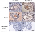 Bovine ovarian follicle BMP15 and GDF9 expression.jpg 874 × 800; 162 KB
 Bovine ovarian follicle BMP15 and GDF9.jpg 1,143 × 810; 219 KB
 Brambell1927a plate28.jpg 1,500 × 1,862; 169 KB
 Brambell1927a plate29.jpg 1,753 × 2,464; 290 KB
 Brambell1927a plate30.jpg 1,732 × 2,386; 451 KB
 Brambell1927a plate31.jpg 1,739 × 2,157; 444 KB
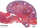
 Cat embryo ovary.jpg 505 × 492; 47 KB
 Corner-table02.jpg 800 × 647; 107 KB
 Corner001.jpg 858 × 356; 44 KB
 Corner002.jpg 873 × 723; 88 KB
 Corner002a.jpg 848 × 307; 49 KB
 Corner002b.jpg 849 × 328; 33 KB
 Corner1920 fig01.jpg 1,000 × 606; 159 KB
 Corner1920 fig02-05.jpg 1,000 × 671; 216 KB
 Corner1920 Plate 1.jpg 756 × 1,000; 222 KB
 Corpus luteum lutein cells.jpg 450 × 600; 104 KB
 Corpus luteum.jpg 450 × 600; 94 KB
 Dalton1851 fig01.jpg 538 × 800; 101 KB
 Dalton1851 fig02.jpg 717 × 800; 77 KB
 Dalton1851 fig03.jpg 681 × 800; 76 KB
 Dalton1851 fig04.jpg 734 × 800; 116 KB
 Dalton1851 fig05.jpg 706 × 800; 105 KB
 Dalton1851 fig06.jpg 599 × 800; 75 KB
 Dalton1851 plate01.jpg 719 × 1,200; 171 KB
 Dalton1851 plate02.jpg 721 × 1,200; 194 KB
 Dalton1851 titlepage.jpg 604 × 1,000; 49 KB
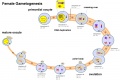 Female gametogenesis.jpg 1,000 × 666; 94 KB
 Fetal adrenal ectopic germ cells 01.jpg 1,092 × 1,280; 358 KB
 Fetal adrenal ectopic germ cells 02.jpg 1,086 × 446; 124 KB
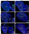
 Fetal gonad retinoid receptor expression 01.jpg 1,004 × 1,000; 226 KB
 Fetal ovary meiosis 01.jpg 1,280 × 410; 132 KB
 Fetal ovary meiosis 02.jpg 496 × 600; 77 KB
 Fetal ovary meiosis 03.jpg 652 × 400; 64 KB
 Gray0589.jpg 900 × 534; 134 KB
 Gray1108.jpg 590 × 400; 73 KB
 Gray1112.jpg 550 × 548; 51 KB
 Gray1113.jpg 600 × 385; 68 KB
 Gray1161.jpg 1,000 × 671; 138 KB
 Gray1163.jpg 600 × 442; 91 KB
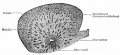
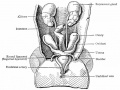
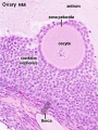
 Historic-ovary.jpg 385 × 283; 34 KB
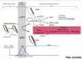
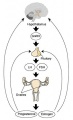
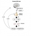 HPG female axis.jpg 600 × 700; 41 KB
 Human corpus luteum - light-and-electron-micrograph.jpg 936 × 711; 208 KB
 Human fetal gonad retinoid receptor expression.jpg 1,004 × 1,000; 447 KB
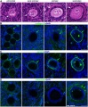
 Human fetal ovary FOXL2.jpg 739 × 1,087; 300 KB
 Human fetal ovary SMAD6 expression.jpg 711 × 535; 167 KB
 Human infant ovary follicle 01.jpg 800 × 800; 107 KB
 Human ovary - corpus luteum 01.jpg 1,024 × 979; 162 KB
 Human ovary - corpus luteum 02.jpg 837 × 800; 119 KB
 Human ovary - corpus luteum 11.jpg 1,024 × 979; 89 KB
 Human ovary - corpus luteum 21.jpg 1,024 × 979; 89 KB
 Human ovary follicle basement membrane 01.jpg 660 × 800; 184 KB
 Human ovary follicle basement membrane EM01.jpg 558 × 697; 100 KB
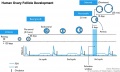 Human ovary follicle development.jpg 700 × 418; 50 KB
 Human ovary follicles light and electron microscopy 01.jpg 586 × 1,080; 225 KB
 Human ovary non-growing follicle model 02.jpg 1,200 × 918; 161 KB
 Human ovary non-growing follicle model.jpg 1,151 × 679; 116 KB
 Human ovary postnatal growth.jpg 800 × 467; 40 KB
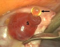
 Human ovulation 01.jpg 1,000 × 799; 175 KB
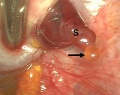
 Human ovulation 02.jpg 734 × 583; 68 KB
 Human ovulation 03.jpg 734 × 583; 76 KB
 Human ovulation 04.jpg 734 × 583; 64 KB
 Human ovulation 05.jpg 734 × 583; 71 KB
 Human ovulation 06.jpg 734 × 583; 82 KB
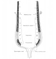
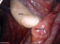 Human right ovary and tube 1.jpg 916 × 680; 32 KB
 Human- adult ovary epithelial cords and primary follicles.jpg 600 × 384; 51 KB
 Human- adult ovary primary follicles.jpg 600 × 639; 56 KB
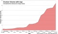
 Human- ovary age primary follicle numbers.jpg 600 × 520; 36 KB
 Infant ovary.jpg 943 × 571; 108 KB
 Keibel Mall 001.jpg 781 × 733; 138 KB
 Keibel Mall 008.jpg 1,200 × 515; 161 KB
 Keibel Mall 2 621.jpg 1,280 × 1,842; 589 KB
 Keibel Mall 2 658a.jpg 1,127 × 1,200; 103 KB
 Keibel Mall 2 658c.jpg 1,000 × 1,019; 90 KB
 Keith1902 fig081.jpg 818 × 800; 113 KB
 Kollmann012.jpg 696 × 437; 31 KB
 Kollmann451.jpg 796 × 826; 102 KB
 Kollmann452.jpg 687 × 606; 74 KB
 Kollmann461.jpg 721 × 528; 100 KB
 Kollmann462.jpg 758 × 429; 63 KB
 Lutein cell glycogen granule em01.jpg 1,149 × 749; 169 KB
 Lutein cell lipid and glycogen em01.jpg 1,156 × 828; 149 KB
 Lutein cell lipid and glycogen em02.jpg 1,109 × 796; 227 KB
 Luteoma of pregnancy 01.jpg 1,200 × 677; 177 KB
 Luteoma of pregnancy 02.jpg 1,200 × 831; 188 KB
 Model of human fetal ovarian cord development 01.jpg 800 × 330; 104 KB
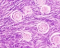
 Monkey- ovary primordial follicle.jpg 1,000 × 800; 292 KB
 Mouse gonad development timeline.jpg 1,200 × 697; 98 KB
 Mouse gonad Gcnf expression 01.jpg 1,947 × 843; 304 KB
 Mouse gonad Gcnf expression E12.5.jpg 331 × 785; 68 KB
 Mouse gonad Gcnf expression E13.5.jpg 332 × 784; 60 KB
 Mouse gonad Gcnf expression E14.5.jpg 334 × 784; 60 KB
 Mouse gonad Gcnf expression E15.5.jpg 338 × 782; 53 KB
 Mouse gonad Gcnf expression E16.5.jpg 325 × 786; 40 KB
 Mouse gonad Gcnf expression E17.5.jpg 328 × 786; 44 KB
 Mouse model of ovarian cord formation 01.jpg 800 × 491; 85 KB
 Mouse model of ovarian cord formation.jpg 800 × 491; 85 KB
 Mouse newborn ovary day 1.mp4 ; 646 KB
- Mouse newborn ovary day 2-3.5.mp4 ; 1.95 MB
 Mouse oogenesis 01.jpg 1,781 × 1,222; 173 KB
 Mouse oogenesis 02.jpg 1,386 × 355; 40 KB
 Mouse ovarian follicle size.jpg 600 × 407; 36 KB
 Mouse ovary 01.jpg 602 × 482; 69 KB
 Mouse ovary normal and polycystic ovary syndrome 01.jpg 913 × 815; 356 KB
 Mouse sex determination genes 01.jpg 1,280 × 923; 73 KB
 Mouse- gonadal supporting cell development.jpg 1,000 × 588; 74 KB
 Mouse-model ovarian cord formation.jpg 600 × 368; 48 KB
 Nelsen1953 fig022.jpg 1,200 × 839; 207 KB
 Nelsen1953 fig030.jpg 1,200 × 995; 414 KB
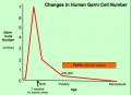 Oocytenumber.jpg 575 × 419; 41 KB
 Ova20he.jpg 450 × 600; 96 KB
 Ova41he.jpg 450 × 600; 113 KB
 Ova44he.jpg 1,280 × 1,024; 315 KB
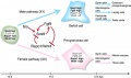
 Ovarian development model.jpg 1,200 × 1,036; 445 KB
 Ovarian developmental genes.jpg 600 × 490; 82 KB
 Ovarian follicle growth in vitro.jpg 1,000 × 769; 73 KB
 Ovarian follicular steroid synthesis.jpg 1,200 × 645; 89 KB
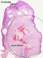
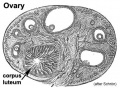
 Ovary corpus luteum.jpg 2,178 × 1,137; 376 KB
 Ovary follicle 01.jpg 450 × 600; 112 KB
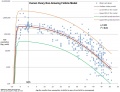
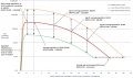
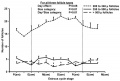
 Ovary follicle size graph.jpg 1,057 × 820; 102 KB
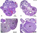
 Ovary Histology - tunica albuginea.jpg 1,280 × 1,024; 336 KB
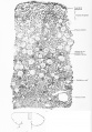
 Ovary histology 001.jpg 1,280 × 1,024; 360 KB
 Ovary histology 002.jpg 1,280 × 1,024; 270 KB
 Ovary histology 003.jpg 1,280 × 1,024; 337 KB
 Ovary histology 004.jpg 1,280 × 1,024; 401 KB
 Ovary histology 005.jpg 1,280 × 1,024; 354 KB
 Ovary histology 006.jpg 1,280 × 1,024; 424 KB
 Ovary histology 007.jpg 1,280 × 1,024; 336 KB
 Ovary histology 008.jpg 1,280 × 1,024; 264 KB
 Ovary histology 061.jpg 1,280 × 1,024; 438 KB
 Ovary histology 061a.jpg 800 × 640; 200 KB
 Ovary histology 061c.jpg 400 × 320; 56 KB
 Ovary histology with chemotherapy.jpg 977 × 872; 232 KB
 Ovary oocyte size graph.jpg 1,057 × 820; 114 KB
 Ovary- atretic follicle 01.jpg 793 × 595; 225 KB
 Ovary- atretic follicle 02.jpg 600 × 450; 139 KB
 Ovary- atretic follicle 03.jpg 790 × 593; 202 KB
 Ovary- atretic follicle 04.jpg 600 × 450; 128 KB
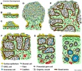
 Ovary- follicle stages.jpg 600 × 337; 45 KB
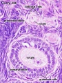
 Ovary- histology overview.jpg 861 × 646; 160 KB
 Ovary- histology secondary follicle 01.jpg 1,000 × 800; 293 KB
 Ovary-human-follicle.jpg 492 × 1,000; 103 KB
 Ovary10x.jpg 480 × 400; 53 KB
 Ovary5x.gif 480 × 400; 162 KB
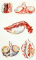
 Pouchet1847 plate03.jpg 1,721 × 2,000; 456 KB
 Pouchet1847 plate04.jpg 1,787 × 2,000; 388 KB
 Pouchet1847 plate05.jpg 1,659 × 2,000; 334 KB
 Pouchet1847 plate06.jpg 1,616 × 2,000; 368 KB
 Rat ovary follicle development 01.jpg 1,200 × 1,035; 201 KB
 Rat ovary histology 01.jpg 1,200 × 938; 274 KB
 Simkins1928 plate01.jpg 1,574 × 2,003; 237 KB
 Simkins1928 plate02.jpg 1,464 × 2,126; 223 KB
 Simkins1928 plate03.jpg 1,551 × 2,086; 293 KB
 Simkins1928 plate04.jpg 1,543 × 2,026; 219 KB
 Simkins1928 plate05.jpg 1,281 × 2,111; 154 KB
 Simkins1928 plate06.jpg 1,587 × 2,088; 277 KB
 Simkins1928 plate07.jpg 1,540 × 2,096; 239 KB
 Simkins1928 plate08.jpg 1,558 × 1,797; 220 KB
 Simkins1928 plate09.jpg 1,548 × 2,096; 263 KB
 Simkins1928 plate10.jpg 1,565 × 1,386; 140 KB
 Ultrasound uterine and ovarian vascularity.jpg 531 × 780; 132 KB
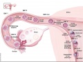 Week1 summary.jpg 1,000 × 747; 107 KB
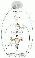 XXhpgaxis.gif 300 × 495; 22 KB
 XXhpgaxis.jpg 300 × 495; 24 KB


