Category:Gastrulation
From Embryology
This Embryology category shows pages and media related to the early embryonic process of Gastrulation. Historically described in the frog by Hans Spemann (1869 - 1941) who identified the primitive node also called "Spemann's organiser". The same region in birds it is known as "Hensen's node" named for Victor Hensen (1835 – 1924) and is also known generally as the primitive node or knot.
| Links: gastrulation | Lecture - Week 3 | Week 3 | Carnegie stage 7 | Carnegie 8 | endoderm | mesoderm | ectoderm | Epithelial Mesenchymal Transition | notochord | ||
|
Subcategories
This category has the following 2 subcategories, out of 2 total.
Pages in category 'Gastrulation'
The following 35 pages are in this category, out of 35 total.
B
G
P
R
Media in category 'Gastrulation'
The following 57 files are in this category, out of 57 total.
 Chicken endoderm origin.jpg 600 × 600; 53 KB
 Chicken neural induction markers.jpg 1,020 × 800; 137 KB
 Chicken primitive streak cell fate 01.jpg 702 × 434; 46 KB
 Chicken primitive streak cell fate 02.jpg 468 × 661; 38 KB
 Chicken primitive streak cell fate 03.jpg 1,200 × 296; 0 bytes
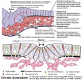 Chicken-gastrulation.jpg 800 × 773; 128 KB
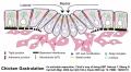 Chicken-gastrulation2.jpg 800 × 443; 55 KB
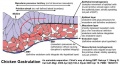 Chicken-gastrulation3.jpg 800 × 432; 71 KB
 Drosophila gastrulation.jpg 714 × 577; 163 KB
 Embryo left-right asymmetry pathway.jpg 800 × 458; 46 KB
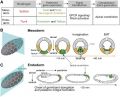
 Gastrulation molecular factors01.jpg 750 × 549; 105 KB
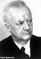 Hans Spemann.jpg 300 × 425; 18 KB
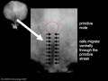
 Primitive streak cell migration.jpg 600 × 420; 92 KB
 Quail HH stage 2 fibronectin movement.jpg 729 × 256; 47 KB
 Rugh 006.jpg 1,000 × 656; 92 KB
 Rugh 063.jpg 1,200 × 755; 173 KB
 Rugh 071.jpg 737 × 1,000; 140 KB
 Stage6 bf02.jpg 638 × 800; 92 KB
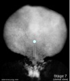
 Stage7 800x700px.jpg 690 × 800; 69 KB
 Stage7 axes.jpg 500 × 375; 9 KB
 Stage7 bf5.jpg 1,712 × 1,206; 875 KB
 Stage7 bf51.jpg 600 × 450; 96 KB
 Stage7 bf52.jpg 600 × 450; 114 KB
 Stage7 bf53.jpg 500 × 375; 74 KB
 Stage7 bf5a.jpg 1,024 × 721; 690 KB
 Stage7 bf5b.jpg 500 × 352; 216 KB
 Stage7 bf6.jpg 347 × 599; 69 KB
 Stage7 bf7.jpg 400 × 341; 22 KB
 Stage7 bf8.jpg 400 × 341; 18 KB
 Stage7 bf9.jpg 1,200 × 1,039; 501 KB
 Stage7 cloacal-oral-membranes.jpg 690 × 800; 70 KB
 Stage7 features.jpg 500 × 375; 9 KB
 Stage7 folding.jpg 500 × 375; 10 KB
 Stage7 intermediate-mesoderm.jpg 690 × 800; 67 KB
 Stage7 lateral-plate.jpg 690 × 800; 65 KB
 Stage7 mesoderm.jpg 690 × 800; 67 KB
 Stage7 notochord.jpg 690 × 800; 70 KB
 Stage7 paraxial-mesoderm.jpg 690 × 800; 69 KB
 Stage7 primitive streak labelled.jpg 500 × 375; 13 KB
 Stage7 primitive-streak-node.jpg 690 × 800; 69 KB
 Stage7 SEM4.jpg 450 × 332; 60 KB
 Stage7-bf1.jpg 600 × 676; 34 KB
 Stage7-bf2.jpg 800 × 579; 53 KB
 Stage7-bf3.jpg 523 × 600; 45 KB
 Stage7-bf4.jpg 800 × 695; 53 KB
 Stage7-sem1.jpg 814 × 600; 100 KB
 Stage7-sem2.jpg 590 × 800; 98 KB
 Stage7-sem3.jpg 1,000 × 664; 89 KB
 Stage7-sem4.jpg 937 × 595; 79 KB
 Stage7-sem5.jpg 1,000 × 735; 202 KB
 Stage8 bf4.jpg 600 × 449; 17 KB
 Stage8 sem2.jpg 822 × 1,000; 142 KB
 Stage8 sem3.jpg 1,000 × 709; 91 KB
 Trilaminar embryo cartoon.jpg 1,029 × 1,000; 107 KB
 Zebrafish ectodermal patterning model.jpg 1,280 × 662; 80 KB


