ANAT2341 Lab 3 - Week 4
| ANAT2341 Lab 3: Introduction | Week 3 | Week 4 | Abnormalities | Online Assessment | Group Project |
Week 4


Key Events of Human Development during the fourth week (week 4) following fertilization or Clinical week 6 (LMP).
These notes cover the fourth week of embryonic development, which is the beginning of organogenesis, (specific tissues and systems are beginning to differentiate) from the trilaminar embryo. With many parallel processes, descriptions begin to get complicated! Many of the described processes begin and extend over a broader range of time. Some developmental processes will be discussed later in the practical to simplify matters.
Ectoderm on the embryo surface undergoes segmentation: The central portion of the embryonic disc forms the neural plate, the edge of this plate forms neural crest and the remainder outside this forms the epitheium of the skin and other structures.
Neurogenesis
- Central Nervous System (CNS) - the neural plate undergoes morphological changes to form the primitive central nervous system (brain, spinal cord). An epithelial layer of cells which contributes all neural (brain, spinal cord, peripheral nervous system) and the external epithelium (surface layer of the skin) of the embryo. Neurogenesis begins towards the end of week 3, when the neural tissues separate from this germ cell layer.
- Peripheral Nervous System (PNS) - the neural crest cells in the body region migrate and spread to different regions of the embryo forming the PNS (dorsal root ganglia, sympathetic ganglia, enteric nervous system) and many other embryonic tissues. Neural crest cells in the head region form skeletal and other structures.
Pharyngeal Arches
In the head region, a series of ventral folds form under the brain in a rostral to caudal sequence, these are the pharyngeal arches.
Placodes
In the head region, ectoderm small patches form pairs of specialised placodes that eventually contribute to specific sensory components, cranial ganglia and the anterior pituitary (adenohypophysis).
Limb Buds
In the body region, limb buds form initially as ectoderm and mesoderm (somitic and somatic) components and are the "paddle-like" projections from the trunk which will form all the upper and lower limb components. (An overview of limb development will be covered in week 8).
Cardiogenesis
Within the embryo mesoderm, the heart tube and vascular development continues. Cardiogenesis will be covered in week 5, when septation begins.
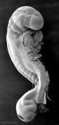
- Week 4 Stages
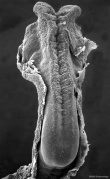
Stage 10
Stage 11
Stage 12
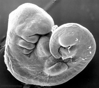
Stage 13


Neurogenesis

Developmental sequence: neural plate -> (day 18-19) neural groove -> neural tube -> Central Nervous System (brain and spinal cord)
- Central Nervous System (CNS) - the neural plate undergoes morphological changes to form the primitive central nervous system. An epithelial layer of cells which contributes all neural (brain, spinal cord, peripheral nervous system) and the external epithelium (surface layer of the skin) of the embryo. Neurogenesis begins towards the end of week 3, when the neural tissues separate from this germ cell layer.
- Peripheral Nervous System (PNS) - the neural crest cells in the body region migrate and spread to different regions of the embryo forming the PNS and many other embryonic tissues. Neural crest cells in the head region form skeletal and other structures.
Human Neuralation - Early Stages
The stages below refer to specific Carneigie stages of development.
- stage 8 (about 18 postovulatory days) neural groove and folds are first seen
- stage 9 the three main divisions of the brain, which are not cerebral vesicles, can be distinguished while the neural groove is still completely open
- stage 10 (two days later) neural folds begin to fuse near the junction between brain and spinal cord, when neural crest cells are arising mainly from the neural ectoderm
- stage 11 (about 24 days) the cranial neuropore (rostral, cephalic or anterior) closes within a few hours; closure is bidirectional, it takes place from the dorsal and terminal lips and may occur in several areas simultaneously. The two lips, however, behave differently.
- stage 12 (about 26 days) The caudal (posterior) neuropore takes a day to close
- the level of final closure is approximately at future somitic pair 31
- corresponds to the level of sacral vertebra 2
- stage 13 (4 weeks) the neural tube is normally completely closed.
![]()
Three primary brain vesicles develop initially due to the neural plate being broader at the cranial (brain) end than the narrower caudal (spinal cord) end. When the plate fuses to form a tube, these 3 initial expansions (vesicles) result.

Neural Patterning
Note that patterning signals are reused throughout the embryo to establish to pattern other parts of embryo (see limb development).
Rostrocaudal Patterning - Hox

|

|
| Fly wild-type head[1] | Fly antennapedia mutant head[1] |
This is the classic mutation that gave rise to the discovery of Hox genes and other genes related to body pattern formation. In this mutant during development the fly embryo incorrectly positioned where (antenna) should have be two legs (pedia)[1]. The discovery of this mutant in Walter Gehring's lab opened up the field of developmental genes and this field has been rewarded with the 1995 Nobel prize in Medicine.
Ventral Patterning - Sonic Hedghog


- SHH is secreted by the notochord, ventralizes the neural tube, inducing the floor plate and motor neurons.
- Regulation of patched by sonic hedgehog in the developing neural tube.[2] "The pattern of PTC expression suggests that Sonic hedgehog may play an inductive role in more dorsal regions of the neural tube than have been previously demonstrated. Examination of the pattern of PTC expression also suggests that PTC may act in a negative feedback loop to attenuate hedgehog signalling."
- Links: Sonic hedgehog
Pharyngeal Arches and Placodes
In the head region, two main components of head development form the pharyngeal arches and sensory placodes.
- Pharyngeal arches form a series of ventral folds under the brain in a rostral to caudal sequence. These arches will form most of the head and neck structures of the embryo and contain all three germ layers (ectoderm, mesoderm and endoderm).
- Small patches of ectoderm form pairs of specialised placodes that eventually contribute to specific sensory components, cranial ganglia and the anterior pituitary (adenohypophysis).


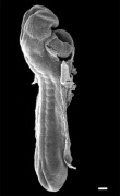
Embryo Stage 13
This EFIC movie shows sagittal sections through the Stage 13 week 4 to 5 human embryo. From your understanding of pdf development to the end of week 4 and general anatomy, how many internal features you can identify within this scan?
<html5media height="820" width="580">File:Stage 13 EFIC_S01.mp4</html5media>
Now have a look at the labelled movie.
Movies - Embryo Carnegie stage 13 - These are rotating animations based upon reconstruction of individual serial slice images of the stage 13 embryo.
| Central Nervous System | Gastrointestinal | Cardiovascular |

|

|

|

|

| ||
| A1L | A2L | A3L | A4L | A5L | A6L | A7L |

|

|

|

|

|

|

|
| B1L | B2L | B3L | B4L | B5L | B6L | B7L |

|

|

|

|

|

|

|
| C1L | C2L | C3L | C4L | C5L | C6L | C7L |

|

|

|

|

|

|

|
| D1L | D2L | D3L | D4L | D5L | D6L | D7L |

|

|

|

|

|

| |
| E1L | E2L | E3L | E4L | E5L | E6L | E7L |

|

|

|

|

|

|

|
| F1L | F2L | F3L | F4L | F5L | F6L | F7L |

|

|

|

|

|

|

|
| G1L | G2L | G3L | G4L | G5L | G6L | G7L |
Week 4 Movies
Note that many of the movies start in week 4 and continue on through later embryonic development.
Note that many of the movies start in week 4 and continue on through later embryonic development.
Ectoderm
|
|
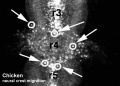
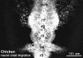
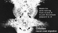
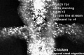
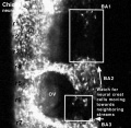
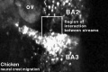
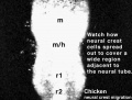
| Neural crest migration Chicken Head (movies overview) | |||||||||||||||||||||||||||
|---|---|---|---|---|---|---|---|---|---|---|---|---|---|---|---|---|---|---|---|---|---|---|---|---|---|---|---|
|
|
|
|
|
|
| |||||||||||||||||||||
Mesoderm
|
|
|
|
|
Endoderm
| Amniotic Cavity |
| Page | Play |
Embryo Stages and Events
| Day | Stage | Event |
| Stage 10 |  
Respiratory System Development
| |
| Heart begins to beat in humans by day 22-23, first functioning embryonic organ formed. | ||
| Stage 11 | 
Thyroid thyroid median endodermal thickening in the floor of pharynx Neural System Development rostral (or cephalic) neuropore closes within a few hours; closure is bidirectional, it takes place from the dorsal and terminal lips and may occur in two areas simultaneously. The two lips, however, behave differently. Optic ventricle appears | |
| Stage 12 | 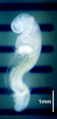
Pituitary Week 4 hypophysial pouch, Rathke’s pouch, diverticulum from roof GIT - Liver septum transversum forming liver stroma and hepatic diverticulum forming hepatic trabeculae PMID: 9407542 Neural System Development caudal neuropore takes a day to close (closure is approximately at future somitic pair 31/sacral vertebra 2) Neural System Development secondary neurulation begins Neural Crest Development cardiac crest, neural crest from rhombomeres 6 and 7 that migrates to pharyngeal arch 3 and from there the truncus arteriosus PMID: 17848161 Neural Crest Development vagal neural crest enter the foregut (20-25 somite stage) | |
| Stage 13 | 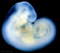 Neural System Development the neural tube is normally completely closed, ventricular system now separated from amniotic fluid. Neural crest at spinal level is segregating, and spinal ganglia are in series with the somites. Spinal cord ventral roots beginning to develop. PMID 3354839 Neural System Development the neural tube is normally completely closed, ventricular system now separated from amniotic fluid. Neural crest at spinal level is segregating, and spinal ganglia are in series with the somites. Spinal cord ventral roots beginning to develop. PMID 3354839
telencephalon cavity appears Liver epithelial cord proliferation enmeshing stromal capillaries PMID 9407542 Sense - Smell Crest comes from the nasal plates PMID 15604533 Skin 4 weeks - simple ectoderm epithelium over mesenchyme Skin 1-3 months ectoderm- germinative (basal) cell repeated division of generates stratified epithelium; mesoderm- differentiates into connective tissue and blood vessels |
| ANAT2341 Lab 3: Introduction | Week 3 | Week 4 | Abnormalities | Online Assessment | Group Project |
References
Glossary Links
- Glossary: A | B | C | D | E | F | G | H | I | J | K | L | M | N | O | P | Q | R | S | T | U | V | W | X | Y | Z | Numbers | Symbols | Term Link
Cite this page: Hill, M.A. (2024, May 8) Embryology ANAT2341 Lab 3 - Week 4. Retrieved from https://embryology.med.unsw.edu.au/embryology/index.php/ANAT2341_Lab_3_-_Week_4
- © Dr Mark Hill 2024, UNSW Embryology ISBN: 978 0 7334 2609 4 - UNSW CRICOS Provider Code No. 00098G