Introduction
This page links to movies of abnormalities of human development as detected by ultrasound. Ultrasound movies or images are now commonly seen by parents in the first trimester as a clinical non-invasive early method of embryo staging (ageing) and early detection of abnormalities. Later in the second and third trimester ultrasound is used as a method for checking fetal growth and detection of developmental abnormalities. Ultrasound can also be used with other techniques to locate both embryo and placenta for other prenatal tests.
The ultrasound technique can be used at any stage during pregnancy for embryo and placenta monitoring. Normal developmental ultrasound and features are listed on a separate page (More? Ultrasound) all content is for educational use only.
Some first trimester (GA 11-14 weeks) diagnostic measurements can include:
- Trisomies - Nuchal translucency (NT), nasal bone length (NBL), prenasal thickness (PT)
- Cardiovascular - tricuspid valve regurgitation (TR), "a"-wave pattern, DV PIV, S-wave (peak systolic velocity), D-wave (peak diastolic velocity), a-wave (atrial contraction in late diastole), time-averaged maximum velocity (TAMXV)
|
The ultrasound movies can be viewed in two ways:
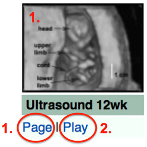
- Clicking either the movie image or "Page" text below the image opens a new page with both the movie and a more detailed text description of features. Embedded movies then play by clicking the play triangle icon lying over the movie image.
- Clicking on the "Play" link will open the MP4 movie version alone on a new page.
Please note that original Quicktime and Flash movie versions will now forward to the MP4 movie page. Also tt the bottom of this current page is further ultrasound information and links to internet ultrasound sites. Abnormal developmental ultrasound and features are listed on a separate page (More? abnormal ultrasound) all content is for educational use only.
Special thanks to Dr Andrew McLennan, Foetal Medicine Unit, Royal North Shore Hospital for many of the original video materials.
|
Ultrasound - Abnormalities
Some Recent Findings
- Subarachnoid space diameter in chromosomally abnormal fetuses at 11-13 weeks' gestation[1] "To examine the subarachnoid space diameters in chromosomally abnormal fetuses at 11-13 weeks' gestation. Stored three-dimensional (3D) ultrasound volumes of the fetal head at 11-13 weeks' gestation from 407 euploid and 88 chromosomally abnormal fetuses (Trisomy 21, n = 40; Trisomy 18, n = 19; Trisomy 13, n = 7; triploidy, n = 14; Turner syndrome, n = 8) were analyzed. The subarachnoid space diameters, measured in the sagittal and transverse planes of the fetal head, in relation to biparietal diameter (BPD) in each group of aneuploidies was compared to that in euploid fetuses. A total of 20 head volumes were randomly selected and all the measurements were recorded by two different observers to examine the interobserver variability in measurements. In euploid fetuses, the anteroposterior, transverse and sagittal diameters of the subarachnoid space increased with BPD. The median of the observed to expected diameters for BPD were significantly increased in triploidy and Trisomy 13 but were not significantly altered in Trisomy 21 and Trisomy 18 or Turner syndrome. In triploidy, the subarachnoid space diameters for BPD were above the 95th centile of euploid fetuses in 92.9% (13 of 14) cases. The intraclass reliability or agreement was excellent for all three subarachnoid space diameters. Most fetuses with triploidy at 11-13 weeks' gestation demonstrate increased subarachnoid space diameters." ventricular
|
References
- ↑ Ferreira C, Rouxinol-Dias AL, Loureiro T & Nicolaides K. (2019). Subarachnoid space diameter in chromosomally abnormal fetuses at 11-13 weeks' gestation. J. Matern. Fetal. Neonatal. Med. , 32, 2079-2083. PMID: 29338474 DOI.
Glossary Links
- Glossary: A | B | C | D | E | F | G | H | I | J | K | L | M | N | O | P | Q | R | S | T | U | V | W | X | Y | Z | Numbers | Symbols | Term Link
Cite this page: Hill, M.A. (2024, June 5) Embryology Ultrasound - Abnormal. Retrieved from https://embryology.med.unsw.edu.au/embryology/index.php/Ultrasound_-_Abnormal
- What Links Here?
- © Dr Mark Hill 2024, UNSW Embryology ISBN: 978 0 7334 2609 4 - UNSW CRICOS Provider Code No. 00098G