Uploads by Z3331556
From Embryology
This special page shows all uploaded files.
| Date | Name | Thumbnail | Size | Description | Versions |
|---|---|---|---|---|---|
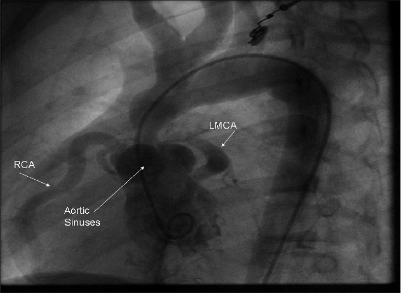
| 16:12, 12 October 2011 | Angiography image indicating Supravalvular aortic stenosis.jpg (file) |  |
133 KB | Original file name: APC-4-213-g001.jpg Left Ventricular Cine angiography picture demonstrating clearly the Supravalvular aortic stenosis in a 12 year old patient. Figure shows pigtail catheter in left ventricle with opacification of aortic sinuses, coron | 1 |
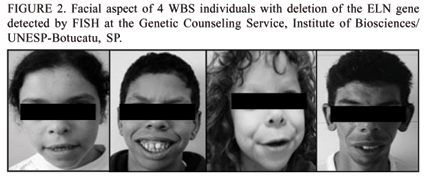
| 16:05, 12 October 2011 | Characteristic facial features of a child with Willams Syndrome.jpg (file) |  |
148 KB | Original file name: APC-4-213-g002.jpg Photograph of a child showing the characteristic facial features of Williams Syndrome This is an open-access article distributed under the terms of the Creative Commons Attribution-Noncommercial-Share Alike 3.0 Unp | 1 |
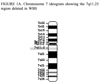
| 19:08, 5 October 2011 | Facial features of four individuals with Willams Syndrome.gif (file) |  |
45 KB | Original file name: En_a13fig02.gif Facial Characteristics of four individuals with Williams Syndrome http://www.scielo.br/scielo.php?pid=S0104-56872006000300013&script=sci_arttext&tlng=en#fig01b All the contents of this journal, except where otherwise | 1 |
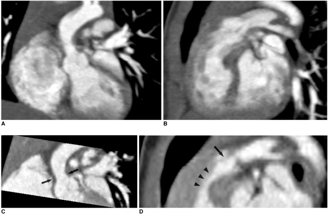
| 18:22, 5 October 2011 | Chromosome 7, indicating 7q11.23 region of Williams Syndrome.gif (file) |  |
11 KB | original file name: En_a13fig1a.gif Image of chromosome 7 indicating the region,7q11.23, that contains the genes which are deleted in Williams Syndrome http://www.scielo.br/scielo.php?pid=S0104-56872006000300013&script=sci_arttext&tlng=en#fig01b All th | 1 |
| 16:41, 17 September 2011 | Elastin (ELN) Gene on Chromosome 7.jpeg (file) | _Gene_on_Chromosome_7.jpeg) |
13 KB | Original file name: ELN.jpeg The ELN gene is located on the long (q) arm of chromosome 7 at position 11.23. More precisely, the ELN gene is located from base pair 73,442,426 to base pair 73,484,236 on chromosome 7. http://ghr.nlm.nih.gov/gene/ELN Gover | 1 |
| 14:52, 17 September 2011 | Scans of Supravalvular Aortic Stenosis and Pulmonary Stenosis.jpg (file) |  |
114 KB | original file name: Kjr-11-4-g009.jpg http://www.ncbi.nlm.nih.gov/pmc/articles/PMC2799649/ Fig. 9 Combo CT scan comprised of non-ECG-synchronized spiral scan with usual scan range (A, B) and prospective ECG-triggered sequential scan with narrow scan ran | 1 |
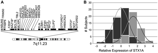
| 13:18, 11 August 2011 | Distribution of quantitative transcription of genes deleted in WS.png (file) |  |
90 KB | Journal.pone.0010292.g001.png http://www.plosone.org/article/info%3Adoi%2F10.1371%2Fjournal.pone.0010292 Figure 1. Distribution of quantitative transcription of genes deleted in WS. a: Map of genes commonly deleted in WS. Genes analyzed in this report a | 1 |