Uploads by Z3291643
From Embryology
This special page shows all uploaded files.
| Date | Name | Thumbnail | Size | Description | Versions |
|---|---|---|---|---|---|
| 05:24, 13 October 2011 | Imprint defect inheritance in Angelman Syndrome.png (file) |  |
21 KB | ===Reference=== Illustration by z3291643. Beginning six months after publication, I (3291643) grant the public the non-exclusive right to copy, distribute, or display the Work under a Creative Commons Attribution-Noncommercial-Share Alike 3.0 Unported | 1 |
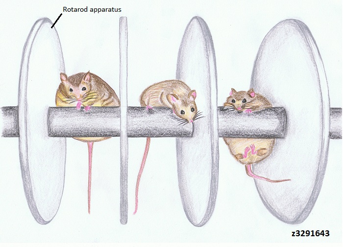
| 10:57, 3 October 2011 | Normal and Angelman Syndrome mice models.jpg (file) |  |
135 KB | Mouse in the center is the normal mouse with UBE3A present. Only the normal mouse exhibits the motor skills required to stay on the rotarod apparatus. In contrast, the mice on the left and right are UBE3A deficient, with the result of slight obesity and m | 1 |
| 10:56, 3 October 2011 | Extent of microcephaly in Angelman Syndrome patients.png (file) |  |
43 KB | Figure 1 Distribution of head circumference (HC) between 20 Angelman Syndrome patients with deletion and 20 Angelman Syndrome patients without deletion. ===Reference=== Reprinted by permission from Macmillan Publishers Ltd: [European Journal of Human Ge | 1 |
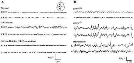
| 10:46, 3 October 2011 | Electroencephalography of Angelman Syndrome.jpg (file) |  |
33 KB | Electroencephalography (EEG) ===Reference=== <pubmed>9763493</pubmed> {{Template:2011 Student Image}} | 1 |
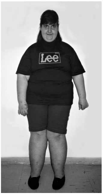
| 10:41, 3 October 2011 | Prader-Willi Syndrome patient.png (file) |  |
149 KB | A 12 year old Prada Willi Syndrome patient with the common characteristic of obesity All the contents of this journal, except where otherwise noted, is licensed under a Creative Commons Attribution License You are free: to Share — to copy, distribute | 1 |
| 10:39, 3 October 2011 | Angelman Syndrome patient.png (file) |  |
88 KB | All the contents of this journal, except where otherwise noted, is licensed under a Creative Commons Attribution License This is a photograph of a 4 year old Angelman syndrome (AS) patient, highlighting the typical facial characteristics of AS, such as b | 1 |
| 16:12, 2 October 2011 | Dr Charles Williams.jpg (file) |  |
101 KB | Photograph of Dr Charles Williams {{Template:2011 Student Image}} | 1 |
| 07:30, 1 October 2011 | Critical region of Angelman Syndrome on chromosome 15.png (file) |  |
356 KB | Prader Willi & Angelman's syndromes - probes Human chromosome highlighted by fluorescent probes that bind to specific sequences of DNA. In this FISH (fluorescence in situ hybridisation) study a probe (red) from the Prader Willi and Angelman's syndromes c | 1 |
| 04:45, 20 September 2011 | Extent of microcephaly in 20 AS patients with deletion and without deletion.png (file) |  |
43 KB | Figure 1 Distribution of head circumference (HC) between 20 AS patients with deletion and 20 AS patients without deletion. http://www.nature.com.wwwproxy0.library.unsw.edu.au/ejhg/journal/v7/n2/pdf/5200258a.pdf Reprinted by permission from Macmillan Pub | 1 |
| 20:31, 17 September 2011 | Role of UBE3A in dendritic spine neuronal synapses.png (file) |  |
94 KB | 1 | |
| 11:02, 15 September 2011 | UBE3A Ubiquitylation Pathway.png (file) |  |
168 KB | 1 | |
| 11:01, 15 September 2011 | Imprinting Inheritance in Familial AS.jpeg (file) |  |
5 KB | 1 | |
| 10:28, 25 August 2011 | A 12 year old PWS patient and a 4 year old AS patient.jpg (file) |  |
141 KB | A_12_year_old_PWS_patient_and_a_4_year_old_AS_patient.jpg All the contents of this journal, except where otherwise noted, is licensed under a Creative Commons Attribution License You are free: to Share — to copy, distribute and transmit the work to Re | 1 |
| 11:13, 15 August 2011 | UBE3A colocalizes with ASPM at the centrosome throughout mitosis.jpg (file) |  |
130 KB | Pone_0020397_g003.jpg UBE3A colocalizes with ASPM at the centrosome. (A) Indirect immunofluorescence of HEK293 cells at interphase and different phases of mitosis stained with antibodies against ASPM and UBE3A (anti-UBE3A-sc-8926). Note colocalization of | 1 |
| 14:06, 11 August 2011 | Abnormal cytokinesis and apoptosis in UBE3A knockdown cells.jpg (file) |  |
138 KB | 110811-abnormal_cytokinesis_and_apop._image.jpg (A) Note cells undergoing cytokinesis with elongated nuclear morphology giving rise to abnormal number of nuclei. An extended midbody (arrowheads) and micronuclei (arrows) can be seen in all the panels. ASP | 2 |