Uploads by S8600021
From Embryology
This special page shows all uploaded files.
| Date | Name | Thumbnail | Size | Description | Versions |
|---|---|---|---|---|---|
| 05:34, 22 February 2012 | Wilson1914-18.jpg (file) |  |
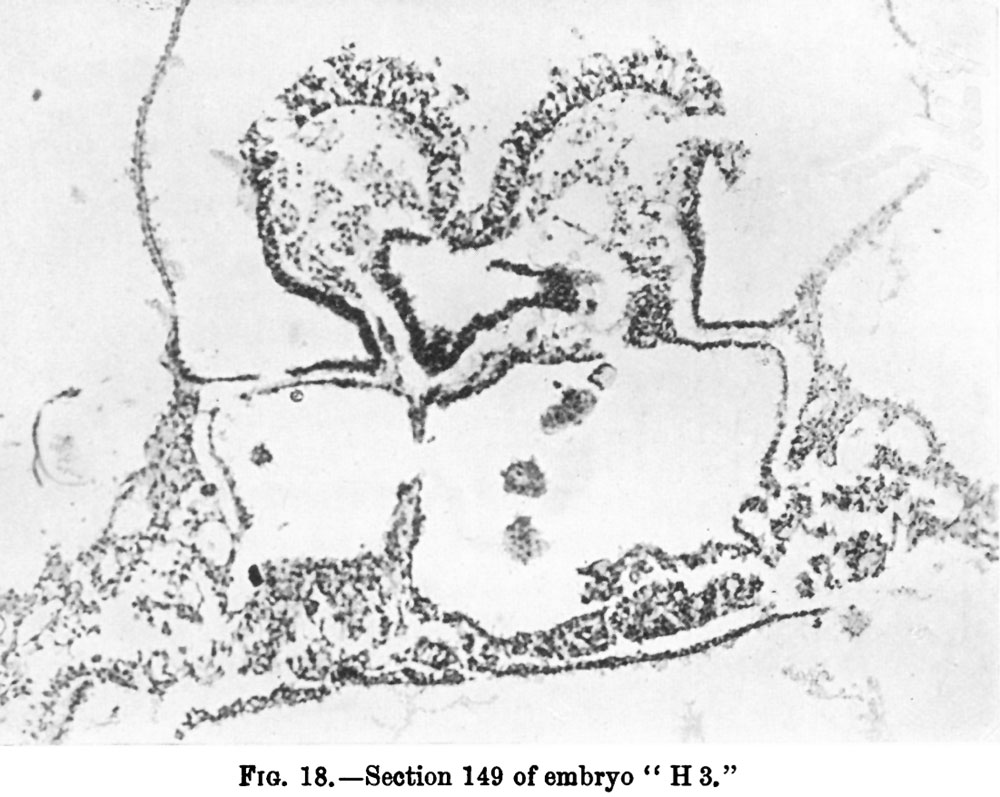
136 KB | ==Fig. 18.== {{Wilson1914}} | 1 |
| 05:33, 22 February 2012 | Wilson1914-17.jpg (file) |  |
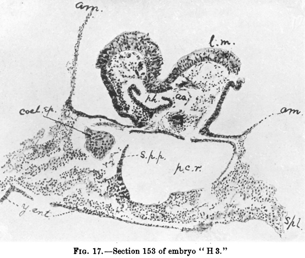
154 KB | ==Fig. 17.== {{Wilson1914}} | 1 |
| 05:33, 22 February 2012 | Wilson1914-16.jpg (file) |  |
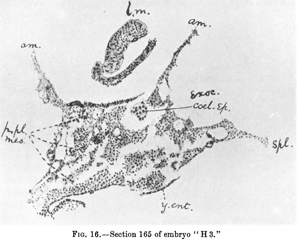
131 KB | ==Fig. 16.== {{Wilson1914}} | 1 |
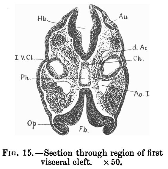
| 05:33, 22 February 2012 | Wilson1914-15.jpg (file) |  |
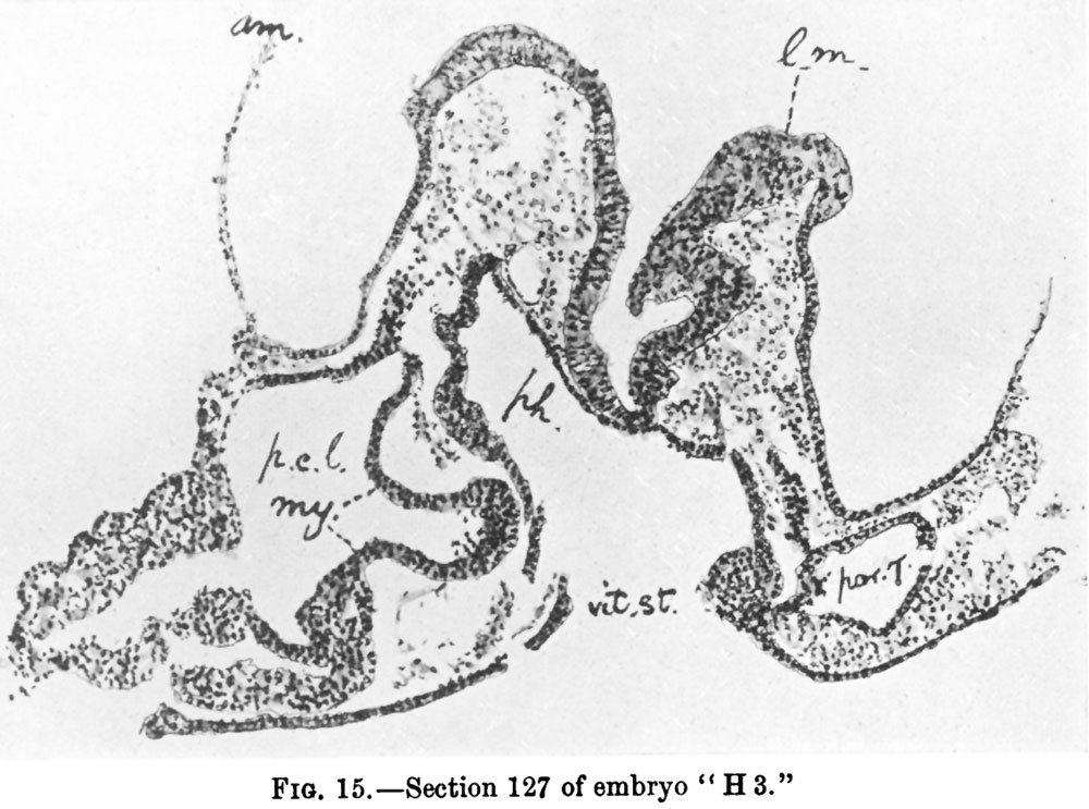
136 KB | ==Fig. 15.== {{Wilson1914}} | 1 |
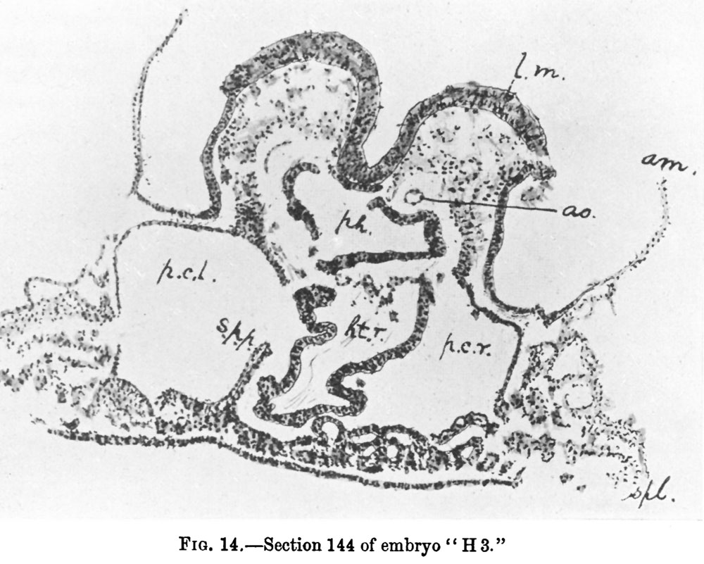
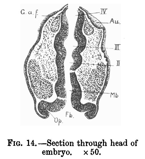
| 05:32, 22 February 2012 | Wilson1914-14.jpg (file) |  |
143 KB | ==Fig. 14.== {{Wilson1914}} | 1 |
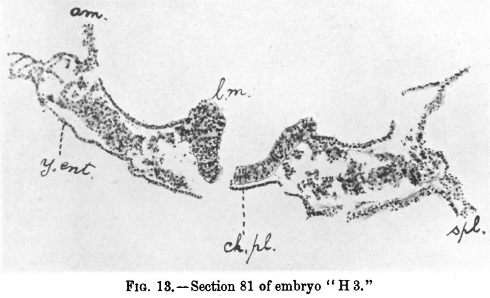
| 05:32, 22 February 2012 | Wilson1914-13.jpg (file) |  |
89 KB | ==Fig. 13.== {{Wilson1914}} | 1 |
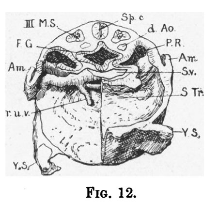
| 05:32, 22 February 2012 | Wilson1914-12.jpg (file) |  |
157 KB | ==Fig. 12.== {{Wilson1914}} | 1 |
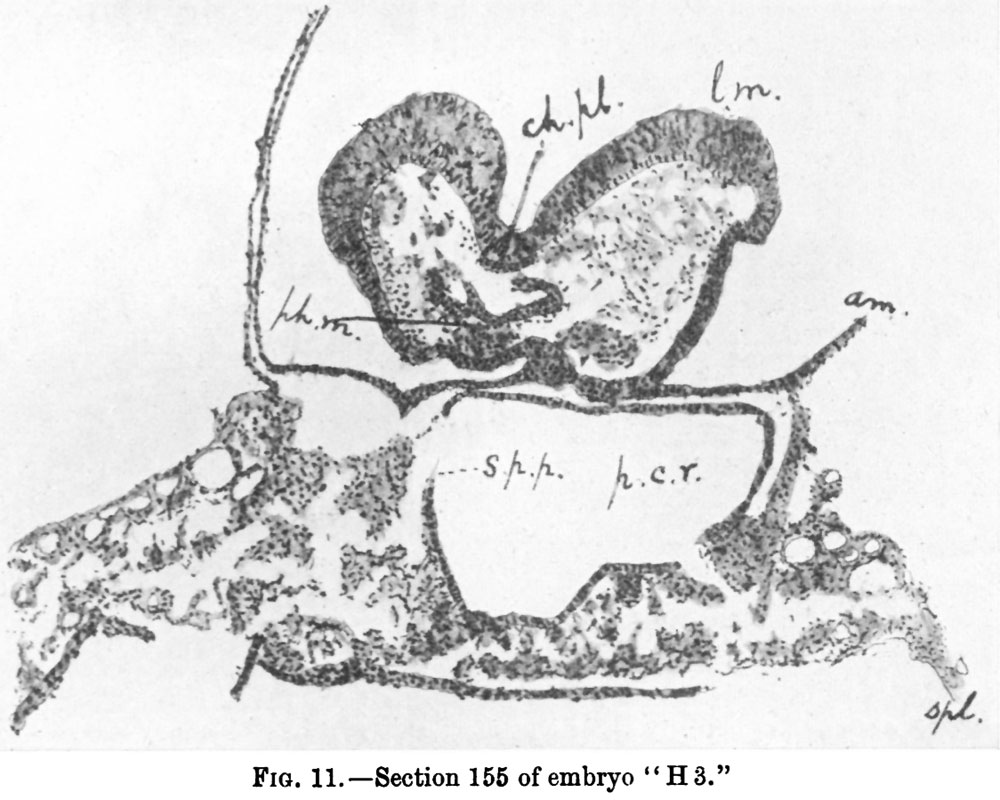
| 05:31, 22 February 2012 | Wilson1914-11.jpg (file) |  |
130 KB | ==Fig. 11.== {{Wilson1914}} | 1 |
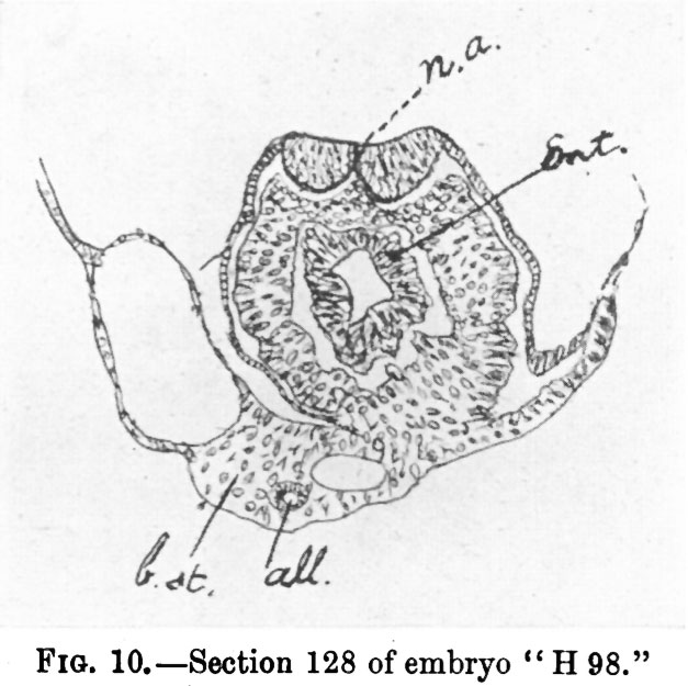
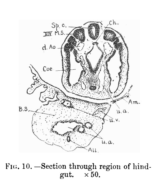
| 05:31, 22 February 2012 | Wilson1914-10.jpg (file) |  |
68 KB | ==Fig. 10.== {{Wilson1914}} | 1 |
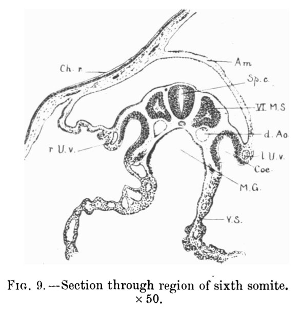
| 05:31, 22 February 2012 | Wilson1914-09.jpg (file) |  |
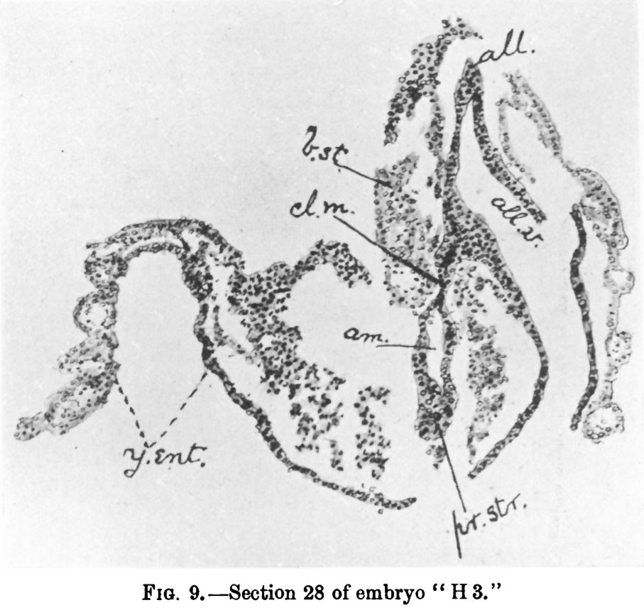
123 KB | ==Fig. 9.== {{Wilson1914}} | 1 |
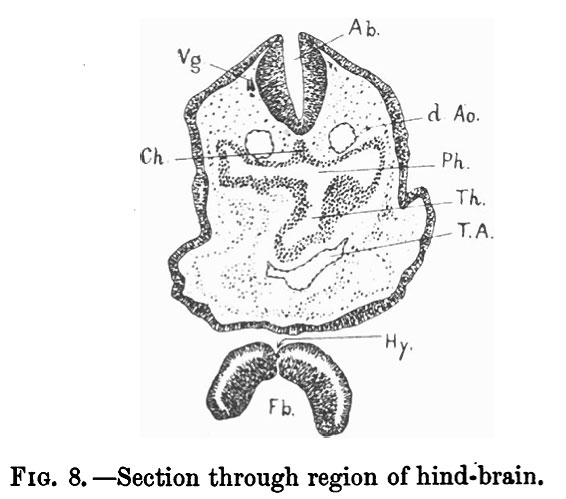
| 05:30, 22 February 2012 | Wilson1914-08.jpg (file) |  |
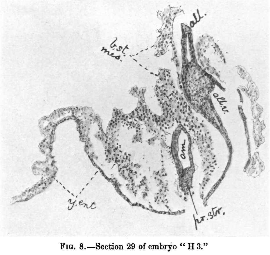
128 KB | ==Fig. 8.== {{Wilson1914}} | 1 |
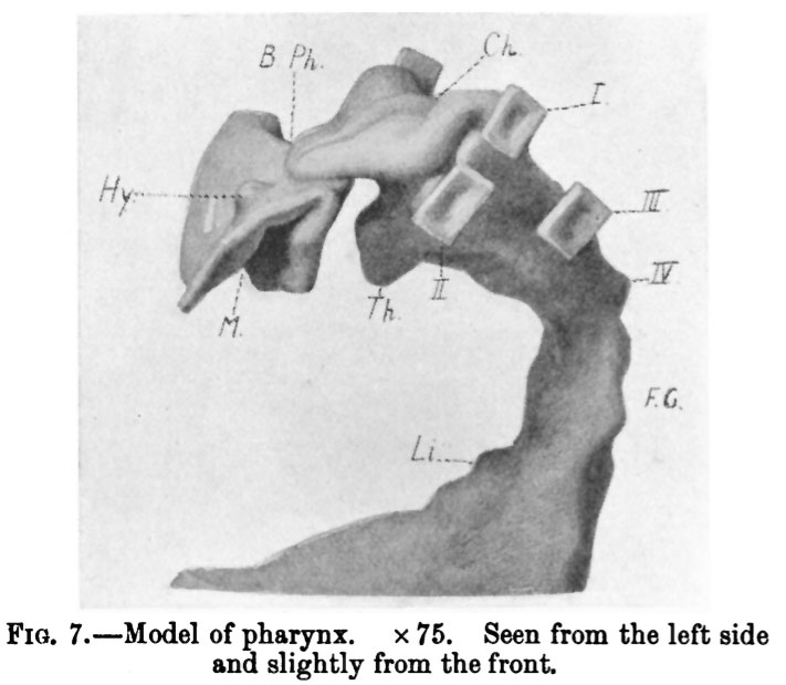
| 05:30, 22 February 2012 | Wilson1914-07.jpg (file) |  |
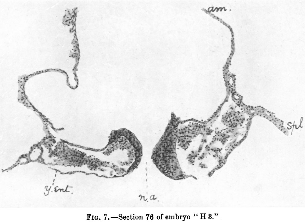
89 KB | ==Fig. 7.== {{Wilson1914}} | 1 |
| 05:30, 22 February 2012 | Wilson1914-06.jpg (file) |  |
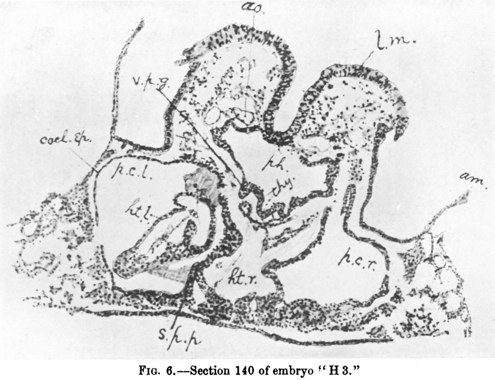
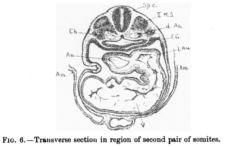
140 KB | ==Fig. 6.== {{Wilson1914}} | 1 |
| 05:29, 22 February 2012 | Wilson1914-05.jpg (file) |  |
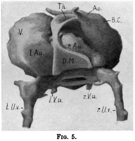
145 KB | ==Fig. 5.== {{Wilson1914}} | 1 |
| 05:29, 22 February 2012 | Wilson1914-04.jpg (file) |  |
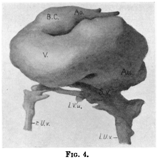
123 KB | ==Fig. 4.== {{Wilson1914}} | 1 |
| 05:29, 22 February 2012 | Wilson1914-03.jpg (file) |  |
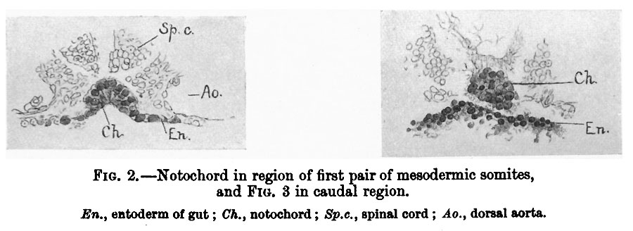
46 KB | ==Fig. 3.== {{Wilson1914}} | 1 |
| 05:28, 22 February 2012 | Wilson1914-02.jpg (file) |  |
62 KB | ==Fig. 2.== {{Wilson1914}} | 1 |
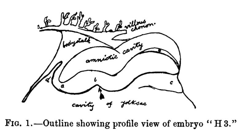
| 05:28, 22 February 2012 | Wilson1914-01.jpg (file) |  |
50 KB | ==Fig. 1.== {{Wilson1914}} | 1 |
| 00:53, 21 February 2012 | Human embryo tomography Carnegie stage 17.mov (file) | 898 KB | ==Human Embryo Carnegie Stage 17 Optical Projection Tomography (OPT) Model== * The embryo was staged as CS17, which is approximately 41 days of development. * The developing central nervous system (CNS) is clearly visible even in the external view of th | 1 | |
| 00:01, 21 February 2012 | Low 15.jpg (file) |  |
73 KB | 1 | |
| 00:00, 21 February 2012 | Low 14.jpg (file) |  |
60 KB | 1 | |
| 23:58, 20 February 2012 | Low 13.jpg (file) |  |
52 KB | 1 | |
| 23:53, 20 February 2012 | Low 12.jpg (file) |  |
43 KB | 1 | |
| 23:53, 20 February 2012 | Low 11.jpg (file) |  |
49 KB | 1 | |
| 23:53, 20 February 2012 | Low 10.jpg (file) |  |
61 KB | 1 | |
| 23:48, 20 February 2012 | Low 09.jpg (file) |  |
50 KB | 1 | |
| 23:48, 20 February 2012 | Low 08.jpg (file) |  |
48 KB | 1 | |
| 23:46, 20 February 2012 | Low 07.jpg (file) |  |
56 KB | 1 | |
| 23:45, 20 February 2012 | Low 06.jpg (file) |  |
59 KB | 1 | |
| 23:44, 20 February 2012 | Low 05.jpg (file) |  |
45 KB | 1 | |
| 23:42, 20 February 2012 | Low 04.jpg (file) |  |
40 KB | 1 | |
| 23:40, 20 February 2012 | Low 02.jpg (file) |  |
58 KB | 1 | |
| 23:38, 20 February 2012 | Low 01.jpg (file) |  |
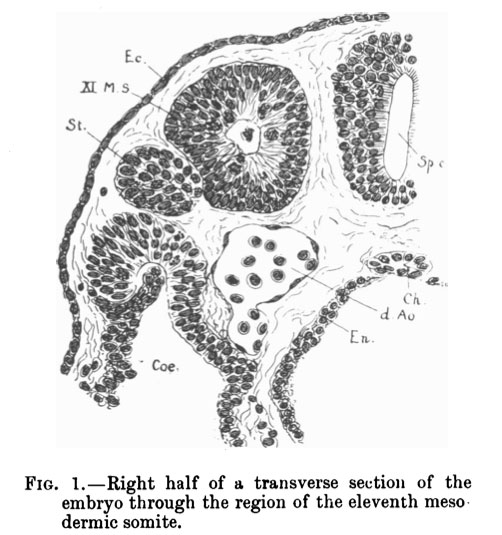
60 KB | ==Fig. 1. - Right half of a transverse section of the embryo through the region of the eleventh mesodermic somite== Ec., ectoderm; XI. M.S., mesodermic somite; St., segmental tubule; Coe., coelom; d.Ao., dorsal aorta; Sp.c., medullary canal; E | 1 |
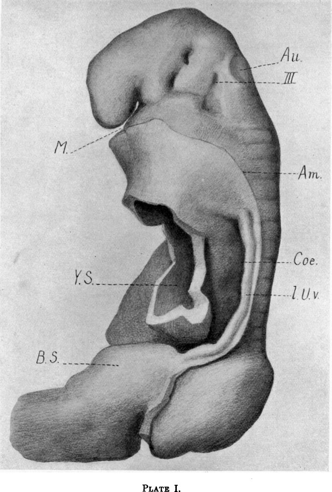
| 23:33, 20 February 2012 | Low plate 02.jpg (file) |  |
118 KB | ==Plate II== Model of the embryo. frontal view (enlarged 60 times). | 1 |
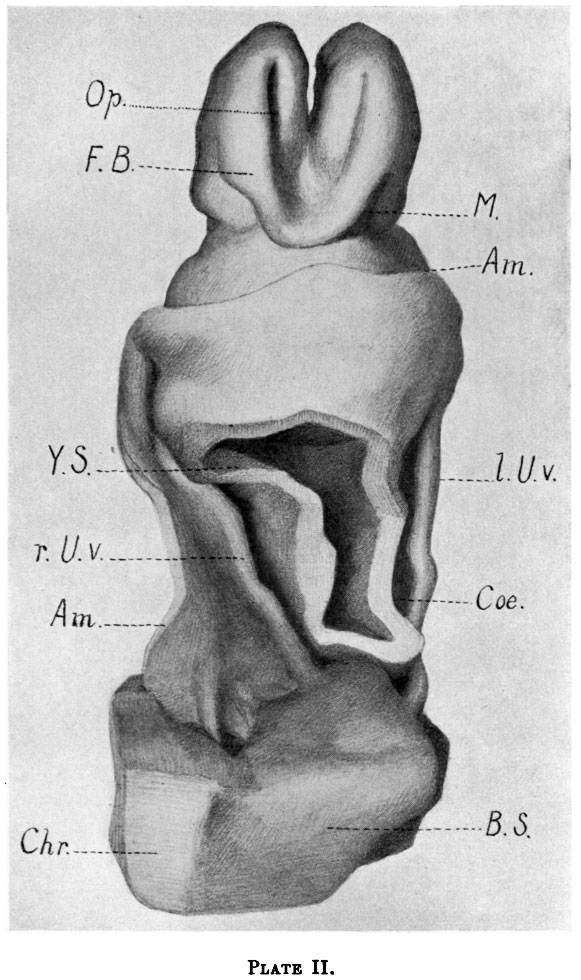
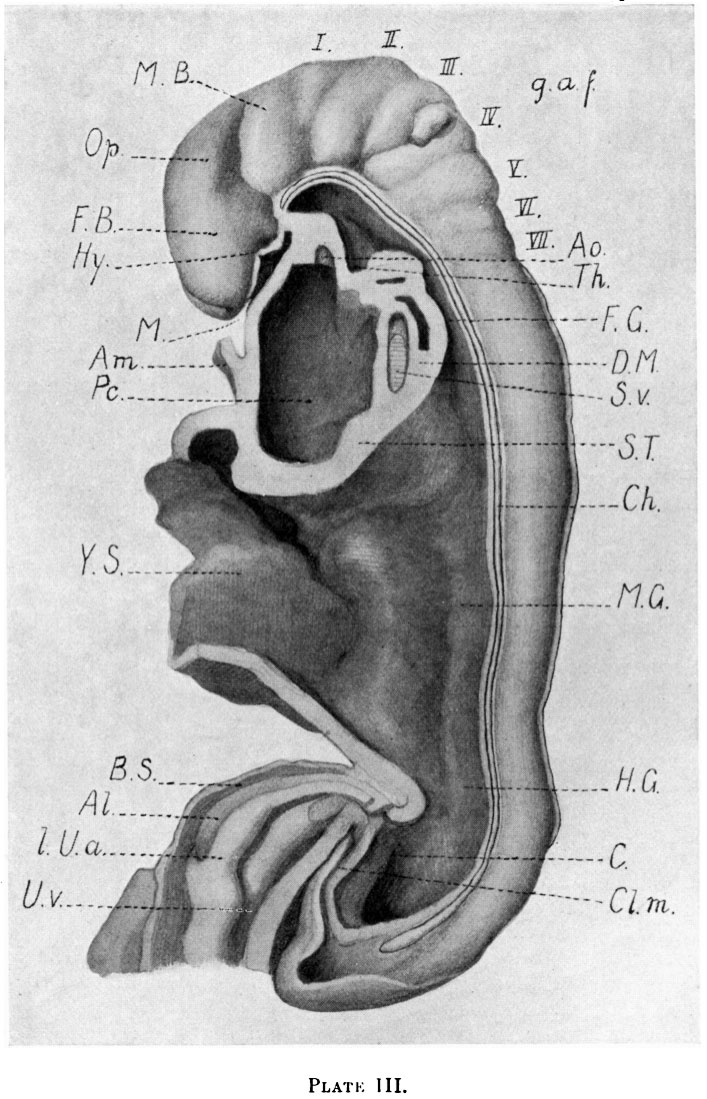
| 23:31, 20 February 2012 | Low plate 03.jpg (file) |  |
166 KB | ==Plate III== The same model in sagittal section, viewed from the left. | 1 |
| 23:28, 20 February 2012 | Low plate 01.jpg (file) |  |
137 KB | ==Plate I== Model of the embryo, left side (enlarged 60 times). | 1 |
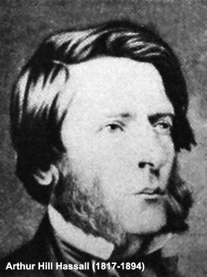
| 14:06, 20 February 2012 | Arthur Hill Hassall.jpg (file) |  |
25 KB | 2 | |
| 13:22, 20 February 2012 | Mouse thymus development 03.jpg (file) |  |
260 KB | ===Reference=== <pubmed>22087235</pubmed>| [http://www.plosone.org/article/info%3Adoi%2F10.1371%2Fjournal.pone.0026795 PLoS One.] © 2011 Wei, Condie. This is an open-access article distributed under the terms of the Creative Commons Attribution Licens | 1 |
| 13:18, 20 February 2012 | Mouse thymus development 02.jpg (file) |  |
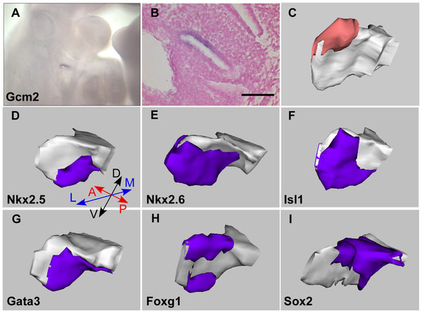
50 KB | ==Expression of Transcription Factors in the pharyngeal region at E10.5== 3D reconstructions of the 3rd pouch reveal differentially regionalized expression patterns of Nkx2-5, Nkx2-6, Isl1, Gata3, Foxg1 and Sox2 at E10.5 days. (A–C) Expression pattern | 1 |
| 13:14, 20 February 2012 | Mouse thymus development 01.jpg (file) |  |
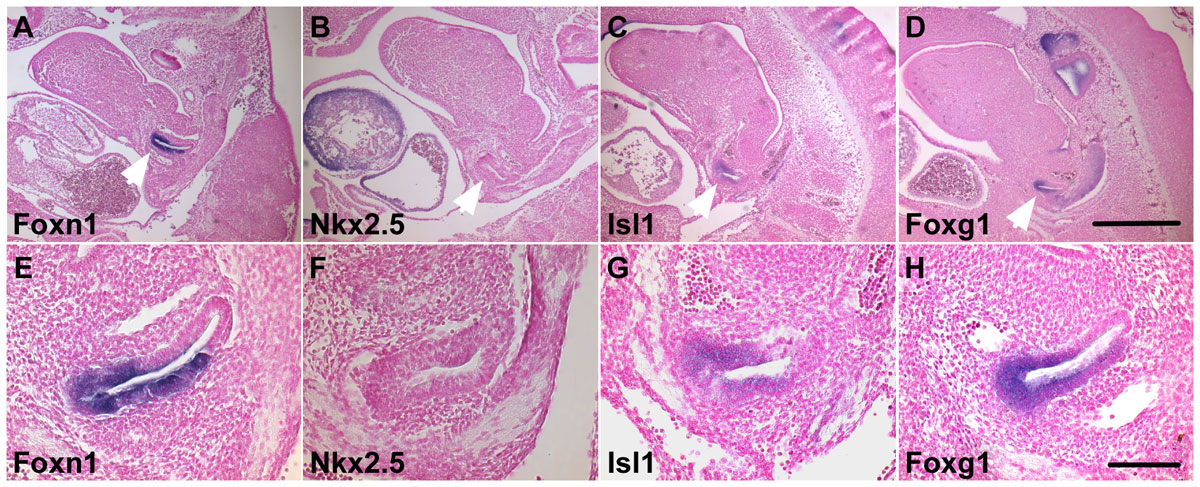
69 KB | ==Expression of Transcription Factors in the pharyngeal region at E9.5 and E10.5== Expression of Nkx2-5, Nkx2-6, Isl1, Gata3, Foxg1 and Sox2 in the pharyngeal region at E9.5 (20–23 somites) and E10.5 (30–33 somites) as detected by whole-mount in situ | 1 |
| 08:26, 16 February 2012 | No smoking sign.jpg (file) |  |
18 KB | ==No Smoking Sign== | 1 |
| 23:34, 15 February 2012 | Keibel Mall 066-071.jpg (file) |  |
58 KB | 1 | |
| 23:33, 15 February 2012 | Keibel Mall 078.jpg (file) |  |
18 KB | 1 | |
| 23:33, 15 February 2012 | Keibel Mall 077.jpg (file) |  |
33 KB | 1 | |
| 23:33, 15 February 2012 | Keibel Mall 075-076.jpg (file) |  |
40 KB | 1 | |
| 23:32, 15 February 2012 | Keibel Mall 074.jpg (file) |  |
41 KB | 1 | |
| 23:32, 15 February 2012 | Keibel Mall 073.jpg (file) |  |
26 KB | 1 | |
| 23:32, 15 February 2012 | Keibel Mall 072a.jpg (file) |  |
9 KB | 1 | |
| 23:32, 15 February 2012 | Keibel Mall 072.jpg (file) |  |
29 KB | 1 | |
| 23:31, 15 February 2012 | Keibel Mall 070-071.jpg (file) |  |
45 KB | 1 |