Respiratory System - Carnegie Stage 22
Introduction
| <html5media height="280" width="290">File:Stage22_GIT3d.mp4</html5media> |
Respiratory SystemBased upon a serial reconstruction from individual embryo slice images (27 mm Human embryo, Carnegie Stage 22 approximate day 56). Note the relative size and position of individual structures and organs at this early stage of development. The respiratory system is endodermal in origin, initially "budding off" the foregut during week 3. This bud forms the respiratory diverticulum, at the level of the glottis between the adult oesophagus and trachea. It continues to bud in week 4, forming a pair of lung buds.
|
| Virtual Slides | |||||||||||
|---|---|---|---|---|---|---|---|---|---|---|---|
|
|
|
- Stage 22 Histology Links: Both lungs overview | Lung and pleural cavity | Right and Left Main Bronchi | Lung Bronchi
| Section | Name | Description |
|---|---|---|

|
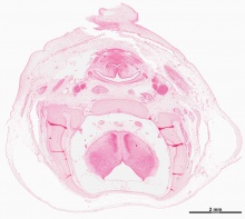
A5L | Bridge of nose.
R and L olfactory bulbs from forebrain. |

|
A6L | Nose. Nasal septum. Nasal capsule.
Olfactory epithelium lining roof of nasal cavity. Orbital part of the developing sphenoid bone (intramembranous ossification). |

|
A7L | Conchae. Nasal capsule and septum. |

|
B1L | Conchae. Optic nerve. |

|
B2L | Description |

|
B3L | Perpendicular plate of ethmoid cartilage. Adenohypophysis. Neurohypophysis. Ant. and post. walls of hypopophysial fossa. Lesser wings of sphenoid cartilage. Internal carotid arteries. |

|
B4L | Dorsum of tongue. Oropharynx communicating with naso-pharynx (cf. B3L - palatal processes not fused). |

|
B5L | Tongue with palatal processes at either side. Transverse (intrinsic) muscle of tongue.
Pharyngotympanic tubes. |

|
B6L | Tongue with transverse muscle, genioglossus muscle (medial) and hyoglossus muscle (lateral).
Palatal processes. Meckel's cartilage. Note teeth enamel organs (dark masses at sides of tongue attachment). |

|
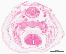
B7L | Transverse caudal pharynx. epiglottis. Hyoid musculature. Pharyngeal constrictor muscle. Submandibular gland. |

|
C1L | Pharynx. Pharyngeal constrictor muscle. laryngeal caecum (ventral). Arytenoid swellings in contact.
Thyroid cartilage laminae (anterolateral), with superior horns (posterolateral). Hyoid cartilage. Internal jugular veins. "Muz's cheshire cat" |

|
C2L | Pharynx. Thyroid cartilage. Smaller laryngeal caecum (cf.C1). Carotid neurovascular bundle. |

|
C3L | Pharynx with its inferior constrictor muscle. Glottis region. |

|
C4L | Oesophagus with muscle layer and trachea with thyroid gland laterally.
Common carotid arteries. Vagus nerve. Internal jugular veins. (Section damaged) |

|
C5L | Oesophagus, smaller than in C4. Trachea.
Thyroid gland (isthmus). Clavicle. Small dark masses near posterolateral borders of thyroid gland are the parathyroid glands from the caudal part of 3rd pharyngeal pouch. |

|
C6L | Trachea.
Clavicles. Dark connecting stalk between parathyroid gland and thymus (rostral end of 3rd pharyngeal pouch). Common carotid artery. |

|
C7L | Trachea.
Oesphagus. Apex of R lung in pleural cavity Sternum. Thymus gland. L brachiocephalic vein. Brachiocephalic trunk. |

|
D1L | Lungs. Visceral and parietal pleurae. Pleural cavities.
Sternum. Thymus. Other contents of superior mediastinum. |

|
D2L | Lungs. |

|
D3L | Tracheal bifurcation. |

|
D4L | Right primary bronchus (torn) and right superior lobe bronchus.
Left primary bronchus. Left and right pulmonary arteries. Ribs joining to sternum. |

|
D5L | R, L primary bronchi. R anterior and posterior segmental bronchi coming off R superior lobe bronchus. L, R pulmonary arteries. Hilar attachments of lungs to mediastinal tissues - note extent of R, L pleural cavities. |

|
D6L | R, L primary bronchi (note left still has not branched). R pulmonary artery. |

|
D7L | R, L primary bronchi: note distinct horizontal course of L, vertical course of R, L pulmonary veins (L empty). R pulmonary artery. |

|
E1L | Pulmonary veins. Azygos, hemiazygos veins. Ribs. Intercostal muscles. |

|
E2L | Pulmonary veins. Azygos, hemiazygos veins. Ribs. Intercostal muscles. |

|
E3L | R dome of diaphragm. R long middle and inferior lobes. L long superior and inferior lobes. Xiphoid process.
Liver. |

|
E4L | Diaphragm (note costal attachment). R lung inferior lobe.
Inferior vena cava, dorsal to diaphragm. |

|
E5L | Inferior lobes of lungs.
Diaphragm with sternal attachments. Inferior vena cava, now ventral to diaphragm (vena caval foramen). Liver. |

|
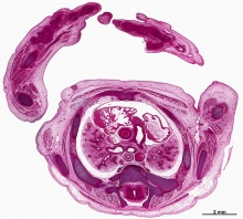
E6L | Liver. Thoracic aorta. Large adrenal glands. |

|
E7L | Lumbar diaphragm. Thoracic aorta. Note ribs 11 and 12 on L and three layers of abdominal muscles extending ventrally. |

|
F1L | Lumbar diaphragm. Thoracic aorta. |

|
F2L | Attachment of lumbar diaphragm near L 1 on R with psoas muscle dorsal to it. Note abdominal aorta giving rise to superior mesenteric artery. |
All Sections

|

|

|

|
|
|
|
| A1L | A2L | A3L | A4L | A5L | A6L | A7L |
|
|
|
|
|
|
|
|
| B1L | B2L | B3L | B4L | B5L | B6L | B7L |
|
|
|
|
|
|
|
|
| C1L | C2L | C3L | C4L | C5L | C6L | C7L |
|
|
|
|
|
|
|
|
| D1L | D2L | D3L | D4L | D5L | D6L | D7L |
|
|
|
|
|
|
|
|
| E1L | E2L | E3L | E4L | E5L | E6L | E7L |
|
|
|

|

|

|

|

|
| F1L | F2L | F3L | F4L | F5L | F6L | F7L |

|

|

|

|

|

|

|
| G1L | G2L | G3L | G4L | G5L | G6L | G7L |
Glossary Links
- Glossary: A | B | C | D | E | F | G | H | I | J | K | L | M | N | O | P | Q | R | S | T | U | V | W | X | Y | Z | Numbers | Symbols | Term Link
Cite this page: Hill, M.A. (2024, May 4) Embryology Respiratory System - Carnegie Stage 22. Retrieved from https://embryology.med.unsw.edu.au/embryology/index.php/Respiratory_System_-_Carnegie_Stage_22
- © Dr Mark Hill 2024, UNSW Embryology ISBN: 978 0 7334 2609 4 - UNSW CRICOS Provider Code No. 00098G