File:ZUltrasound Image of Fetal Aorta.jpg
ZUltrasound_Image_of_Fetal_Aorta.jpg (640 × 480 pixels, file size: 47 KB, MIME type: image/jpeg)
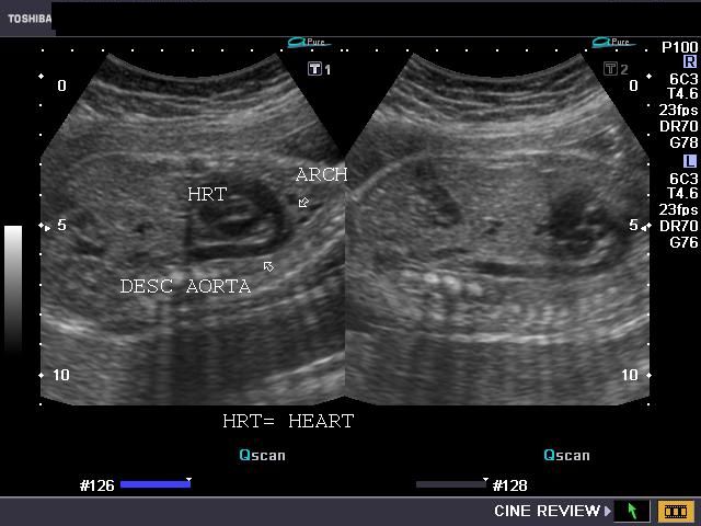
What am I looking at?
This is a 2D ultrasound image of a fetal chest in sagittal section. Marked with arrows on the image are the fetal heart, aortic arch and descending aorta.
Copyright
Image obtained at: http://www.ultrasound-images.com/fetal-chest.htm
Author (content provider): Dr. Joe Antony
This image is not classed as a public domain image, but has been reproduced here with the kind permission of Dr. Joe Antony, who controls the website Ultrasound Images, "a free gallery of high-resolution, ultrasound, color Doppler and 3D images". Correspondence attesting to this fact will be cheerfully provided upon request.
- Note - This image was originally uploaded as part of an undergraduate science student project and may contain inaccuracies in either description or acknowledgements. Students have been advised in writing concerning the reuse of content and may accidentally have misunderstood the original terms of use. If image reuse on this non-commercial educational site infringes your existing copyright, please contact the site editor for immediate removal.
File history
Click on a date/time to view the file as it appeared at that time.
| Date/Time | Thumbnail | Dimensions | User | Comment | |
|---|---|---|---|---|---|
| current | 19:10, 10 September 2010 | | 640 × 480 (47 KB) | Z3252833 (talk | contribs) | ===What am I looking at?=== This is a 2D ultrasound image of a fetal chest in sagittal section. Marked with arrows on the image are the fetal heart, aortic arch and descending aorta. ===Image Copyright Information=== Image obtained at: http://www.ul |
You cannot overwrite this file.
File usage
The following page uses this file: