File:Wyndham1943 fig05.jpg
From Embryology
Size of this preview: 511 × 599 pixels. Other resolution: 632 × 741 pixels.
Original file (632 × 741 pixels, file size: 120 KB, MIME type: image/jpeg)
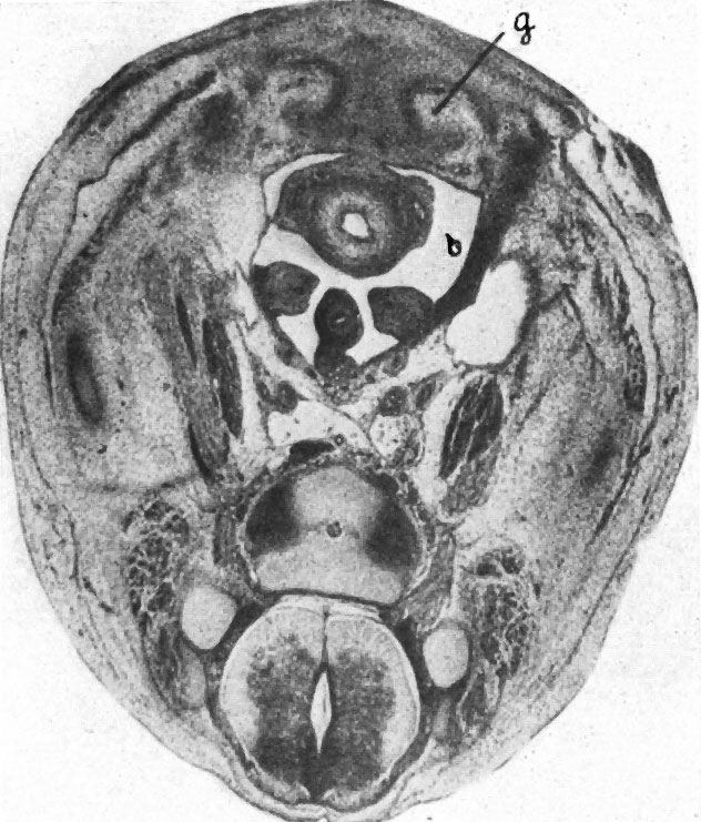
Fig. 5. Transverse section through caudal region of a 20 mm embryo H 304
x 20. b, plica. inguinalis; g, pubic anlage.
| Historic Disclaimer - information about historic embryology pages |
|---|
 |
- Links: Plate 1 | Fig 1 | Fig 2 | Fig 3 | Fig 4 | Plate 2 | Fig 5 | Fig 6 | Fig 7 | Fig 8 | Plate 3 | Fig 9 | Fig 10 | Fig 11
Reference
Wyndham NR. A morphological study of testicular descent. (1943) J Anat., 77(2):179-188.3. PMID 17104926
Cite this page: Hill, M.A. (2024, May 19) Embryology Wyndham1943 fig05.jpg. Retrieved from https://embryology.med.unsw.edu.au/embryology/index.php/File:Wyndham1943_fig05.jpg
- © Dr Mark Hill 2024, UNSW Embryology ISBN: 978 0 7334 2609 4 - UNSW CRICOS Provider Code No. 00098G
File history
Click on a date/time to view the file as it appeared at that time.
| Date/Time | Thumbnail | Dimensions | User | Comment | |
|---|---|---|---|---|---|
| current | 11:12, 5 September 2015 | | 632 × 741 (120 KB) | Z8600021 (talk | contribs) |
You cannot overwrite this file.
File usage
The following 2 pages use this file: