File:West09.jpg
Original file (403 × 806 pixels, file size: 44 KB, MIME type: image/jpeg)
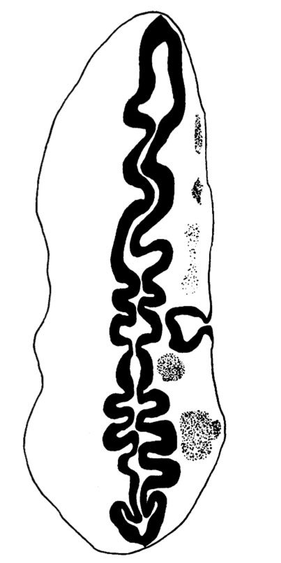
Fig. 9. Otocyst and Neural Tube
Neural Tube
The neuromeres resemble closely those described by other observers, but they show more clearly than in other specimens (Text-fig. 9); there are ten of these segments which involve the lateral wall of the brain. The 2nd, 4th, 6th and 7th involve also the flor, to which they give an irregular outline;itisthe grooves between the projections of the floor caused by these neuromeres, and especially the 4th, which are referred to by Thompson (1907), Bremer (1906), and van den Broek (1911),as having given rise to a difficulty in their interpretation, and van den Broek suggested that they might have some association with the Vth and VIlIth ganglia, which is indirectly true in view of the relation of these ganglia to the neuromeres which are responsible for the production of these grooves. The ganglion of the trigeminal nerve lies lateral to segments 2 and 3; the acoustico-facial ganglion is lateral to segment 4; the otocyst is lateral to segment 5; the glossopharyngeal ganglion is lateral to segment 7, and the vagus ganglion is lateral to segments 7 and 8 (Text-figs. 8 and 9), but the glossopharyngeal and vagus ganglia are difficult to define and appear only as rather diffuse clumps of cels.The first somite is opposite the 8th neuromere, the 2nd somite is opposite the 9th neuromere, and the 3rd is opposite the 9th and 1oth neuromeres and the intervening groove.
Otocyst
This is a hollow, oval vesicle, measuring 0-18 mm. in a craniocaudal direction, and 0.14 mm. from side to side; itisthus of much the same size as the otocyst of embryos described by Davis (1923), Watt F (1915), and van den Broek (1911). The sac is open on to the exterior (Text-fig. 9) through an orifice that measures 0*02mm. in diameter, and which is, as in van den Broek's specimen, nearer the cranial pole than thecaudalpoleofthevesicle. From the ventromedial surface of the sac there projects forwards a hollow diverticulum for a distance of 0 04 mm. Waterston (1914) shows a rather similar diverticulum, but in his specimen the-projection is in a dorsomedial direction.The picture presented by my specimen is very like Arey's (1930) Fig. 488, of a section through a 6-mm. pig embryo, and he refers (p. 492) to the ventromedial diverticulum as "a median outpocketing where the endolymph duct attaches"; yet though the vesicle in this pig embryo antinomy specimen are so alike,mine must be much the younger,and it is stil open to the outside, and it is not til after the closure of the opening that there arises, from the point of closure, the endolymph duct which then shifts in a ventromedial direction,so that however tempting it maybe to interpret it otherwise,it is probable that this diverticulum is a folding due to shrinkage. The vesicle as a whole overlies the 5th neuromere and corresponds almost exactly with it in size; the diverticulum referred to projects towards the groove between the 4th and 5th neuromeres and towards the acoustico-facial ganglion; at the more cranial part of the vesicle, at the level of its opening to the exterior, the relationship between the vesicle and the acoustico-facial ganglion is a very close one, the ganglion appearing to be moulded round the ventral surface of the vesicle. The structure of the vesicle is essentially similar to that of the adjacent wall of the hindbrain and it shows the same arrangement of mantle and marginal zones.
| Historic Disclaimer - information about historic embryology pages |
|---|
 |
- Links: Fig 1 | Fig 2 | Fig 3 | Fig 4 | Fig 5 | Fig 6 | Fig 7 | Fig 8 | Fig 9 | Fig 10 | Fig 11 | Plate 1 | Plate 1 Fig 1 | Plate 1 Fig 2 | Plate 1 Fig 3 | Plate 1 Fig 4 | Plate 1 Fig 5 | Plate 1 Fig 6
Reference
West CM. A human embryo of twenty-five somites. (1937) J. Anat., 71(2): 169-200.1. PMID 17104635
Cite this page: Hill, M.A. (2024, April 26) Embryology West09.jpg. Retrieved from https://embryology.med.unsw.edu.au/embryology/index.php/File:West09.jpg
- © Dr Mark Hill 2024, UNSW Embryology ISBN: 978 0 7334 2609 4 - UNSW CRICOS Provider Code No. 00098G
File history
Click on a date/time to view the file as it appeared at that time.
| Date/Time | Thumbnail | Dimensions | User | Comment | |
|---|---|---|---|---|---|
| current | 15:22, 28 January 2012 | | 403 × 806 (44 KB) | S8600021 (talk | contribs) | ==Fig. 9 == {{Template:West1937}} {{Historic Disclaimer}} {{Historic Papers}} ===Reference=== <pubmed>17104635</pubmed>| [http://www.ncbi.nlm.nih.gov/pmc/articles/PMC1252340 PMC1252340] Category:Carnegie Stage 12 |
You cannot overwrite this file.
File usage
The following page uses this file: